Aliasghar Yarmohamadi, MD;
*Ali Reza Akhavan Rezayat, MD;
*Bahram Memar, MD;
†Hamid Reza Rahimi, MD,
PhD Cand.
§*Department of Urology, Ghaem Educational General Hospital, School of Medicine, Mashhad University of Medical Sciences, Mashhad, Iran; †Surgical Oncology Research Center, Imam Reza Hospital, Faculty of Medicine, Mashhad University of Medical Sciences, Mashhad, Iran; §Student Research Committee,Department of Modern Sciences and Technologies, School of Medicine, Mashhad University of Medical Sciences, Mashhad, Iran
Cite as: Can Urol Assoc J 2014;8(3-4):e282-6. http://dx.doi.org/10.5489/cuaj.1207 Published online April 14, 2014.
Abstract
A black kidney has 3 major differential diagnoses: hemosiderosis, lipofuscin pigment and melanotic renal cell carcinoma. Excluding lipofuscin, the other 2 are accompanied by an abnormal renal func-tion. We report on a 25-year-old man who intended to donate a kidney to his cousin. On the operating room table when we incised the left flank region and exposed the kidney, we found a firm and black kidney so the operation was cancelled due to potential vas-cular injuries. Days after the incomplete procedure, we reviewed the donor’s biochemistry and imaging to reassess his renal func-tion, but the results showed quite normal renal function again. The result of Ham test was also negative. Two weeks later, we began the operation, removed the same left kidney and found that it was in the same conditions as it was before. We took the opportunity to send needle biopsies of the kidney for histopathologic analy-sis. The analysis showed a melanotic kidney without pathological changes in glomeruli and interstitium and vessels. A black kidney may result in hemosiderin, lipofuscin or melanin deposits in the kidney, which can confirm the diagnosis; however, special tests for underlying disease and renal function should be considered. Some causes of black kidney lead to abnormal function, but our patients’s kidney returned to normal.
Introduction
Kidney transplantation is preferred for patients with end-stage renal disease (ESRD) or chronic kidney disease
(CKD).1,2 Organ sources are limited to live and cadaveric
human donors. The first kidney transplant in Iran was in
1967; in 2002, Iran was ranked fifth in kidney transplants.2
Mashhad University of Medical Sciences, in north east part of Iran, started performing kidney transplants in 1988. So far, as one of the pioneering universities in the region, it has recorded about 3000 kidney transplants and has become the transplant centre for our neighbouring states.
Black kidney has 3 major differential diagnosis: hemosid-erin deposits,3 lipofuscin pigment4 and melanotic renal cell
carcinoma (RCC); all cases are very rare.5 The differential
diagnosis for melanin in cancer cell usually leads to the diagnosis of melanoma, melanotic neurofibroma, melanot-ic dermatofibrosarcoma protuberans, pheochromocytoma, basal cell carcinoma, seborrhetic keratosis or breast cancer.6
Hemosiderosis is a form of iron overload disorder result-ing in the accumulation of hemosiderin in different organs. This disorder is a disease, like sickle cell anemia and thalas-semia, in which chronic blood loss requires frequent blood transfusions (though beta minor thalassemia has been associ-ated with hemosiderin deposits in the liver in patients with non-alcoholic fatty liver disease independent of any transfu-sions).7,8 Also, renal hemosiderosis is a complication of chron-ic intravascular hemolytchron-ic states, such as hemolytchron-ic anemia, paroxysmal nocturnal hemoglobinuria (PNH) and mechani-cal hemolysis after inserting a prosthetic cardiac valve9,10 or black-water fever as well.3 Renal hemosiderosis (blue kidney) is the anatomic indicator of severe intravascular hemolysis.3
The exact pathogenesis of renal failure in blue kidney is unknown, but the iron chemical activity in hemosiderin
may cause tubular damage and, eventually, cell death.11 The
role of hemosiderin in acute renal toxicity remains contro-versial.12 Calazans and colleagues reported on a 37-year-old woman with renal hemosiderosis and sickle cell anemia,
which caused renal failure.13 In another case, a
68-year-old non-diabetic male, with a history of metastatic colon cancer, was evaluated for a rising serum creatinine level after clinical evaluations and imaging; in this case, the renal hemosiderosis was considered the cause of the renal failure.9
Lipofuscinoses occur due to abnormal accumulation of this pigment.14,15 Pathologic lipofuscin can lead to blue
kidney,4 macular degeneration and other diseases.16-19 In
melanotic RCC, dark-brown endogenous pigments can be found; the kidney may appear black one, and the differential diagnosis may be hemosiderin, homogentisic acid and lipo-fuscin.5 Black or blue kidney is difficult to diagnose without advanced paraclinical studies.
We report the case of a 25-year-old male kidney donor who was referred to our centre for an unusually coloured kidney with normal function, but with a diagnosis of mela-notic kidney. Written informed consent was obtained from the patients (both donor and recipient) for publication.
Case presentation
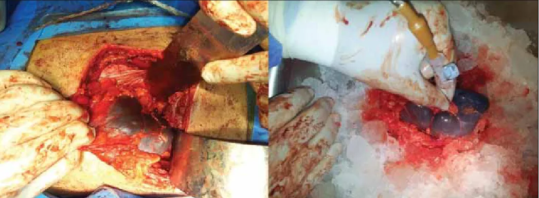
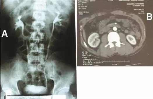
A 25 year old man, who wanted to donate a kidney to his cousin, was referred to our organ transplantation centre (Montaserieh Educational Hospital). On October 2012, we arranged a full biochemistry, imaging (Fig. 1), psychological and histocompatibility tests, followed by physical examina-tions. On the operating room table when we incised the left
flank region and exposed the kidney, we found a firm and black kidney (Fig. 2). After consulting with the transplant team, we stopped the operation for further assessment. Days after the incomplete procedure, we reviewed the donor’s biochemistry (blood urea nitrogen, creatinine, 24-hour urine collection) and imaging to reassess his renal function, but the results were normal again (Fig. 3, parts A and B). The result of Ham test (to rule out PNH) was also negative. Two weeks later we repeated the operation and removed the same kidney. We found the left kidney in the same condition it was before (black, firm with good dieresis) and we sent needle biopsies of the kidney for histopathologic analysis. The analysis showed a melanotic kidney without patho-logical changes in glomeruli and interstitium and vessels. Based on pathology report, glomeruli are normal and there were coarse, tubular intracytoplasmic granules (Fig, 4, part A). These brown granules were positive for Fontana (black) (Fig. 4, part B) and negative for modified Ziehl-Neelsen20,21 for lipofuscin (Fig. 4, part C). They were also was negative for other specific staining (repeat periodic acid Schiff [PAS], Pearl and trichrome). These findings were associated with heavy renal melanin pigmentation. One week after the sec-ond operation, the donor and recipient were checked by blood urea nitrogen and creatinine; both values were normal postoperation and the patients were discharged.
Discussion
The normal size of a kidney is 10 to 12 cm in length, 5 to
7 cm in width and 2 to 3 cm in thickness.22,23 Its normal
colour is reddish-brown due to the number of small capil-laries.24 Our patient had a black kidney. After the renal vas-cular and renal function tests, we ruled out acute vasvas-cular disorders. After the transplant, the pathology reports showed
Fig. 1. Normal renal computed tomography angiography was done before operation.
black-brown pigment deposits in the patient’s renal cells. One of the differential diagnoses of black pigment in renal cells is renal hemosiderosis. Renal hemosiderosis is a rare cause of renal failure that can occur in diseases character-ized by chronic intravascular hemolysis and lead to a black kidney. Free hemoglobin released in sickle cell anemia is filtered by the glomeruli and reabsorbed by proximal
con-voluted tubules, leading to renal hemosiderosis.13
Siddappa and colleagues presented a rare case of renal hemosiderosis appearing with renal failure due to repeated blood transfusion (about 2 per month) over past 2 years for
chronic refractory anemia.25 Three cases of paroxysmal
noc-turnal hemoglobinuria with renal hemosiderosis and renal failure were reported by Nair and colleagues.10 Most patients with renal hemosiderosis also have renal failure.
Although the pathogenesis of renal failure in a blue kid-ney is unknown, iron depositing in glomeruli and intersti-tium is a trigger of renal malfunction. Additional tests, like sucrose hemolysis test and Ham’s test for acid hemolysis,
were recommended by Siddappa and colleagues.25 These
tests were negative in our patient.
In 1987, Mocelin and colleagues reported on a 29-year-old living donor with a blue kidney. At the 6-month
follow-up, renal function of the donor and recipient were normal, but pigmentation in this case was lipofuscin;4 lipofuscin pig-mentation has been called the aging pigment or phenacetin
analgesic nephropathy.26,27 In 1964, Biava and colleagues
reported on 114 patients with lipofuscin-like granules in renal vascular smooth muscle cells. Patient age ranged from 2 to 70 years. The authors found that the number of granules, such as lipofuscin, in different patients correlated with age
and hypertension.28
In 2001, Lei and colleagues reported an unusual case of melanin-pigmented clear cell RCC with melanocytic differ-entiation.29 In their case, upon gross examination, they found a solid yellow and tan tumour measuring 8 × 7 × 6 cm occu-pying the upper pole of the kidney. Upon light microscopic
evaluation, they found abundant black pigment.29 Moreover,
many cases were melanocytic differentiation, which led to RCC.5
Conclusion
Black or blue kidney may be caused by vascular or non-vascular factors. Non-non-vascular black pigments in light micro-scopic examination may result in hemosiderin, lipofuscin or
melanin deposits in the kidney. A histopathological study can confirm the diagnosis; moreover, special tests for under-lying disease and renal function should be considered.
Competing interests: Dr. Yarmohamadi, Dr. Rezayat, Dr. Memar and Dr. Rahimi all declare no competing financial or personal interests.
This paper has been peer-reviewed.
References
1. Aghighi M, Heidary Rouchi A, Zamyadi M, et al. Dialysis in Iran. Iran J Kidney Dis 2008;2:11-5. 2. Ghods AJ, Mahdavi M. Organ transplantation in Iran. Saudi J Kidney Dis Transpl 2007;18:648-55. 3. Roberts WC. Renal hemosiderosis (blue kidney) in patients with valvular heart disease. Am J Pathol
1966;48:409-19.
4. Mocelin AJ, Brandina L, Fraga AM, et al. A blue kidney in a living donor. Transplantation 1987;44:169. http://dx.doi.org/10.1097/00007890-198707000-00039
5. Shetty J, Chandrika, Laxman P. Renal cell carcinoma with melanin pigment. Indian J Urol 2010;26:292-3. http://dx.doi.org/10.4103/0970-1591.65407
Fig. 4. A: Coarse tubular intracytoplasmic brown granules, glomeruli are completely spared (hematoxylin and eosin stain 400×); B: Coarse Fontana positive cytoplasmic granules (100×); C: Intratubular granules negative in modified Ziehl Neelsen for lipofuscin (100×).
!
6. Nobukawa B, Fujii H, Hirai S, et al. Breast carcinoma diverging to aberrant melanocytic differentiation: a case report with histopathologic and loss of heterozygosity analyses. Am J Surg Pathol 1999;23:1280-7. http://dx.doi.org/10.1097/00000478-199910000-00015
7. Valenti L, Canavesi E, Galmozzi E, et al. Beta-globin mutations are associated with parenchymal siderosis and fibrosis in patients with non-alcoholic fatty liver disease. J Hepatol 2010;53:927-33. http://dx.doi. org/10.1016/j.jhep.2010.05.023
8. Stickel F, Hampe J. Dissecting the evolutionary genetics of iron overload in non-alcoholic fatty liver disease.
J Hepatol 2010;53:793-4. http://dx.doi.org/10.1016/j.jhep.2010.06.010
9. Relia N, Kaushik C. Renal hemosiderosis: A case of black kidneys causing renal failure. J Postgrad Med
2010;56:216-7. http://dx.doi.org/10.4103/0022-3859.68635
10. Nair RK, Khaira A, Sharma A, et al. Spectrum of renal involvement in paroxysmal nocturnal hemoglobinuria: Report of three cases and a brief review of the literature. Int Urol Nephrol 2008;40:471-5. http://dx.doi. org/10.1007/s11255-008-9356-5
11. Zhou XJ, Laszik Z, Wang XQ, et al. Association of renal injury with increased oxygen free radical activity and altered nitric oxide metabolism in chronic experimental hemosiderosis. Lab Invest 2000;80:1905-14. http://dx.doi.org/10.1038/labinvest.3780200
12. Ackermann D, Vogt B, Gugger M, et al. Renal haemosiderosis: An unusual presentation of acute renal failure in a patient following heart valve prosthesis. Nephrol Dial Transplant 2004;19:2682-3. http:// dx.doi.org/10.1093/ndt/gfh429
13. Calazans LM, de Souza Santos RF, de Souza Goncalves M, et al. Renal hemosiderosis complicating sickle cell anemia. Kidney Int 2012;81:709. http://dx.doi.org/10.1038/ki.2011.470
14. Terman A, Brunk UT. On the degradability and exocytosis of ceroid/lipofuscin in cultured rat cardiac myo-cytes. Mech Ageing Dev 1998;100:145-56. http://dx.doi.org/10.1016/S0047-6374(97)00129-2 15. Elleder M, Drahota Z, Lisa V, et al. Tissue culture loading test with storage granules from animal models
of neuronal ceroid-lipofuscinosis (Batten disease): Testing their lysosomal degradability by normal and Batten cells. Am J Med Genet 1995;57:213-21. http://dx.doi.org/10.1002/ajmg.1320570220 16. Mann DMA, Yates PO, Marcyniuk B. Changes in nerve cells of the nucleus basalis of Meynert in Alzheimer’s
disease and their relationship to ageing and to the accumulation of lipofuscin pigment. Mech Ageing Dev
1984;25:189-204. http://dx.doi.org/10.1016/0047-6374(84)90140-4
17. Meredith GE, Totterdell S, Petroske E, et al. Lysosomal malfunction accompanies alpha-synuclein aggrega-tion in a progressive mouse model of Parkinson’s disease. Brain Res 2002;956:156-65. http://dx.doi. org/10.1016/S0006-8993(02)03514-X
18. Allaire J, Maltais F, LeBlanc P, et al. Lipofuscin accumulation in the vastus lateralis muscle in patients with chronic obstructive pulmonary disease. Muscle Nerve 2002;25:383-9. http://dx.doi.org/10.1002/ mus.10039
19. Samorajski T, Ordy JM, Rady-Reimer P. Lipofuscin pigment accumulationin the nervous system of aging mice. Anat Rec 1968;160:555-73. http://dx.doi.org/10.1002/ar.1091600305
20. Talbot IC, Mowat AP. Liver disease in infancy: histological features and relationship to alpha-antitrypsin phenotype. J Clin Pathol 1975;28:559-63. http://dx.doi.org/10.1136/jcp.28.7.559
21. Moller JC, Kristensen IB. Xanthogranulomatous pyelonephritis. A clinico-pathological study with special reference to pathogenesis. Acta Pathol Microbiol Scand A 1980;88:89-96.
22. National Kidney and Urologic Diseases Information Clearinghouse (NKUDIC). http://kidney.niddk.nih. gov/Kudiseases/pubs/yourkidneys/#kidneys. Accessed February 4, 2014.
23. McDougal WS, Wein AJ, Kavoussi LR, et al. Campbell-Walsh Urology. 10th ed. Review E-Book: Elsevier Health Sciences; 2011.
24. Taylor JJ, Cohen BJ. Structure & Function of the Human Body. Baltimore, MD: Lippincott Williams & Wilkins; 2012.
25. Siddappa S, Mythri KM, Kowsalya R, et al. Refractory anemia leading to renal hemosiderosis and renal failure. Indian J Pathol Microbiol 2011;54:379-80. http://dx.doi.org/10.4103/0377-4929.81648 26. Rubenstein AH, Abrahams C, Stables DP, et al. Acetophenetidin nephritis and papillary necrosis: A clinical
and pathological study of six cases. Arch Intern Med 1964;113:378-94. http://dx.doi.org/10.1001/ archinte.1964.00280090064011
27. Elseviers MM, De Broe ME. Analgesic nephropathy: is it caused by multi-analgesic abuse or single sub-stance use? Drug Saf 1999;20:15-24. http://dx.doi.org/10.2165/00002018-199920010-00003 28. Biava C, West M. Lipofuscin-like granules in vascular smooth muscle and juxtaglomerular cells of human
kidneys. Am J Pathol 1965;47:287-313.
29. Lei JY, Middleton LP, Guo XD, et al. Pigmented renal clear cell carcinoma with melanocytic differentiation.
Hum Pathol 2001;32:233-6. http://dx.doi.org/10.1053/hupa.2001.22009