A GENETIC ANALYSIS
OF TETRAHYMENA T H A T HAVE
ABORTED NORMAL DEVELOPMENT
STEVEN B. SCHOLNICK' A N D PETER J. BRUNS2
Section of Genetics and Development, Cornell University, Ithoca, New York 14853
Manuscript received January 27, 1982 Revised copy accepted June 1,1982
ABSTRACT
Conjugating Tetrahymena can abort the sexual cycle before the production of progeny somatic (macro-) nuclei and return to vegetative growth. We have analyzed the germinal (micronuclear) genotypes of these cells in order to determine the stage at which they aborted conjugation. Our data demonstrate that nearly all of these cells attempt meiosis, but that very few reach the successful completion of cross-fertilization. The resulting micronuclear geno- types suggest that either germinal chromosomes or entire nuclei are lost during an unsuccessful attempt at meiosis or cross-fertilization. We conclude that the decision to develop progeny macronuclei is made during meiosis and is depend- ent on the completion of some step necessary for successful cross-fertilization.
ONJUGATION in the ciliated protozoan Tetrahymena thermophila may be
C
a useful model for studying a simple nuclear differentiation. Tetrahymena is binucleate, having separate germinal and somatic nuclei: the transcriptionally silent, diploid micronucleus and the transcriptionally active, large (45C amount of DNA) macronucleus, respectively. The macronucleus is solely responsible for the phenotype of the cell (GOROVSKY and WOODWARD 1969; BRUNS and BRUSSARD1974a). Conjugation results in the production of new, recombinant micro- and macronuclei. Both of these nuclei are mitotically derived from the same zygotic nucleus formed during conjugation (RAY 1956). The development of a new macronucleus involves both amplification and sequence rearrangement of the germinal genome (YAO and GALL 1979).
During conjugation, cells are confronted with a major developmental decision: after meiosis and the production of a new germinal nucleus, mating cells can either develop this new genome into a new somatic (macro-) nucleus or abort conjugation and return to vegetative growth. Cells that have aborted conjugation retain their parental macronuclei and therefore their parental phenotypes. These cells have been termed nonconjugants by NANNEY (1957).
In a previous paper (SCHOLNICK and BRUNS 1982), we demonstrated that nonconjugants can be selected by allowing the cells to mate at 40°. This treatment is lethal to all cells producing new macronuclei and only the noncon- jugants survive (SCHOLNICK and BRUNS 1980; SCHOLNICK and BRUNS 1982). In this paper we demonstrate that the nonconjugants selected from 40' matings are genetically similar if not identical to their 30' nonconjugant counterparts. We have found that nonconjugation is associated with a limited set of genetic
Present address: Department of Biological Chemistry, Harvard Medical School, Boston, Massachusetts 02215. *Author to whom all correspondence should be addressed.
30 S. B. SCHOLNICK AND P. J. BRUNS
abnormalities almost always associated with an early step in conjugation. These results reveal both the timing of, and a possible mechanism for, the events leading to the subsequent abortion of macronuclear development.
MATERIALS AND METHODS
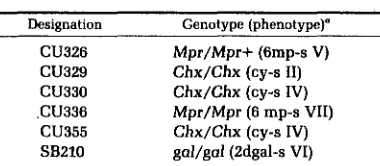
Strains: The strains used in this study are fully described in Table 1. All of these strains are derived from inbred strain B1868. SB210 was provided by Dr. EDUARDO ORIAS, University of California, Santa Barbara. T. thermophila was formerly known as Tetrahymena pyriformis, syngen 1 (NANNEY and McCoy 1976).
Media: The growth medium (PPYS) was 1% proteose peptone (Difco), 0.1% yeast extract (Difco), and 0.003% Sequestrene (Geigy). The starvation medium was 10 mM Tris-HC1, pH 7.4 (Sigma). Starved cells were fed with 10% proteose peptone to a final concentration of 1%. Stocks of strains were maintained in 1% proteose peptone at room temperature. All media were sterilized by autoclaving.
Matings in flasks: As previously described (BRUNS and BRUSSARD 1974b) cells were prepared for mating by washing once in 10 mM Tris and resuspending in Tris at a final concentration of 1.4 x IO6 cells/ml. Cells were allowed to starve for 18-24 hr. Matings were made by mixing equal numbers of the prestarved parents in an Erlenmeyer flask at a final concentration of 1.4 X
lo5
cells/ml. Cell numbers were determined using a Coulter Counter (Coulter Electronics) with a 200-pm aperture. The flasks used to contain the matings had a capacity of at least 10 times the volume of the mating mix. Unless otherwise noted, the matings were fed 6 hr after mixing in order to stop further pairing. Pair isolation, subcloning and microtiter plate manipulation: Pairs were isolated by micropipette starting 30 min after refeeding the mating mix. Pairs were transferred to individual drop cultures arranged in 6 x 8 array in a 100- x 15-mm disposable Petri dish. This array matches the pattern of wells found on microtiter plates (Dynatech Costar). The pairs were allowed to grow for 3 days at 30' or 40'. They were then transferred to microtiter plates by using a custom &-prong stainless steel replicator (Lansing Industries). These master plates were replicated to other plates for drug testing or further matings by using a 96-prong stainless steel replicator (Lansing Industries). Subclones of the strains used in these experiments were established by isolating single cells from vegetative cultures. These cells were manipulated in the same manner as isolated pairs.Matings in microtiter plates: Microtiter plate cultures were grown overnight at 30' in 0.1 ml/well PPYS. All of the cultures on the plate were pelleted by gentle centrifugation in an IEC model UV centrifuge equipped with microtiter plate carriers (Dynatech). The medium was removed with a custom-made 96-channel aspirator. The cells were resuspended in 0.1 ml/well 10 mM Tris using a 12-channel manifold (Dynatech) and a Cornwall repeating syringe. The cultures were pelleted once more and resuspended in 0.05 ml/well 10 mM Tris. These cultures were allowed to starve overnight at 30'. Mating was initiated by adding 0.05 ml/well of a prestarved culture of the appropriate strain. Matings used for the isolation of pairs were fed with 5% proteose peptone to a final concentration of 1% peptone 6-8 hr after mixing the parents. Matings used for mating-type testing were not refed; 6-8 hr-old matings were examined for the presence of pairs, using a dissecting microscope with darkfield illumination.
Drug selection: The drug doses used for selecting cycloheximide- and 6-methylpurine-resistant cells were 25 pg/ml and 15 pg/ml, respectively. These drugs were maintained as 5 0 0 ~ stocks in 95% ethanol or distilled water, respectively, at -20'. The dose used for selecting Z-deoxygalactose- resistant cells was 2.5 mg/ml. This drug was maintained as a 50X stock in distilled water at -20'. All three drugs were diluted with PPYS just before use. Cells were maintained in the drug media for 4-5 days (cycloheximide and 6-methylpurine) or 7-8 days (2-deoxygalactose) before resistance was scored.
RESULTS
Experimental rationale: We compared the genetic consequences of noncon- jugation at 30' and 40' by analyzing the micronuclear genotypes of nonconju- gants collected from the cross Mpr/Mpr+ (6mp-s) x Chx/Chx (cy-s) (CU326 X
ABORTED TETRAHYMENA DEVELOPMENT
TABLE 1
Strains used in this study
Designation Genotype (phenotype)"
CU326 Mpr/Mpr+ (6mp-s V)
CU330 Chx/Chx (cy-s IV) CU336 Mpr/Mpr (6 mp-s VII)
SB210 @/gal (adgal-s VI) CU329 Chx/Chx (CY-s 11)
cu355 Chx/Chx (CY-s IV)
As previously suggested (BRUNS and BRUSSARD 1974a), the letters preceding the parentheses desig- nate the micronuclear genotype of the cell; the letters inside the parentheses represent the phenotype ex- pressed by the macronucleus, and the Roman numer- als stand for the mating type. Chx is a dominant mutation conferring resistance to cycloheximide (BYRNE, BRUSSARD and BRUNS 1978). Mpr is a domi- nant 6-methylpurine resistance mutation (BYRNE, BRUSSARD and BRUNS 1978). gal is a recessive muta- tion conferring resistance to 8-deoxygalactose (ROB- ERTS and MORSE 1980). CU329 is therefore a hetero- karyon with a germline homozygous for the Chx mutation. The cell's phenotype is cycloheximide sen- sitive and its mating type is 11. See BRUNS and BRUS-
SARD (1974a) for a description of how these strains were constructed.
conferring resistance to either 6-methylpurine or cycloheximide, their macro- nuclei express the wild-type drug-sensitivity phenotype. The parents and non- conjugants are therefore sensitive to both drugs. All of the progeny are resistant to cycloheximide and one-half are resistant to 6-methylpurine. We assayed the micronuclear genotypes of the clones derived from each parent of the noncon- jugant pairs (termed the exconjugant clones) by a testcross to SB210, a hetero- karyon with a micronucleus homozygous for the gar1 mutation. This strain is a homozygous homokaryon for the wild-type
Chx+
and Mpr+ alleles (Table 1) (ROBERTS andMORSE
1980).The data generated by this experimental approach allowed us to test the validity of our temperature selection for nonconjugants and to gain some understanding of the mechanism of the developmental decision to abort ma- cronuclear differentiation.
Genetic analysis of the nonconjugants' micronuclear genotypes detects the types of genetic abnormalities, if any, associated with nonconjugation. This allows a first approximation of the step in the pathway at which the cells abort conjugation. Conjugation before macronuclear development can be divided into three distinct periods: 1) premeiosis, 2) postmeiosis-precross-fertilization and 3) postcross-f ertilization.
32 S. B. SCHOLNICK AND P. J. BRUNS
t
t
t
FIGURE 1.-Schematic showing the major events of conjugation. The heavy black arrows indicate the three genetically distinguishable stopping points as outlined in the text (Experimental rationale). The diagram shows a cross between two homozygous homokaryons.
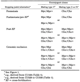
TABLE 2
Predicted micronuclear genotypes of nonconjugants
Exconjugant clones
Stopping point/aberration" Mating type Vb Mating type I1 or IV'
Premeiosis Mpr/Mpr+ C h x / C h x
Postmeiosis/pre-XFd Mpr/Mpr Chx/Chx or
Mpr+/Mpr+
Post-XF Mpr/Mpr+ Mpr/Mpr+
Chx/Chx+ Chx/Chx+ or
Mpr+/Mpr+ Mpr+/Mpr+ Chx/Chx+ Chx/Chx+
Genomic exclusion Mpr/Mpr Mpr/Mpr Chx+/Chx+ Chx+/Chx+
or
Mpr+/Mpr+ Mpr+/Mpr+ Chx+/Chx+ Chx+/Chx+
or
Mpr+/Mpr+ . Mpr+/Mpr+ Chx/Chx Chx/Chx
See Figure 1.
e.g., derived from CU326 (Table 1).
e.g., derived from CU329 or CU330 (Table 1). XF = cross-fertilization.
clones because they have retained their parental macronuclei and therefore their parental mating types.
Round I. If a Round I genomic exclusion-like event is associated with noncon- jugation in wild type crosses it would result in a class of nonconjugants with its own predictable and distinct set of micronuclear genotypes. These are also listed in Table 2.
Collection of the nonconjugants: CU326 and either CU329 or CU330 (Table 1) were grown and starved separately and then mixed for mating as described in
Nonconjugants were collected from 30' matings in the following fashion. The mating cells were fed 6 hr after mixing the prestarved parents and pairs were isolated in drops of growth medium (PPYS) in Petri dishes (MATERIALS AND METHODS). These cultures were incubated at 30' for 3 days and then replicated to microtiter plates containing PPYS medium. After 3 days of growth these master plates were replicated to media containing cycloheximide and 6-meth- ylpurine. The parental strains are sensitive to both drugs. Half of the progeny are resistant to 6-methylpurine and all are resistant to cycloheximide. Noncon- jugant pairs are those that did not produce new, progeny macronuclei. They give rise to clones that have retained their parental mating type and drug- sensitive phenotype. Their status as nonconjugants was confirmed in each case by the fact that these cells were still capable of mating (mature) and still expressing their parental mating type. Tetrahymena which have produced new macronuclei are incapable of mating for approximately 40 fissions (NANNEY and
CAUGHEY 1953).
The nonconjugants identified by this procedure were subcloned by isolating single cells from the master culture. These subclones were tested for mating type by crossing them to the parental strains (MATERIALS AND METHODS), allowing us to identify the two exconjugant clones from each pair. It was not always possible to recover both exconjugant clones. This may result from the death of one of the exconjugants during conjugation or to one of the exconjugant clones outgrowing the other during subsequent vegetative growth. The exconjugant clones were transferred to stock tubes and saved for further analysis.
Nonconjugants were isolated from 40' matings in a similar fashion. CU326 and CU330 were grown, starved and mated at 30'. The mating mixes were fed at either 4.5 or 5.5 hr and pairs were isolated into drop cultures as above. These drop cultures were transferred to 40' at 5.5 or 7.5 hr (designated series 40'E and 4OoL, respectively) and incubated at that temperature for 3 days. We have previously demonstrated that only pairs that have aborted conjugation before macronuclear development can survive this treatment (SCHOLNICK and BRUNS
1980; SCHOLNICK and BRUNS 1982). The survivors of these crosses were subcloned and tested for mating type in the same fashion as their 30' counterparts. Their nonconjugant nature was confirmed in every case by their maturity and the retention of their parental mating types. Exconjugant clones descended from each member of the nonconjugant pairs were transferred to stock tubes as before. We were again unable to obtain both exconjugant clones in all cases.
Genetic analysis of the nonconjugants: All of the nonconjugant clones were prepared for genetic analysis by transferring the stocks to individual wells on a microtiter plate. Three replicates of each master plate were prepared for mating as described in MATERIALS AND METHODS. Two of these replicates were
mated to the parental strains in order to confirm the mating type and, therefore, the origin of the clones. The clones on the remaining plate were crossed to
34 S. B. SCHOLNICK A N D P. J. BRUNS
SB210, a heterokaryon for the gal mutation (ROBERTS and MORSE 1980), which confers resistance to 2-deoxygalactose (Table 1); 44 pairs were isolated from each of the testcrosses. These pairs were grown at 30' for 3 days and then replicated to microtiter plates. These master plates were incubated for 3 days and then replicated to media containing 2-deoxygalactose, cycloheximide or 6-
methylpurine in order to identify the testcross progeny and ascertain their phenotypes.
Table 3 lists the results obained from the analysis of only those nonconjugants for which we recovered both exconjugant clones. The analysis of those noncon- jugants represented by only one exconjugant clone is consistent with these results (data not shown). However, the recovery of only one of the exconjugant clones does not allow the resolution of certain types of cytogenetic events: the recovery of a mating type
I1
nonconjugant clone (i.e., one derived from CU329)with the micronuclear genotype Chx/Chx Mpr+/Mpr+ could have been the result of three possible cytogenetic events: abortion of conjugation before meiosis, after meiosis but before cross-fertilization, or genomic exclusion (Table
2).
Although some pairs were recovered in each category, by far the most common class (64.4% of the nonconjugants recovered) appeared to be the result of a process similar to Round I genomic exclusion. Thus, in most cases both of the exconjugant clones from a given pair are homozygous for the marker inherited from only one of the parents (Table 2). The genome of the Mpr
heterozygote, CU326, was recovered more frequently than that of the Chx homozygotes, CU329 and CU330 (31:9). The Mpr and Mpr+ alleles segregated with a 1:l ratio (15 Mpr:l6 Mpr+) when all of the genomic exclusion events are taken into account.
We also recovered four nonconjugant pairs that appear to have undergone a self-fertilization. This is indicative of the abortion of conjugation after meiosis but before cross-fertilization (Tables 2 and 3). An additional nine pairs did not fit any of the predicted classes described above nor could they be reconciled with the normal segregation of meiotic products (Table 3). The micronuclear genotypes of these pairs are listed in Table 4.
Origin of the genomic exclusion events: The genomic exclusion events asso-
TABLE 3
Distribution of nonconjugant classes
Treatment
~
Stopping point/aberration" 30' 40°Eb 4OoL'
Premeiosis NR' NR 3
Postmeiosis/pre-XF NR 2 2
Post-XF 1 NR NR
Genomic exclusion 16 15 9
Abnormal segregationd 2 3 4
Infertile 2 1 2
Total 21 21 20
a See Figure 1 and Table 2.
'
Transferred to 40' at 5.5 hr.e Transferred to 40° at 7.5 hr. e NR = not recovered.
TABLE 4
Micronuclear genotypes of clones showing abnormal segregation
Exconj ugant clones *
Source" Mating tvpe V' Mating type I1 or IVd
30° 30'
Chx/Chx+ Mpr+/Mpr+ Chx/Chx Mpr+/Mpr+ Chx+/Chx+ Mpr/Mpr Chx+/Chx+ Mpr/Mpr+
40°E Chx+/Chx+ Mpr/Mpr Chx/Chx+ Mpr+/Mpr+
40°E Chx+/Chx+ Mpr/Mpr+ Chx+/Chx+ Mpr/Mpr+ 40"E Chx/Chx Mpr+/Mpr+ Chx+Chx+ Mpr/Mpr
40"L 40°L 4OoL 40°L
Chx+/Chx+ Mpr/Mpr Chx+/Chx+ Mpr+/Mpr+ Chx/Chx Mpr+/Mpr+ Chx+/Chx+ Mpr+/Mpr+ Chx/Chx Mpr+/Mpr+ Chx+/Chx+ Mpr/Mpr Chx+/Chx+ Mpr/Mpr Chx+/Chx+ Mpr/Mpr+
a See text.
*
Each line in the table represents both exconjugant clones derived from a single nonconjugant pair.e.g., derived from CU326 (Table 1).
e.g., derived from either CU329 or CU330 (Table 1).
ciated with nonconjugation could be caused by star-like cells within the parental strains. Alternatively, genomic exclusion could be the result of a cytogenetic accident during mating between normally fertile cells. We differentiated be- tween these possibilities by subcloning CU329 and testing the fertility of these subclones in mass crosses to CU336, a homozygous heterokaryon for the Mpr mutation (Table 1). Fertile subclones of CU329 will yield progeny resistant to both cycloheximide and 6-methylpurine. Star-like subclones will yield excon- jugants either sensitive to both drugs (as in Round I genomic exclusion), or
resistant to only 6-methylpurine (as in Round
I1
genomic exclusion). Completely infertile subclones will also yield exconjugants sensitive to both drugs.Subclones of CU329 were established by cell isolation as described in MATE- RIALS AND METHODS. Replicates of these subclones were transferred to microtiter
plates and mated to CU336 (MATERIALS AND METHODS). These matings were
allowed to proceed for 24 hr at 30", which is sufficient time for both rounds of genomic exclusion. This allowed us to distinguish between star-like and com- pletely infertile subclones. The mating mixtures were then fed and allowed to grow at 30" for 1 day. They were then replicated to media containing cyclohex- imide and 6-methylpurine.
Fertility tests of 257 subclones of CU329 did not detect any star-like or infertile clones by the criteria described above. A retest of 258 new subclones approxi- mately 8 mo later revealed the existence of 25 star-like clones, indicating that the breeding performance of CU329 is degenerating. We also tested 258 sub- clones of CU326 during this second set of fertility tests by crossing them to CU355, a homozygous heterokaryon for the Chx mutation. We did not detect any star-like clones in these tests.
DISCUSSION
36 S. B. SCHOLNICK AND P . J. BRUNS
identical to those recovered from 30' controls and from matings transferred to 40" at 7.5 hr (series 30' and 40"L in Table 3). This suggests that the elevated temperature does not affect cross-fertilization, which occurs at approximately 6 hr after mixing (MARTINDALE, ALLIS and BRUNS 1982). We did not recover all classes of nonconjugants in all three treatments, possibly because of the small sample sizes involved. A problem in this study was the isolation of a sufficient number of nonconjugants from the 30' matings. We optimized our procedure for collecting nonconjugants at both temperatures by using older heterokaryons: CU326, CU329 and CU330. Although these strains are still fertile, crosses between them yielded more nonconjugants than crosses between younger strains, e.g., CU336
x
CU355 (approximately 10% and 3% of the pairs isolated, respectively). The ability to select for nonconjugants directly should allow a much more extensive analysis of nonconjugation using a greater variety of strains.These data also indicate that the vast majority of nonconjugants that were tested (54/57, since 5/62 were infertile and thus untestable) aborted conjugation at some step after the onset of meiosis but before the fusion of the gametic nuclei. This suggests that in many cases the decision to abort conjugation may be associated with aberrant micronuclear events before the completion of cross- fertilization. These data also suggest that almost all pairs are committed to attempt meiosis.
Four of the pairs examined appear to have undergone a self-fertilization similar to the micronuclear events of osmotically-induced cytogamy (ORIAS, HAMILTON and FLACKS 1979). Both of these variations of normal conjugation generate two different whole-genome homozygotes from each pair. However, elements of osmotically-induced cytogamy must be different from that of nonconjugation as the former results in the development of progeny macronu- clei.
has suggested that germinal ageing is a consequence of the occurrence of random defects in the micronucleus and that the rate of ageing can vary from strain to strain (NANNEY 1974; SIMON and NANNEY 1979).
The restricted range of micronuclear genotypes detected in this study suggests that several classes of nonconjugants may be the result of similar cytogenetic events. Both the cytogamous micronuclear events and genomic exclusion may involve the loss of one or more of the gametic nuclei. It is possible that the decision to produce a new macronucleus is in part dependent on the successful completion of some event leading to cross-fertilization or to the successful completion of cross-fertilization itself. The former possibility seems to be the more likely of the two as the osmotic induction of cytogamy does not lead to nonconjugation (ORIAS, HAMILTON and FLACKS 1979). This hypothesis is con- sistent with our recovery of the class of nonconjugants labeled “abnormal segregation” in Table 4. The micronuclear genotypes of these pairs are not consistent with normal meiotic segregation of markers or with any of the cytogenetic abnormalities discussed above. However, these genotypes could be generated by the loss of one or more of the gametic nuclei after nuclear transfer but before the fusion of the nuclei. Alternatively, some of these micronuclear genotypes could be the result of the loss of one or more of the genetically marked germinal chromosomes. A strain monosomic for a dominantly marked germinal chromosome would behave like a heterozygote in a testcross to a diploid strain, i.e., one-half of the progeny would express this dominant phe- notype and one-half would not. Such monosomic cells could arise as a result of an aberrant meiosis. However, monosomy is not sufficient to cause the cells to abort conjugation. Monosomic and even nullisomic strains are capable of completing conjugation when crossed to a diploid strain (BRUNS and BRUSSARD
1981). We have not karyotyped our collection of nonconjugants to see if chromosome loss is associated with nonconjugation. The infertile nonconjugant pairs listed in Table 3 might also result from meiotic accidents. In this case part of the germinal genome might have sustained much greater damage. An extreme possibility is that these strains have haploid micronuclei, so the production of progeny is below the resolution of these experiments.
Ample precedent exists for the involvement of cytogenetic abnormalities with nonconjugation. Meiosis in the classic, highly aneuploid, star strains, A*III, A*V, and C*III, appears to result in the loss of the micronucleus (ALLEN 1967; WEINDRUCH and DOERDER 1975). PITTS (1979) has reported several star strains that complete meiosis but fail to associate a meiotic product with the attachment membrane between the members of the pair; all of the meiotic products are subsequently destroyed. Random accidents of this nature during the conjugation of otherwise normal strains could be responsible for some of the abnormalities we have detected and for many incidents of nonconjugation. Our demonstration that high temperature matings faithfully select for nonconjugants provides an efficient way of assaying the number of cells in a mating mix that have aborted conjugation. This should allow the experimental dissection and manipulation of this developmental decision in a manner not previously possible.
38 S. B. SCHOLNICK AND P. J. BRUNS
thank JOE ARDIZZI, JEAN FINLEY, and RON PITTS for helpful discussions and critical commentaries on the manuscript.
LITERATURE CITED
ALLEN, S. L., 1967 Cytogenetics of genomic exclusion in Tetrahymena. Genetics 55: 797-822. BRUNS, P. J. and T. B. BRUSSARD, 1974a Positive selection for mating with functional heterokaryons
in Tetrahymena pyriformis. Genetics 7 8 831-841.
BRUNS, P. J. and T. B. BRUSSARD, 1974b Pair formation in Tetrhaymena pyriformis, a n inducible developmental system. J. Exp. Zool. 188: 337-344.
BRUNS, P. J. and T. B. BRUSSARD, 1981 Nullisomic Tetrahymena: eliminating germinal chromo- somes. Science 213 549-551.
BYRNE, B. C., BRUSSARD, T. B. and P. J. BRUNS, 1978 Induced resistance to 6-methylpurine and cycloheximide in Tetrahymena. I. Germline mutants of T. thermophila. Genetics 89: 695-702. GOROVSKY, M. A. and J. WOODWARD, 1969 Studies on nuclear structure and function in Tetrahy-
mena pyriformis. I. RNA synthesis in macro- and micronuclei. J. Cell Biol. 42: 673-682.
a temporal analysis of cytological stages. Exp. Cell Res. In press. MARTINDALE, D. W., ALLIS, C. W. and P. J. BRUNS, 1982
NANNEY, D. L., 1957 Inbreeding degeneration in Tetrahymena. Genetics 42: 137-146.
NANNEY, D. L., 1974 Ageing and long-term temporal regulation in ciliated protozoa: a critical review. Mech. Ageing Dev. 3: 81-105.
NANNEY, D. L. and P. A. CAUGHEY, 1953 Mating type determination in Tetrahymena pyriformis. Proc. Natl. Acad. Sci. 39 1057-1062.
NANNEY, D. L. and J. W. McCoy, 1976 Characterization of the species of the Tetrahymena
ORIAS. E., HAMILTON, E. P. and M. FLACKS, 1979 Osmotic shock prevents nuclear exchange and
PITTS, R. A., 1979 Age-associated micronuclear defects of Tetrahymena thermophila: genetic and
RAY, C., 1956 Meiosis and nuclear behavior in Tetrahymena pyriformis. J. Protozool. 3: 88-96.
ROBERTS, J. R. and D. E. MORSE, 1980 Galactokinase-deficient mutants of Tetrahymena thermo-
SCHOLNICK, S. B. and P. J. BRUNS, 1980 Effect of elevated temperatures on Tetrahymena conjuga-
SCHOLNICK, S. B. and P. J. BRUNS, 1982 Conditional lethality associated with macronuclear
SIMON, E. M. and D. L. NANNEY, 1979 Germinal ageing in Tetrahymena thermophila. Mech. Ageing
WEINDRUCH, R. H. and F. P. DOERDER, 1975 Age-dependent micronuclear deterioration in Tetra-
YAO, M-C. and J. G. GALL, 1979 Alteration of the Tetrahymena genome during nuclear differen-
Corresponding editor: S. ALLEN Conjugation in Tetrahymena thermophila:
pyriformis complex. Trans. Am. Micros. Soc. 95: 664-682.
produces whole-genome homozygotes in conjugating Tetrahymena. Science 203: 660-663.
cytological studies. M. A. Thesis, University of Pittsburgh, Pittsburgh, PA.
phila: selection and characterization. Mol. Gen. Genet 180 129-134.
tion. Genetics 94: s93.
development in Tetrahymena. Dev. Biol. In press.
Dev. 11: 253-268.