ISOLATION
OF
PLAQUE-FORMING, GALACTOSE-TRANSDUCING STRAINS OF PHAGE LAMBDA1MICHAEL FEISS,2 SANKAR ADYHA,3 A N D DONALD L. COURT4
Department of Biological Sciences, Stanford University, Stanford, California 94305
Manuscript received November 8, 1971 Revised copy received January 11, 1972
ABSTRACT
Plaque-forming, galactose-transducing lambda strains have been isolated from lysogens in which bacterial genes have been removed from between the
galactose operon and the prophage by deletion mutation.-A second class has been isolated starting with a lysogenic strain which carries a deletion of the
genes to the right of the galactose operon and part of the prophage. This strain was lysogenized with a second lambda phage to yield a lysogen from which galactose-transducing, plaque-forming phages were obtained. These plaque- forming phages were found to be genetically unstable, due to a duplication of part of the lambda chromosome. The genetic instability of these partial diploid strains is due to homologous genetic recombination between the two identical copies of the phage DNA comprising the duplication. The galactose operon and the duplication of phage DNA carried by these strains is located between the phage lambda P and Q genes.
acteriophage lambda chromosome integrates into the E. coli chromosome T:sEab specific site. In lysates obtained from lysogenic culturq, transducing variants are found at low frequency. These variants carry bacterial genes, but only those that are closely linked to the prophage. Each of these transducing variants appears t o result from a single non-homologous recombination event which excises a DNA segment from the lysogenic chromosome.-This segment includes bacterial genes and phage genes; the sequence of genes in the lysogenic chromosome is maintained in transducing variants. The only requirement for phage material carried by transducing variants seems to be that the cohesive end site of the phage be present
(KAYAJANIAN
and CAMPBELL 1966;SATO
and CAMPBELL 1970). Whether such transducing variants need be defective (unable to form plaques) or not seems to be determined by a limitation on the size of a transducing fragment (KAYAJANIAN and CAMPBELL 1966). This size limitation is in all probability the capacity of the phage lambda capsid. Thus, among galactose-transducing variants of lambda, isolated from a wild-type lysogen, the amount of bacterial DNA is so large that some essential phage genes must be ' This research was supported by National Institutes of Health Grants AI 8573 and 5T01-GM158-12. D. L. C. was e Present address and reprint address: Department of Microbiology, College of Medicine, University of Iowa, Iowa City, Present address: Laboratory of Molecular Biology, National Cancer Institute, National Institutes of Health, Bethesda, Present address: Department of Molecular Biology, University of California, Berkeley, California 94.720.USPHS predoctoral trainee of the Department of Biology, University of Rochester.
Iowa 52240.
Maryland 20014.
190 M. FEISS, S . ADHYA A N D D. L. COURT
discarded to produce molecules that can be packaged into mature phage parti- cles. These galactose-transducing strains have been the subject of much research and a phage from this class will be referred to as a hdgal: the d indicates the phage is defective, gal denotes the ability to transduce galactose markers. The phsg:! material which is replaced by bacterial genes in hdgal strains is a con- tiguous block from the right prophage end (as drawn in Figure 1 a) and includes the b region and essential genes. The amount of essential material deleted varies from isolate to isolate but always includes genes K , I and J . A typical Xdgal has the following vegetative gene order: A gal chlD pgl N cl 0 P
Q
S R.
Recently, methods have been developed which allow selection of deletion mu- tations on either side of lambda (ADHYA et aZ. 1968; SHAPIRO and ADHYA 1969). Such deletions can alter the proximity and sequence of bacterial genes and lambda prophage genes. W e describe here non-defective (plaque-forming) galactose-transducing variants obtained from two types of deletion lysogens. The first contains a deletion which removes bacterial DNA between the galactose genes and the prophage. In the second type, a deletion obtained in a lambda lysogen removes bacterial DNA between the galactose operon and the prophage and also removes part of the prophage. A second phage chromosome inserted in tandem into this latter type yields upon induction galactose-transducing variants. In these, the galactose genes are located in a different position than in hdgal
strains. These strains are plaque-forming and, in addition, contain duplications of phage DNA, a result consistent with the proposed structure of the deletion lysogen. Our studies indicate a vegetative gene order of: A J N c l 0 P gal Q S R.
MATERIALS A N D METHODS
Strains: These are listed in Table 1.
Media: Tryptone broth, tryptone broth agar (1.0% agar), and tryptone soft agar (0.65% agar) were used for growth and titration of bacteria and phage; eosin methylene blue plus galac- tose agar (EMBG) was used for selection of transductants able to utilize galactose (Gal+) and testing the Gal character; these complex media have been described by CAMPBELL (1957 and 1961). Eosin methylene blue agar lacking sugar (EMBO) was used during selection of the lyso- gen SA307 (XcI857b221). Synthetic agar, appropriately supplemented, was used for biotin, nico- tinic acid, and moG transduction. P l k c transduction was carried out as described by ROTHMAN
(1965). The test for the pgl character has been described by ADHYA and SCHWARTZ (1971).
Transductions: Gal- bacteria (W3805, W3350, etc.) were grown to 4 x l O S / m l , mixed with phage lysates, allowed to absorb and plated on indicator agar. Transduction of aroG was tested as described by WALLACE and PITTARD (1967), using AB3250. Transduction of nicA, using GG30, was as described by SATO and CAMPBELL (1970).
Preparation of lysates: Induction was carried out by heating growing cultures of Xc1857
lysogens a t 4.3% for 15 min. After heating, aeration was continued at 37°C until lysis.
LAMBDA T R A N S D U C I N G STRAINS
TABLE 1A E. coli K12 strains
191
Strain Description Source o r reference
SA500 W3805 W3350 R5W 138-2 MSA151 MSA79 MS226 MS60 W3101 R865 SA307 C600 c24'6 KLIG99 SA472 112-12 GG30 p3478 AB3250
his, str, lysogenic for Xc1857 galEl6
galK2, galTl
W3350 str galKl38 galT15I galT79 galT226 galT60 galTl
W3350 (Ximm434) (chlD
.
..
P ) A , str, his leu, thr, thi, sull+(galT
.
. .
bioD) A, str, his, pro Hfr, thi, recAl; transfers recAl early(aroG .
. .
ch1A)A gal, cys, su112-12+(nicA
. .
, chlD) A, his, strW3110 thy, polA1
thi, his, pro, arg, ilv, gal, aroG
This laboratory
E. LEDERBERG
This laboratory This laboratory
BUTTIN
SHAPIRO and ADHYA (1969) SHAPIRO and ADHYA (1969) SHAPIRO and ADHYA (1969) SHAPIRO and ADHYA (1969) SHAPIRO and ADHYA (1969)
This laboratory This laboratory This laboratory
P. CLEARY Low (1968)
This laboratory
THOMAS et al. (1967) This laboratory
DFLUCIA and CAIRNS (1969) WALLACE and P I ~ A R D (1967)
his = histidine requirement; str = resistant to streptomycin; c1857 == heat-sensitive X repressor mutation; galK,T,E = inability to ferment galactose, mutations in kinase, transferase, epimerase, respectively; CUD and chlA = mutations conferring resistance to anaerobic growth on chlorate;
imm4s4 = +unity region of phage 444 recombined into A; I = genes of A; lam2 = particular amber mutabon in I gene of A; s u l l f =amber suppressor; sul12-12f = ochre suppressor; poZA = absence of DNA polymerase; arcG = requirement for tryptophan when grown in presence of tyrosine and phenylalanine; nicA = requirement for nicotinic acid. A symbolizes a deletion of the region in parentheses. Other genotypic symbols as proposed by TAYLOR (1970).
TABLE 1B
Phage strain
Strain he1857 Xc26b221 Plkc Xc1857red270 Xc1857Nam7am53 himmhJ4 hb2imm4J4c Ximm4s4b221 Xc1857Nam7am53nin5
Description S o m e or reference This laboratory
D. BERG: DAVIS and DAVIDSON (1968) P. CLEARY
D. SCANDELLA E. SIGNER This laboratory This laboratory This laboratory This laboratory
DNA content = 0.78*
am mutation in p cistron
DNA content = 0.97* DNA content = 0.85* DNA content = 0.75*
192 M. FEISS, S. ADHYA A N D D. L. C O U R T
scored by measuring the stability of phage particles to incubation in the presence of a chelating agent. As shown by PARKINSON and HUSKEY (1971), Ab221 is inactivated by chelating agents very much more slowly than A+. SA307(AcI857b221) was prepared by spotting AcI857b221 on a lawn of SA307 at 30°C and the resulting spot of lysis was streaked at 30°C on EMBO agar seeded with about IO9 hc26b221 as described by GOTTESMAN and YARMOLINSKY (1968). Clones of immune cells were purified. An isolate yielding only turbid plaque-formers (at 30°C) was selected and designated SA307 (hcI857b221).
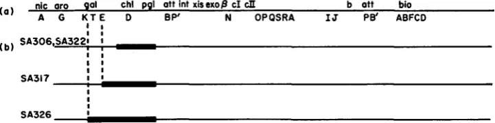
Isolation of bacterial deletions: Using techniques described previously (AWYA e t al. 1968), chl
bacterial mutants of SA500 were isolated and screened f o r deletions entering adjacent genes. Five deletion mutants were used in this study. Four are shown in Figure Ib: SA306, SA322, SA317, SA326. The fifth, SA307, is shown in Figure 2.
C d l gradients: 4.8 m l of a CsCl solution in 0.01 M MgSO, were mixed with phage in a nitro- cellulose tube, overlayed with mineral oil and spun for 20 hr at 29K rpm in the Spinco SW39
rotor a t 20°C. Fifty to 100 fractions were collected into 0.01 M MgSO, and analyzed for phage
titers. Densities and DNA contents were calculated from gradients in which two marker phages were present (most often Ximm434 and hb2imm434c) using the equation of DAVIS and DAVIDSON
(1968).
R E S U L T S
Isolation of plaque-forming Xgal: Strains SA322 and SA306 carry independ- ently isolated deletions of the chLD and PgL genes (Figure l b )
.
The i d 8 5 7 pro- phage in strain SA322 has not been altered by the deletion as judged by induci- bility, phage yield, and buoyant density of phage released. Lysates of SA322 and SA306 were used to infect W3805 at a multiplicity of 10 and plated on EMBG plates to select transductants. Gal+ colonies appeared at a frequency of per( a ) nic or0 gal chl pgl att int xisexop cI cII b att bio A G U T E D B P' N OPQSRA IJ Pe' ABFCD
I I SA317 1 0
I
I
I SA326 I
FIGURE 1.-Orientation of prophage h and neighboring genes. nicA = nicotinic acid biosyn- thesis; ard: = 3-deoxy-D-arabino-heptulosonic acid-7-phosphate synthetase (tyrosine-repressible enzyme) ; galK = galactokinase; galT = galactose-I-phosphate uridyl-transferase; galE = uri-
dine diphosphogalactose Cepimerase, the operator-promoter proximal gene in the galactose op- eron; chlA,chlD = reduction of chlorate, nitrate; pgl = 6-phosphoglucon~lactonase; bioA, B,F,C,D = biotin biosynthesis. Capital letters on the prophage map indicate cistrons defined by nonsense (am) mutations. Not all lambda cistrons are indicated. There are 15 essential genes be-
tween the A and I genes of lambda. The termini of the prophage map are delineated by the two hybrid attachment regions attBP' o n the left and attPB' at the right (see GUERRINI 1969). If the prophage were to be cured, the bacterial attachment site alone would remain (attBB'). int = a function necessary for site specific integration and excision, xis is required only for excision. ex0 and /3 are cistrons comprising red, the phage generalized recombination system. cl specifies the
LAMBDA TRANSDUCING STRAINS
TABLE 2
Gal content of non-defective gal-transducing phages
193
Frequency of Galactose markers present
occurrence Gal DNA
among Gals strain numbers K-2 T-79 T-226 T-151 T-l T-60 E-16 content’
1/21 8 c c C c c c c 0.97
16/21 1,2,5,9,11,12,
+
c c C C c c 0.95 (#25)2/21 24527 C
1 /21 28
-
_
+ + + + c+ + + c
1 /21 26
- 317
+ + + + + + -
- 326
+ + + - - - -
13,14,16,17,18, 19,20,21,22,25
__ C C C c c
-
_
-Galactose markers were assayed by spot test for transducing activity of diluted Agal lysates. Markers were ordered previously by SHAPIRO and ADHYA (1969). c = complementation of the gene in question;
+
= allele present by recombination;-
= allele absent by recombination.* DNA content of A+ = 1.00.
plaque-forming particle; 37 transductants were purified by restreaking. Thirty- five of these segregated Gal- clones, and on heat induction gave high frequency transducing (HFT) lysates for Gal. Twenty-one of these
HFT
lysates, when plated on EMBG plates for single plaques on W3805 at 30°C produced both normal turbid plaques and (at a frequency of about 0.05 to 0.2) plaques with a central Gal+ turbid area. All the Gal+ plaques when purified gave lysates con- taining only phages that can transduce W3805. Various of these phage variants differed in density from the parental phage when banded in CsCl gradients (Table 2 and unpublished). The gal endpoints, determined by transduction of various gal point mutants, vary among isolates (Table 2).W e have also isolated plaque-forming Xgal phages carrying the distal gene (kinase) of the galactose operon. In these experiments hosts were used i n which the proximal part of the galactose operon, together with the pgl and
chlD
genes, was deleted (strains SA326 and SA317; Figure l b ) .Xgal segregation and lysogeny: According to the CAMPBELL (1962) model of transducing phage genesis, Xgal particles should carry the “left” prophage attach- ment region, attBP’. Since some of the “by’ region is not essential for phage growth, and since the densities of some plaque-forming hgal particles are lower than the parental (Table 2), we assume that the Xgal particles must have lost some phage genetic material from the “right” prophage end-presumably the
“b” region. One property of such a phage is that it lysogenizes at extremely low frequencies (GUERRINI 1969). W e will show next that this is also true for the hgal studied.
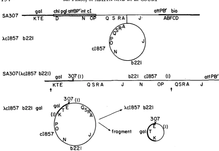
194 M. FEISS, S. ADHYA A N D D. L. COURT gal chl pgl att6Pint cI ottPB' bio
D N 06 Q S R A l J. ABFCD
S A 3 0 7 K T E
X c I 8 5 7 b221
gal 397(1) b221 c I 8 5 7 (1) att PB'
SA307(Xc1857 b221)
KT E Q S R A J N OP Q S R A J
t t
kc1857 b221
( ; (
-J 307 ( I )
cI857
N
b221
/
xc1857 b221\
fragment gal&')FIGURE 2.-Lysogenization of SA307 by XcI857b221. The deletion of SA307 is indicated by the hatched bar in the diagram of SA307; thereafter this deletion is noted by a check with the strain number: 307. Heavy lines denote bacterial genetic material. The numbers (I) indicate identical stretches of X DNA between P and Q. Abnormal excision of the prophage (at the ar- rows) with the left break point to the left of gal and the right break point to the right of the region ( I ) generates a galactose-transducing phage which is diploid for the region ( I ) . Recom- bination in the duplicated region, if reciprocal, would generate the hypothetical fragment shown and XcI857b221.
V
Stabilized Gal+ transductants can be isolated by coinfection with both the
Xgal and a A+ or Ximm4'4 helper phage. These stable lysogens segregate Gal- cells at a frequency comparable to that observed with lysogens of ordinary
Adgal.
A Xga2 single lysogen can be isolated by selection for Gal' on minimal galac- tose medium, or by plating on EMBG agar in the presence of antiserum to pre- vent lytic phage growth.
To summarize in part, a hgal phage does not form a stable lysogen except in rare instances detectable by strong selection. It readily lysogenizes with a helper phage.
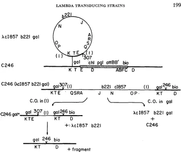
Transducing particles from SA307 (XcI857b221) : SATO and CAMPBELL (1970) reported a system for isolating lambda transducing variants containing deletions
LAMBDA T R A N S D U C I N G S T R A I N S 195
not be exceeded. We have used this system to look for plaque-forming trans- ducing phages. The bacterial strain used was SA307 which contains a deletion removing chlD and pgl and prophage genes including the P gene, as shown in Figure 2. The galactose operon in SA307 appears unaltered by the deletion.
SA307 was lysogenized with hcI857b221. The b221 marker is a large deletion of non-essential D N A and was used to increase the amount of relevant bacterial and phage D N A which might be carried by transducing particles.
A
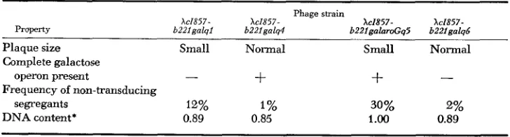
lysate of SA307(kZ857b221) was prepared by heat induction of a culture in tryptone broth. The lysate was used as a donor (at a multiplicity of I O ) to transduce the Gal- strain R594 to Gal+. R594 is galK2galT1, so a transducing phage must contain the wild-type galK2+gaZTI+ alleles. Since the normal inte- gration system is missing in strains carrying the b221 deletion, integration should require homologous recombination between bacterial D N A of the transducing particle and the recipient chromosome.The frequency of transduction of the lysate was about 1 X IO-? transductions per plaque-forming unit. Thirty-one transductants were examined. Of these, 8 released no plaque-forming phages and were not examined further. The remain- ing 23 transductants were heat-induced to prepare lysates. The lysates were diluted and plated at 30°C on R594 on EMBG agar. All 23 lysates yielded some plaques which contained a Gal+ center. When Gal+ clones derived from some of these Gal+ plaques were purified and induced, the resulting lysates again con- tained galactose-transducing plaque-forming phages when assayed on R594 on EMBG plates at 30°C. Phage isolated from plaques containing Gal+ centers were purified by single plaque isolation and high titer lysates prepared from con- fluent lysis plates. Such lysates contained mostly plaque-forming phage particles which could transduce R594 to Gal+. Therefore it was concluded that plaque- forming galactose-transducing particles had been isolated. Several properties of four such strains are presented in Table 3. Two of the four strains studied make somewhat smaller plaques than XcZ857b221. The strains were also tested for ability to transduce all of the galactose operon by using as a recipient a strain,
SA472, containing a deletion of the whole operon. Isolates XcI857b221galql and
XcZ857b221galqb must be broken in the galK gene, since they contain the wild-
TABLE 3
Properties of galactose-transducing phages from SA307 (AcI857b221)
Property
Phage strain
XcI857- XcI857- Xc1857- Xc1857-
b 2 2 1 ~ a l ~ 1 - . b221salo4 - . b221eaIaroGa5 b22lnala6
- .
Plaque size Small Normal Small Normal
Complete galactose
+
operon present -
Frequency of non-transducing
DNA content* 0.89 0.85
segregants 12% 1 %
-
+
30% 2%
1.00 0.89
~~ ~ ~
196 M. FEISS, S. ADHYA A N D D. L. CO U RT
type galK2+ allele but don’t transduce SA472 to Gal+. The two isolates capable of transducing all of gaZ were tested for the ability to transduce the adjacent bac- terial genes aroG and nicA. Isolate hcI857b221galq5, hereafter designated
hcI857b221galaroGq5, transduced the aroG marker; hcI857b221galq4 did not. Neither strain transduced nicA. The letter q which is included in the designation of these isolates serves to distinguish them from other galactose-transducing strains of 1 from which they differ in a number of respects. The letter q was chosen because the Q gene is the first h gene to the right of the gal insertion in these strains, as shown below.
Such plaque-forming, galactose-transducing strains of h presumably contain all the genes essential for the growth of wild-type h. Therefore the abnormal ex- cision events giving rise to the transducing strains must have occurred to the right of the P gene in the prophage contained in SA307(XcI857b221) (see Figure
2 ) .
plaque-forming units) derived from single plaques of galactose-transducing strains contained a fraction, from 1
%
to 30% (Table 3) of the plaque-forming particles which did not transduce the recipientR594
to Gal+. Repeated cycles of purification of galactose-transducing, plaque- forming phage resulted in similar lysates which contained a fraction of non- transducing particles.It
was postulated at this point that these phage strains are genetically unstable because of a duplication of phage genetic material bracket- ing the bacterial genes carried by the phage strains. Such a structure can be generated by a single abnormal excision event in SA307(XcI857b221) if the right-hand break occurs to the right of the genetic site between P and Q defined by the SA307 deletion. The origin of this structure is indicated in Figure 2, where (1) denotes the phage material in the duplication and the arrows indicate the sites of breakage in the excision event. Such a phage strain is expected to be un- stable because homologous recombination can promote crossing over between the two homologous copies of phage genetic material comprising the duplication. Such recombination results in a chromosome which has lost the galactose genes and the duplication.The fraction of non-transducing segregants is presumably a function of the rate of recombination in the duplication and the growth rate of the segregants relative to the growth rate of the galactose-transducing particles.
The DNA content of the four strains was determined by CsCl centrifugation with appropriate marker strains. The DNA content of these strains, as noted in Table 3, varies from 0.85 for hcI857b22Zgalq4 to 1.00 for kI857b22ZgalaroGq5,
where h+ has a DNA content of 1.00 and the parental phage, hc1857b221, has a
DNA content of 0.78 (DAVIS and DAVIDSON 1968).
As mentioned above, recombination between the two segments of DNA com- prising the duplication results in loss of the bacterial genes and the duplication- thus the non-transducing segregants should be identical to the parental phage,
XcI857b221. This prediction was tested by banding a lysate of XcI857b221galq4
(DNA content = 0.85) in CsCl with the marker phage AimmJ34b221 (DNA con- tent = 0.75). The non-transducing segregants were estimated to have a DNA
LAMBDA T R A N S D U C I N G STRAINS 197
XcI857 b221 gal
q4
p =
1.490
t
I I I I I0 10 20 30 40
1051
I
I IDROP NUMBER
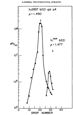
FIGURE 3.-CsCl gradient analysis of non-transducing segregants in a lysate of AcI857b221galq4:
A-A-A = AcI857b221galq4; 0-0-0 = non-transducing segregants; the arrow marks the position of the marker phage Ximm4s4b221.
content of 0.78 which agrees exactly with the DNA content of the parental phage
Xc1857b221 (see Figure 3 ) .
Lysogens of AcI85 7b221galq strains: Lysogens of the four phage duplication strains were prepared using as a host the strain C246 which contains a deletion extending from gal (the break in gal is either in galT or galE) through most of the biotin operon. This strain was transduced to Gal+ by the four galactose- transducing strains. Lysogenization of C246 by these phage strains was infre- quent. hcZ857b221galq4, for example, was classed as integration defective in the test of GOTTESMAN and YARMOLINSKY (1968). Lysogens of hcZ857b221galq4 and
198 M. FEISS, S. ADHYA A N D D. L. COURT
ration of lysogens of the galactose-transducing strains from SA322 and SA306. Rare transductants were easily found with XcI857b221galql and kcI857b221- galq6 since transduction required recombination between the two fragments of the galactose operon carried by the phages and C246. C246 was chosen because the gal mutation it carries, being a large deletion, cannot be picked up by the transducing phages and thus cannot segregate from the host.
These C246 lysogens are useful f o r obtaining information about the phage strains they carry, as discussed below. We sought first to show for one of the lysogens, C246 (XcI857b221galq4)
,
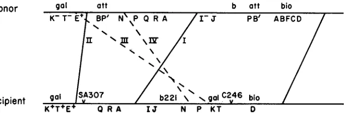
that the transducing phage was integrated into the bacterial chromosome by crossing over between the galactose genes car- ried by the phage and the homologous part of the galactose operon present in C246. This sort of integration is diagrammed in Figure 5, which shows the pro- posed structure of a C246 (hcZ857b221galq) lysogen. A PI transduction experi- ment has been performed to test this proposed structure, and thus eliminate other possibilities, such as plasmid formation (SIGNER 1969) and integration at sites in the C246 chromosome other than gal. Also note that the structure drawn for a C246 (XcZ857b221gaZq) lysogen is dependent on the presumed structure of theXcZ857b221galq phage strains. The P1 transduction experiment (diagrammed in Figure 4) also tests the order of lambda genes in the prophage which is a test of the proposed structure of the XcI857b221galq phages.
The donor strain used is W3350(XIam2) ; it is Gal- and Bio+ and contains the defective prophage XIam2. As shown in Figure 4, the wild-type prophage gene order in W3350(hIam2) differs from the gene order of the XcI857b221galq4 pro- phage in the recipient. The recipient is Gal+ and Bio-. GalfBiof recombinants are expected to arise in several ways in this cross. Since the P1 transducing phage carries a linear fragment of donor DNA, a double crossover is required for inte- gration of donor genetic material into the recipient chromosome. For the recipi- ent to become Bio+ the C246 deletion must be crossed out; this requires the right- most crossover indicated in Figure 4. There are four possibilities for the second crossover; these are indicated in Figure 4. Of these four possible crossover types, two yield non-defective lysogens (recombinant strains which upon induction pro-
Donor gal att b att bio
PB’ ABFCD
Recipient
K+T+E+ Q R A I J N P K T D
LAMBDA T R A N SD U C IN G STRAINS 199
XcI857 b221 gal
C 2 4 6
C 2 4 6 (XcI857 b221 gal) galx>7(I)
b221 c1857 (I) gaI2t6 bio
K T E ' QSRA J N O P . KT D
C.O. in (11
f---/
C O , in gal~ 2 4 6 gal+ gal 3Q7 (I gal 2f bio XcI857 b221 gal
KTE KT D
+
+s XcI857 b221 C 2 4 6
1
gal 2$6 bio
+fragment
KT
FIGURE Ei.-Lysogenization of C246 by Xc1857b22igalq isolates. The deletion i n C246 is shown for the diagram of C246 as a hatched bar; thereafter the (346 deletion is noted by a check with the strain number: 246. Heavy lines denote bacterial genetic material. Segregants which have lost the prophage can arise in two ways. Crossing over in the gal regions, noted as C.O. i n gal,
is expected to yield a gal bacterial strain identical to C246. Recombination in the phage duplica-
tion, noted as C.O. in ( I ) , is expected to yield a bacterial strain that is a gaZ/gaZ+ heterozygote
and which contains one copy of the phage genetic material comprising the duplication. The
gaZ/gal+ heterozygote is expected to give gal segregants identical to C246.
V
duce plaque-forming phage particles). These two types are indicated by dashed lines in Figure
4
and are labeled I11 and IV. The other two crossover types yield defective lysogens and are indicated by solid lines and are labeled I and 11. For technical reasons we have only studied Gal+ Bio+ recombinants that are defec- tive lysogens. The expected crossovers of type I yield defective lysogens contain- ing lambda genes from Q to J (as in SA307) which are either I+ o r Zam2. Type I1 crossovers yield lysogens containing XZam2. Both classes of recombinants are expected only if XcZ857b221galq4 has integrated at gal in C246. Recombinants of the sort described for type I crossovers are only expected if the presumed struc- ture for XcZ857b221galq4 is correct.Bacteriophage Plkc was grown on W3350 (hZam2) to produce a donor lysate.
200 M. FEISS, 5 . ADHYA A N D D. L. C O U R T
transductants were purified and 12/35 were found to be defective lysogens. The defective lysogens were of the type expected-of 12 examined, one was a type I1 recombinant containing XIam2 and ten were type I recombinants. Three of the type I recombinants were Iam2+ and seven were Iam2. The remaining trans- ductant carried no phage genes and perhaps arose by transduction of a non- immune recipient cell from which the XcI857b221 prophage has segregated, re- sulting in excision and segregation of the XIam2 prophage from the donor frag- ment. The fact that the two predicted types of Bio+ transductants were obtained strongly supports the structure of C246 (hcI857b221galq4) indicated in Figure 4. Integration of XcI857b221galql and XcI857b221galqb in the galactose region of C246 is very likely since generation of Gal+ cell requires crossing over be- tween the two fragments of the galactose operon carried by the phage and bacte- rial chromosomes.
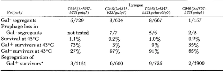
Several properties of the four lysogens are presented in Table 4. As shown in Figure 5 , the lysogens have two duplications; the duplication of phage material and a duplication of bacterial genes including the galactose region. T h e lysogens are thus gal+/gal heterozygotes and should segregate gal cells. Cultures of the lysogens plated on EMBG agar at 30°C contain gal segregants. Such gal segre- gants have also become non-lysogenic, as expected from the structure indicated in Figure 5 .
Segregants which have lost the XcI857b221 prophage can also be selected by plating a culture (grown at 30°C to allow segregation) at 43°C with anti-X serum to prevent reinfection. Cells carrying the prophage are killed when the phage is induced by heating. Two types of segregants are expected; recombination within the duplication of phage DNA will generate a gal+/gal cell line, whereas recom- bination within the duplication of bacterial genes will generate a gal cell as shown in Figure 5. The total frequency of cured segregants and the percentage of segregants that are Gal+ and Gal- are presented in Table 4. I n cultures of each of the four lysogens both Gal+ and Gal- segregants were obtained. The Gal+ segregants still contain the duplication of galactose material and one copy of the
TABLE 4
Properties of C246 lysogens of transducing phages from SA307 (XcI857b221)
Lysogen
C246 (XcIX57- C246( Xc1857- C246(Xc1X57- Property b221galql) b221galq4) b22lgalaroGq5)
Gal- segregants 5/729 3/604 8/667
Prophage loss in
Gal- segregants not tested 7/7 5 / 5
Survival at 43°C 1.1% 0.2% 1.0%
Gal+ survivors at 43°C 73 % 3% 9%
Gal- survivors at 43°C 27% 97% 91%
Segregation of
Gal+ survivors* 3/1131 6/600 9/726
C246(XcIX57- b221 galqb )
1/157
2/2 0.2% 35%
65%
2/1900
LAMBDA T R A N S D U C I N G STRAINS 20 1
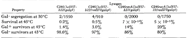
TABLE 5
Properties of C246(XcI857b221galq4): effect of rec and red mutations
Lysogen
C246(Xc1857- C246(Xc1857- C246recA(Xc1857- C246recA(Xc1857-
property b221galq4) b221red270galq4) b221gaIq4) 221red270galq4)
Gal- segregation at 30°C 2/1550 4/910 0/2000 0/1750 Survival at 43°C 0 2% 0.5% 7 x 1 1 ~ 5 % 5 x 1@5%
Gal+ survivors at 43°C 1.4% 3.0% 14% 20% Gal- survivors at 43°C 98.6% 97% 86% 80%
phage material originally duplicated. These gal+/gal heterozygotes are expected to be genetically unstable, segregating gal clones. As noted in Table 4 a Gal+ survivor from each lysogen was unstable. Since the Gal+ survivors of C246 lyso- gens still contain one copy of the phage material present in the duplication one can ask if the duplication contains any known h genetic markers. This is done
for the Gal+ survivors of C246 (hZ857b22Igalq6). If any phage markers are car- ried one expects that Q gene markers would be most likely to be present. How- ever, no
Q
markers could be rescued.Role of generalized recombination systems in segregation properties of lyso- gens: The effect of mutations in the bacterial (rec) and phage (red) generalized recombination systems on segregation properties of a C246 lysogen of one of the transducing strains: hcI857b221galq4 is shown in Table
5.
The phage red system has little if any role in the production of segregants: C246(hcZ857b22Ired270- galq4) has about the same survival frequency as C246 (hcZ857b22Igalq4) when plated at 43"C, and the percentages of Gal+ and Gal- survivors are not signifi- cantly changed. This result is expected since the phage red system is under the control of the h repressor and is therefore not expressed during growth of a lyso- genic culture at 30°C. A dramatic effect is observed when the bacterial rec sys- tem is inactive. With C246recAI (hcZ857b22Igalq4) survival decreased by a fac- tor of I O 4 to 7x
and an increased fraction of Gal+ survivors ( 1 4 % ) was obtained. These results are independent of the state of the red system, since sim- ilar values were obtained with C246recAl (hcZ857b22Ired270galq4). Surviv- ors obtained from rec lysogens could occur by a number of mechanisms: (1) reversion of the rec mutation, followed by segregation, ( 2 ) deletions, ( 3 ) phage mutations allowing survival of the lysogen, ( 4 ) residual recombination ac- tivity present in reCA lysogens which allows a low rate of segregation. Both the Gal+ and Gal- survivors obtained from rec lysogens are still rec, as determined by the test of UV sensitivity, indicating that the survivors are not rec+ revert- ants. The rec character was further tested for two Gal- survivors by matings with202 M. FEISS, S. ADHYA A N D D. L. COURT
endpoints in the galactose region. Thus some survivors should not be transduced by Xc1857ga128 which contains the galE gene and part of the galT gene. In fact, 24/24 (in two independent groups of 12) Gal- survivors of C246recA1 (hc1857b- 221gaZq4) were transduced to Gal+ by Xc1857gaZ28. This transduction result in- dicates that the C246 deletion endpoint remains in the survivors and that dele- tions are not the source of the Gal- survivors of recA lysogenes. I n sum, these tests indicate that a residual generalized recombination activity remains in the recA lysogens.
The increased fraction of Gal+ survivors in recA derivatives of C246 (XcI857b- 221galq4) is possibly due to a significant fraction of cells which survive the 43°C treatment because the prophage has lost (by mutation) the ability to kill the host a t 43"C, but this possibility has not been tested.
The duplication strains of h described here have the E. coli galactose operon situated between the X P and
Q
genes. One might expect such an addition to have a radical effect on normal phage development. Transcription of the regionc I I . O . P ~ Q ~ S R proceeds in a rightward direction (KOURILSKY et al. 1968; TAYLOR,
HRADECNA
and SZYBALSKI 1967), whereas the galactose operon is transcribed leftwards (GUHA, TABACZYNSKI and SZYBALSKI 1968). The opposite direction of transcription of the galactose operon is expected to interfere (LEVINTHAL and MIKAIDO 1969) with transcription from the P gene to theQ
gene (if it occurs). Such speculation raises the question of how transcription of theQ
gene is accomp- lished in strains carrying galq additions. Although this question remains open, we have compared the galq4 addition with another chromosomal abnormality,nin5, which maps between P and Q and alters normal lambda transcription. The
nin5 mutation, isolated and described as pf by COURT and SATO (1969), is a dele- tion of 5.4% of the h chromosome (COURT 1970). The nin5 mutation renders strains of N-independent; that is, able to grow in the absence of N gene product.
hcI857b221 Nam53galq4 was prepared, and its ability to grow in the non-permis- sive host R594 was compared with control strains.
R594 was infected at a multiplicity of 2.5 phage/cell. The yield of phage/in- fected cell was determined 90 min after infection. The yield of XcI857b221galq4
was 84 ;the yield of Xc1857Nam7am53nin5 was 24. Both hcI857b221Nam53- galq4 and XcI857Nam7am53 gave yields of less than one phage/cell, indicating that the galq4 addition does not confer the nin character.
DISCUSSION
Isolation of plaque-forming hgal strains has been described. We note that these strains have already been useful in genetic studies (SIGNER 1969; ECHOLS 1970) and galactose transformation of mammalian cells ( MERRILL, GEIER and PETRIC- CIANI 1971).
The transducing phages isolated from SA307 (hcI857b221) carry additions which contain various amounts of bacterial and phage genetic material. The smallest addition, that carried by hcI857b221galq4, is estimated to be 7% of the
LAMBDA T R A N S D U C I N G STRAINS 203
mated from the sum of the number of amino acid residues in the three galactose enzymes. This sum is 1088
(WILSON
andHOGNESS
1969a,b; SAITO, OZUTSUMI andKURAHASHI
1967). The sum of 1088 X 3 base pairs per amino acid equals 3264 base pairs as the minimum size of the galactose operon. 3264 base pairs + 50,000 base pairs per X+ chromosome gives a n estimate for the galactose operon of 6.5% of the length of the h+ chromosome. Thus the size of the galq4 addition agrees well with the minimum size of the galactose operon. One expects that the size of the duplication of phage DNA in this strain is at most 1%
of the A + length. The genetic data are consistent with this expectation; only 3% of the non- lysogenic survivors of C246 (hcZ857b221galq4) are Gal+ and arose by recombi- nation in the phage duplication.XcI857b221galql and XcI857b221galq6 both contain 11
%
additions. Both are broken in the galactose operon between the amino terminal end of the ga!K gene and the galK2 site. They contain less bacterial DNA than hcI857b22lgalq4,which contains the complete galactose operon. Since the gala6 and galql addi- tions are both estimated to be larger than the galq4 addition, the excess DNA must be phage DNA comprising the duplication. The difference is a minimum estimate of the size of the duplications carried by hcZ857b22lgalql and
AcI857b221galq6; for both strains this estimate is 11
%
-
7% = 4%.As discussed above, the Gal+ survivors of C246 lysogens of these duplication phages contain the addition which includes the phage DNA in the duplication. This phage DNA is at least 4% of the A+ chromosome length in the case of the
galq6 addition. Since no Q gene markers could be rescued from the Gal+ sur- vivors of C246(hcZ857b221galq6) the Q gene of X is not included in the phage duplication carried by hcI857b221galq6. This means that the deletion mutation in SA307 has left intact a stretch of prophage DNA between the P and
Q
genes equal in length to at least 4% of the A+ length. Recent studies have resulted in a physical map of the vegetative lambda chromosome ( DAVIDSON and SZYBALSKI1971). The lambda chromosome length is defined as 100% and the end of the chromosome near the A gene as 0% and the end near the R gene as 100%. On this map a site in the
Q
gene is at 91.7 and a site in the P gene is at 81.4. The SA307 deletion endpoint is at least 4% to the left of theQ
gene and thus must be at or to the left of 87.7. nin5 deletes DNA from 83.8 to 89.2. COURT (1970) was unable to recover the nin+ allele from SA307, so the SA307 deletion endpoint probably does not map to the left of 83.8.204 M. FEISS, S. ADHYA A N D D. L. COURT
of the elements comprising the addition: the duplication, bacterial transcription units including promoters and stop signals, and entirely new base sequences created by the deletion and abnormal excision event.
Other examples of partial diploid strains of bacteriophage exist. WEIL et al.
(1965) have described tandem duplication strains of T 4 in which duplications were specifically selected in the rZZ region of the T 4 chromosome. BERG (1 970) has described tandem duplication strains of h formed by recombination between
h and the h-derived plasmid hdu. BELLETT, BUSSE and BALDWIN (1971) have pre- sented evidence that tandem duplication strains can be detected in stocks of a A-880 hybrid phage. ANDOH and OZEKI (1968) isolated a n su3+-transducing de- rivative of 880 which was genetically unstable. This phage segregated non-- transducing variants which also contained less DNA than the transducing par- ticles. It seems likely, as ANDOH and OZEKI suggested, that 880su3+ phage has a structure analogous to the structure of the partial diploid galactose-transducing phages described here.
We thank Dr. ALLAN CAMPBELL for advice and suppxt during the course of this work. W e thank DOROTHEA SCANDELLA for helpful discussions and those mentioned for gifts of strains. A preliminary account of part of this research has appeared: FEISS and CAMPBELL (1970).
LITERATURE CITED
ADHYA, S., P. CLEARY and A. CAMPBELL, 1968 A deletion analysis of prophage lambda and ad jacent genetic regions. Proc. Nat. Acad. Sci. U.S. 41 : 956-962.
ADHYA, S. and M. SCHWARTZ, 1971 Phosphoglucomutase mutants of Escherichia coli K-12.
J. Bacteriol. 108: in press.
ANDOH, T. and H. OZEKI, 1968 Suppressor gene su3+ of E. coli, a structural gene f o r tyrosine TRNA. Proc. Nat. Acad. Sci. US . 59: 792-799.
BELLETT, A. J. D., H. G. BUSSE and R. L. BALDWIN, 1971 Tandem genetic duplications in a de- rivative of phage lambda. pp. 501-514. In: The Bacteriophage Lambda. Edited by A. D. HERSHEY. Cold Spring Harbor Laboratory, Cold Spring Harbor, New York.
BERG, D. E., 1970 CAMPBELL, A., 1957
Plasmid mutants of bacteriophage lambda. Genetics fj4: s6.
Transduction and segregation in Escherichia coli. Virology 4 : 366-384. -, 1961 Sensitive mutants of bacteriophage lambda. Virology 14: 22-32. -, 1962
Studies of bacteriophage lambda: I. Helping effect in lambda gal transduc- tion, 11. Regulation of lambda development: The isolation of two new regulatory gene mu- tants of Lambda. Ph.D. Thesis, University of Rochester.
Studies of novel transducing variants of lambda: dispensability of genes N and Q. Virology 39: 348-352.
Physical and chemical characteristics of lambda DNA. pp. 45-82. In: The Bacteriophage Lambda. Edited by A. D. HERSHEY. Cold Spring Harbor Laboratory, Cold Spring Harbor, New York.
Electron-microscopic visualization of deletion mutations Proc Nat. Acad. Sci. U.S. 60: 243-250.
Deletion mutants of bacteriophage lambda. 111. Phys- ical structure of att@. J. Mol. Biol. 5f5: 403-42.3.
Episomes. Advan. in Genetics 11: 101-146. COURT, D., 1970
COURT, D. and K. SATO, 1969
DAVIDSON, N. and W. SZYBAISKI, 1971
DAVIS, R. W. and N. DAVIDSON, 1968
LAMBDA TRANSDUCING STRAINS 205
DE LUCIA, P. and J. CAIRNS, 1969 Isolation of an E. coli strain with a mutation affecting DNA polymerase. Nature 224: 1164-1166.
ECHOLS, H., 1970
excision-specific recombination protein. J. Mol. Biol. 47: 575-583.
FEISS, M. and A. CAMPBELL, 1970 lambda. Genetics 64.: s19.
GOTTESMAN, M. E. and M. B. YARMOLINSKY, 1968 lambda. J. Mol. Biol. 31 : 487-505.
GUERRINI, F., 1969
GUHA, A., M. TABACZYNSKI and W. SZYBALSKI, 1968
Integrative and excisive recombination by bacteriophage A: Evidence f o r an Isolation and characterization of partial diploid strains of
Integration-negative mutants of bacteriophage
oh the asymmetry of lambda integration sites. J. Mol. Biol. 46: 523-542. Orientation of transcription f o r the galac- tose operon as determined by hybridization of gal mRNA with the separated DNA strands of coliphage lambda dg. J. Mol. Biol. 35: 207-213.
The relationship between heritable physical and ge- netic properties of selected gal- and gal+ transducing lambda dg. Virology 30: 482-492.
Studies on the messenger RNA of bacteriophage lambda. I. Various species synthesized early after induction of the prophage. Proc. Nat. Acad. Sci. U.S. 60: 1013-1020.
Consequences of deletion mutations joining two operons of opposite polarity. J. Mol. Biol. 42: 511-520.
Formation of merodiploids in matings with a class of rec- recipient strains of
Escherichia coli K12. Proc. Nat. Acad. Sci. U.S. 60: 16CL167.
Bacterial gene expression in mammalian cells. Nature 233 : 398400.
Genetics of the left arm of the chromosome of bacteriophage lambda. Genetics 59: 311-325.
-
, 1971 Deletion mutants of bacteriophage lambda. 11. Ge- netics properties of aft-defective mutants. J. Mol. Biol. 56: 385-401.Deletion mutants of bacteriophage lambda. I. Iso- lation and initial characterization. J. Mol. Biol. 56 : 369-384.
ROTHMAN, J., 1965 Transduction studies on the relation between the prophage and host chromo-
SAITO, S., M. OZUTSUMI and K. KURAHASHI, 1967 Galactose-1-phosphate uridylyltransferase of
Escherichia coli, 11. Further purification and characterization. J. Biol. Chem. 242 : 2362- 2368.
KAYAJANIAN, G. and A. CAMPBELL, 1966
KOURILSKY, P., L. MARCAUD, P. SHELDRICK, D. LUZZATI and F. GROS, 1968
LEVINTHAL, M. and H. NIKAIDO, 1969
Low, B., 1968
MERRILL, C. M., M. GEIER and J. PETRICCIANI, 1971
PARKINSON, J. S., 1968
PARKINSON, J. S. and R. J. HUSKEY, 1971
some. J. Mol. Biol. 12: 892-912.
SATO, K. and A. CAMPBELL, 1970
SHAPIRO, J. A. and S. L. ADHYA, 1969
Specialized transduction of galactose by lambda phage from
The galactose operon of E . coli K12. 11. A deletion
Plasmid formation: a new mode of lysogeny by phage A. Nature 233: 158- a deletion lysogen. Virology 41 : 474-487.
analysis of operon structure and polarity. Genetics 62: 249-264..
160.
SIGNER, E. R., 1969
TAYLOR, A. L., 1970
TAYLOR, K., Z. HRADECNA and W. SZYBALSKY, 1967
Current linkage map of Escherichia coli. Bacteriol. Rev. 32: 155-175. Asymmetric distribution of the transcribing regions on tha camplementary strands of coliphage lambda DNA. Proc. Nat. Acad. Sci. U.S.
57: 1618-1635.
THOMAS, R., C. LEURS, C. DAMBLY, D. PARMENTIER, L. LAMBERT, P. BRACHET, N. LEFEBVRE,
206 M. FEISS, S. ADHYA A N D D. L. COURT
SZYBALSKI, W., 1970 Genetic and molecular map of Escherichia coli bacteriophage lambda (X).
Handbook of Biochemistry, 2nd Ed. 1-35-1-38. The Chemical Rubber Co., Cleveland, Ohio. WALLACE, B. J. and J. P I ~ A R D , 1967 Genetic and biochemical analysis of the isoenzymes con-
cerned in the first reaction of aromatic biosynthesis in Escherichia coli. J. Bacteriol. 93:
237-2444.
WEIL, J., B. TERZAGHI and J. CRASEMANN, 1965 Partial diploidy in phage T4. Genetics 52:
683-693.
WILSON, D. B. and D. S. HOGNESS, 1969 The enzymes of the galactose operon in Escherichia