pISSN 2320-1770 | eISSN 2320-1789
Research Article
Association between ophthalmoscopic changes and obstetric outcomes
in pre-eclampsia and eclampsia
Sujoy Dasgupta*, Payal Banerjee Ray
INTRODUCTION
Hypertensive disorder in pregnancy is a major concern among the obstetricians. It is a major cause of maternal and perinatal mortality and morbidity world-wide, especially in low and middle income countries where they account for 10-25% of maternal deaths.1 The Working Group of the National High Blood Pressure Education Program (NHBPEP) defined hypertension as blood pressure ≥140/90 mmHg using Korotkoff V sound for diastolic blood pressure.2 The same Working Group classifies hypertensive disorders in pregnancy into four groups- (i) Gestational Hypertension; (ii) Preeclampsia and Eclampsia syndrome; (iii) Superimposed
preeclampsia on chronic hypertension; (iv) Chronic hypertension.2 Preeclampsia is defined as presence of hypertension and proteinuria (300 mg or more in 24 hour urine).2
Preeclampsia is a multisystem disorder of vascular function, specific to pregnancy. It arises from placenta, probably as a result of ischemia, and propagates throughout the maternal vascular tree such that virtually all organ systems can be affected. Unfortunately, it continues to be a disease of theories and no one theory, alone, can explain its pathogenesis. Roberts et al proposed that maternal endothelial dysfunction is the key event resulting in diverse clinical manifestation of Department of Obstetrics & Gynaecology, KPC Medical College and Hospital, IF Raja S. C. Mullick Road, Jadavpur, Kolkata, India
Received: 30 September 2015
Accepted: 02 November 2015
*Correspondence:
Dr. Sujoy Dasgupta,
E-mail: dr.sujoydasgupta@gmail.com
Copyright: © the author(s), publisher and licensee Medip Academy. This is an open-access article distributed under the terms of the Creative Commons Attribution Non-Commercial License, which permits unrestricted non-commercial use, distribution, and reproduction in any medium, provided the original work is properly cited.
ABSTRACT
Background: Hypertensive disorder is a major cause of maternal and perinatal mortality and morbidity. The widespread endothelial dysfunction associated with preeclampsia can affect the choroid and the retina leading to characteristic ophthalmoscopic changes. So, we tried to find out the association between ophthalmoscopic changes and obstetric outcomes in women with preeclampsia-eclampsia.
Methods: In a comparative, prospective study carried out from July 2011 to July 2015 in Medical College, Kolkata
and KPC Medical College, Kolkata we included antenatal women with pre-eclampsia and eclampsia. Based on the ophthalmoscopic findings they were divided into two groups; group A (no ophthalmoscopic abnormalities) and group B (having ophthalmoscopic abnormalities). The results were analyzed by standard statistical methods.
Results: Out of the total 200 women included, 102 women belonged to group A and 98 to group B. Majority of the
patients were ophthalmologicaly asymptomatic. Most common fundoscopic abnormality in group B was focal arteriolar narrowing. Women in group B had significantly higher level of blood pressure, proteinuria and thrombocytopenia. The need of multiple drugs, incidence of HELLP syndrome and oliguria, rate of Caesarean section and CCU admission were significantly higher in group B compared to group A. There was significantly higher incidence of perinatal complications in group B, viz: IUGR, low birth weight, low Apgar score and NICU admission.
Conclusions: Ophthalmoscopic changes correlate positively with adverse feto-maternal outcomes in preeclampsia. Fundoscopy should be carried out in all preeclamptic mother irrespective of the visual symptoms.
Keywords: Pregnancy, Preeclampsia, Hypertension, Retinopathy, Ophthalmoscopy, Obstetric outcome
preeclampsia.3 So, it is not surprising that this endothelial dysfunction may affect the choroid and the retina leading to characteristic ophthalmoscopic changes.
Ophthalmological examination may provide clinical evidence of vasoconstriction, which otherwise may be difficult to establish. The findings may correlate with severity of preeclampsia. The retinal vascular changes are present in 30-100% of preeclampsia cases, the most frequent being the vasoconstriction of the retinal arterioles.4,5 The exudative retinal detachment is a rare cause (under 1%) of visual loss in the preeclampsia-eclampsia syndrome, being produced by the involvement of the choroidal vascularization.4 Retinal infarction or ischemia (Purtscher retinopathy) may lead to sudden onset blindness.6 Asymptomatic retinal detachment is also common.6 In most cases, these changes resolve after delivery.
Preeclamptic women may have visual complaints. The most common symptoms are scotoma, a transient perception of bright or black spots. There may be sudden inability to focus blurred vision and in severe cases, complete blindness. The visual symptom is considered as of the criteria for defining severe preeclampsia.2 The NICE clinical guidelines consider visual disturbance or papilloedema in the background of hypertension and proteinuria as severe preeclampsia and recommend consideration of prophylactic magnesium sulphate therapy in such conditions.7
Keeping all these in mind, our study was done to find out the association between ophthalmological changes and obstetric outcomes in women with preeclampsia and eclampsia.
The aims of our study are (1) to find out the factors associated with retinopathy in preeclampsia-eclampsia; and 2) to detect the correlation between ophthalmoscopic changes and maternal and fetal outcomes in preeclampsia-eclampsia.
METHODS
A comparative prospective study was carried out simultaneously in pregnant women attending the Emergency and the Out Patient Department of the Department of Obstetrics and Gynecology, Medical College and Hospital, Kolkata and KPC Medical College and Hospital, Kolkata from July 2011 to July 2015. Hypertension was defined according to NHBPEP criteria, i.e., women having BP ≥140/90 mm Hg in at least two occasions, six hours apart were included in the study. Antenatal women with preeclampsia and eclampsia-were all included. Excluded were women having gestational hypertension, chronic hypertension, preeclampsia superimposed on chronic hypertension, pre-existing diabetes, known myopia or other known retinopathy, thyrotoxicosis, anaemia or other haematological disorders, renal disorders and women presenting in
postpartum period. Total 200 women were studied in the study period.
After getting the clearance from the Institutional Ethical Committee (IEC), informed consent was obtained from all the participants. Detailed history taking and systemic and obstetric examination were done. Proteinuria was provisionally diagnosed by dipstick urine test method (≥1+ or ≥300 mg/ litre) and confirmed by presence of ≥300 mg protein in 24 hour urine collection. Laboratory investigations were done with special reference to haemoglobin and platelet count, lactate dehydrogenase (LDH), liver enzymes (SGOT- Serum glutamate oxaloacetate transferase and SGPT- Serum glutamate pyruvate transferase) and creatinine, obstetric ultrasound to detect fetal growth and liquor volume and colour Doppler studies whenever indicated.
Detailed ophthalmological check-up was done including detailed history (especially blurred vision, scotoma, diplopia, blindness), examination of visual acuity followed by detailed choroidoretinal check up with the help of direct ophthalmoscope using tropicamide as mydriatic, soon after the diagnosis of hypertension.
Treatment was started as indicated, using antihypertensive drugs, antenatal corticosteroids and magnesium sulphate when indicated. All the women were followed up regularly to note control of BP, worsening of clinical and/or laboratory parameters, development of eclampsia and fetal growth and well-being. The labour events, mode of delivery, gestational age at delivery and birth weight, Apgar score, admission to NICU (Neonatal Intensive Care Units) were also studied. The maternal conditions particularly noted were eclampsia, pulmonary oedema, HELLP syndrome (Haemolysis, Elevated Liver enzymes and Low Platelet counts), oliguria, haemorrhage, caesarean section rate, admission to CCU (Critical care unit) and maternal mortality.
The women were divided into two groups based on the findings of ophthalmological examination. Those without ophthalmoscopic changes were included into the “Group A” and those with such changes, into the “Group B”.
Both the groups were compared in terms of demographic variables, clinical and laboratory parameters and maternal and fetal outcomes. The results were analysed by standard statistical methods like two sample „t‟ test (both tailed or one tailed accordingly) and Fisher‟s exact test (using 2 x 2 contingency table) and p value <0.05 was considered to be significant.
RESULTS
women with hypertension had retinal changes at ophthalmoscopy (Figure 1).
Table 1: Distribution of the women according to the visual symptoms.
Group A (n = 102)
Group B (n =98)
Blurred Vision,
Scotoma 21 (20.59%) 49 (50.00%) Diplopia 0 (0.00%) 2 (2.04%) Blindness 0 (0.00%) 1 (1.02%) Asymptomatic 81 (79.41%) 46 (46.94%)
[image:3.595.52.286.139.229.2]In group A, i.e., women without any ophthalmoscopic abnormalities, majority (79.41%) were ophthalmologicaly asymptomatic and only few women (20.59%) had mild symptoms like blurred vision or scotoma. In contrast half (50.00%) of the women in group B, i.e., women with ophthalmoscopic changes, had complaints of visual blurring and/ or scotoma. One women (1.02%) developed sudden onset blindness (due to central serous retinopathy) that resolved after delivery. Two (2.04%) complained of diplopia. It is important to note that 46 women with ophthalmoscopic abnormalities (46.94%) did not have any visual complaints (Table 1).
Table 2: Ophthalmoscopic findings in women in group B.
Number of women (n = 98)
Percentage (%)
Arteriolar narrowing 68 (69.39%) Cotton wool exudates 7 (7.14%) Retinal oedema 8 (8.16%) Retinal haemorrhage 2 (2.04%) Macular changes 5 (5.01%) Retinal detachment 4 (4.08%)
CSR 1 (1.02%)
Papilloedema 0 (0.00%)
In group B, the most common ophthalmoscopic finding (in 69.39% cases) was focal arteriolar narrowing leading to decreased arterio-venous (A:V) ratio (from 2:3 to 1:3). Retinal oedema, cotton wool exudates, retinal haemorrhage and macular oedema and macular stars were also common findings. Central Serous Retinopathy (CSR) was noted in one woman. However, not a single case of papilloedema was reported (Table 2).
Both the groups were comparable in terms of demographic variables like maternal age, gravida, Body mass index (BMI) and gestational age at diagnosis of hypertension. Women with group B had significantly higher levels of both systolic blood pressure (SBP) and diastolic blood pressure (DBP) compared to those in group A. Also, in group B, the severity of proteinuria and
thrombocytopenia were significantly higher than in group A. However, there was no significant statistical correlation between ophthalmoscopic findings (group B) and laboratory parameters like serum LDH, liver enzymes and serum creatinine (Table 3).
Table 3: Factors associated with ophthalmoscopic changes.
BMI- body mass index; SBP- systolic blood pressure; DBP- diastolic blood pressure; LDH – lactate dehydrogenase; SGOT- Serum glutamate oxaloacetate transferase; SGPT- Serum glutamate pyruvate transferase;
a
Measured by dipstick, confirmed by 24 hours urinary protein excretion; *Significant p-value
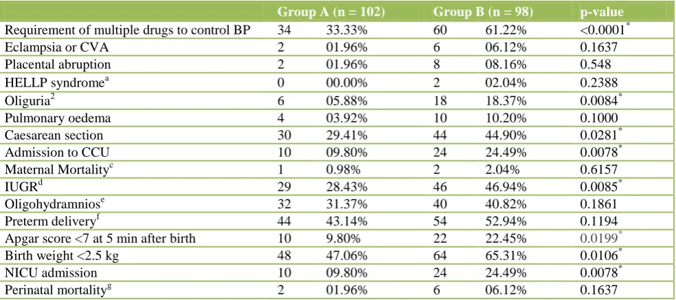
While carefully analysing the maternal outcomes we found that requirement of multiple drugs to control BP, incidence of oliguria, Caesarean section rate, rate of admission to CCU and maternal mortality were higher in group B compared to group A and this difference was statistically significant (p value <0.05 was considered significant). In contrast, there was no significant difference between these two groups in terms of development of eclampsia, placental abruption and pulmonary oedema. Regarding perinatal outcomes, the incidence of intrauterine growth restriction (IUGR), low Apgar score at birth, low birth weight and NICU admission rate were significantly higher in the babies of women in group B than those in group A. However, incidence of oligohydramnios and perinatal mortality were similar in both the groups (Table 4).
DISCUSSION
Ophthalmological changes are common in hypertensive disorders in pregnancy. The fundoscopic changes may reflect the global endothelial dysfunction that characterizes preeclampsia. More than fifty years ago, Pollak VE and Nettles JB reported that retinal vascular
Group A (n = 102) Mean ± SD
Group B (n = 98) Mean ± SD
p-value
Maternal Age
(years) 24.52±2.98 24.06±3.31 0.3025 Gravida 1.23±1.05 1.08±0.98 0.2980
BMI (Kg/m2) 22.89±1.53 23.09±1.78 0.3946 Gestational age at
diagnosis (weeks)
28.82±8.32 29.50±6.25
0.5155
SBP (mm Hg) 150.00±10.7 158.80±11.3 <0.0001* DBP (mm Hg) 112.56±10.2 116.95±10.8 0.0036* Proteinuriaa 1.69±0.32 2.10±0.21 <0.0001* Platelet count
(lacs/cmm) 1.56±0.32 1.34±0.21 <0.0001
*
Serum LDH
(IU/L) 675.45±94.8 672.93±82.9 0.8419 SGPT (IU/L) 48.76±10.75 51.85±12.79 0.0655 SGOT (IU/L) 57.92±7.74 59.75±6.38 0.0702 Serum creatinine
[image:3.595.311.544.173.406.2]changes correlate with renal biopsy findings in preeclampsia.8
Some studies reported the incidence of retinopathy may be as high as 85% in cases of pregnancy-hypertension.8 A recent study of Uto M et al showed incidence of choroidoretinal changes in pregnancy-hypertension was 43.2%. In our study we found this incidence 49%.9
Abu Samara K mentioned that 25% of women with preeclampsia may have visual symptom and in
[image:4.595.53.545.246.464.2]ophthalmoscopy 50% of the women may have abnormalities.5 Similarly, in our study, out of the 200 cases 127 women had no visual symptoms whereas 98 women had fundoscopic abnormalities. Thus, it is worthy to note that even without any visual symptoms, retinal changes may be present, which may carry adverse prognosis from obstetric point of view. So, in all women with preeclampsia, fundoscopic examination should be considered irrespective of the symptoms.
Table 4:Maternofetal complications associated with ophthalmological changes.
Group A (n = 102) Group B (n = 98) p-value
Requirement of multiple drugs to control BP 34 33.33% 60 61.22% <0.0001*
Eclampsia or CVA 2 01.96% 6 06.12% 0.1637
Placental abruption 2 01.96% 8 08.16% 0.548
HELLP syndromea 0 00.00% 2 02.04% 0.2388
Oliguria2 6 05.88% 18 18.37% 0.0084*
Pulmonary oedema 4 03.92% 10 10.20% 0.1000
Caesarean section 30 29.41% 44 44.90% 0.0281*
Admission to CCU 10 09.80% 24 24.49% 0.0078*
Maternal Mortalityc 1 0.98% 2 2.04% 0.6157
IUGRd 29 28.43% 46 46.94% 0.0085*
Oligohydramniose 32 31.37% 40 40.82% 0.1861
Preterm deliveryf 44 43.14% 54 52.94% 0.1194
Apgar score <7 at 5 min after birth 10 9.80% 22 22.45% 0.0199*
Birth weight <2.5 kg 48 47.06% 64 65.31% 0.0106*
NICU admission 10 09.80% 24 24.49% 0.0078*
Perinatal mortalityg 2 01.96% 6 06.12% 0.1637
BP- Blood Pressure; CVA- Cerebro-Vascular Accident; HELLP- Haemolysis; Elevated Liver enzymes; Low Platelet count; CCU- Critical care unit; IUGR- Intrauterine Growth Restriction; NICU- Neonatal Intensive Care Unit; aDefined as per NICE guidelines; bas urine output <30 ml/hour or <100 ml/ 4 hour; cOne mother died of coagulopathy, one of renal failure, one of embolism; d Defined as estimated fetal weight less than 10th percentile with respect to gestational age; eDefined as single largest vertical pocket <2 cm or Amniotic Fluid Index (AFI) <5 cm; fBirth before 37 completed weeks, spontaneous or iatrogenic; gInclude still birth (>28 weeks of gestation and early neonatal (<7 days) death; *Significant p-value
Figure 1:Distribution of the women according to the ophthalmoscopic findings.
[image:4.595.52.286.560.724.2]severe forms of retinopathy should be immediate termination of pregnancy along with conservative measures like control of blood pressure to prevent permanent visual damage.
Hong J et al showed that he degree of retinopathy correlated positively with the degree of gestosis and the degree of hypertension and they concluded that fundus examination in gestosis patients is an significant guideline for clinical treatment.12 In their retrospective study on hypertensive women who delivered at term, Gupta A et alfound that the severity of retinopathy might be independent of systemic BP.13 In contrast, Kalliaperumal S et al reported that the severity of retinopathy was more closely related with diastolic BP rather than systolic BP and the significance of this correlation increased in severe preeclampsia.14 In our study, however, we found that both SBP and DBP were significantly higher in women having ophthalmoscopic abnormalities, compared to women having normal fundus. Again, we found that severity of both proteinuria and thrombocytopenia was significantly higher in women having retinopathy than women without retinopathy. This can be explained by the fact that retinopathy represents the widespread multi-system vasospasm occurring in preeclampsia.
Retinopathy may serve as an independent indicator of severe preeclampsia. Araújo J et al reported a case of a woman with malignant hypertensive retinopathy who was discovered to bear a dead fetus.15 Our study found that obstetric complications were common in women having retinal changes compared to those without the changes. Some of these complications might be life threatening, viz: HELLP syndrome and oliguria. The rate of Caesarean section, admission to CCU and the need of more than one drugs to control BP were significantly higher in women having retinal abnormalities. So, we can easily argue that the ocular changes can predict the severity of preeclampsia.
Both Gupta A et al.and Kalliaperumal S et al.suggested that retinopathy in preeclampsia may indirectly indicate the level of placental insufficiency and thus can predict IUGR.13,14 In our study also, we found that the women having retinopathy had significantly higher incidence of IUGR than women having no retinopathy.
Karki P et alperformed a prospective cohort study in 153 subjects and found that ophthalmoscopic changes were associated with low birth weight and low Apgar score.16 In our study also, the incidence of low birth weight, low Apgar score and NICU admission rate were significantly higher in the mothers having fundoscopic changes compared to those without changes.
Garg A, et al, demonstrated subclinical retinal and choroidal thickening in the setting of severe preeclampsia, which may reflect rising levels of vascular endothelial growth factor.17 Therefore; retinal and
choroidal markers could serve as novel predictive markers of severe preeclampsia. This further establishes the need of routine ophthalmoscopic examinations in all preeclamptic mothers irrespective of visual complaints.
CONCLUSIONS
Ophthalmoscopic examination should be done in all cases of hypertensive disorders of pregnancy, even if the patient is asymptomatic. The choroidoretinal changes may indicate ongoing vascular damages and correlate with obstetric complications and poor perinatal outcomes. They may be the first warning sign of impending feto-maternal complications. DIPSI criteria was also found to be effective and comparable method of screening with respect to GCT/WHO GTT with 75 gm glucose, but larger studies are required to further validate its importance.
ACKNOWLEDGEMENTS
The authors acknowledge the contribution of Mr Abhyuday Chanda, Banaras Hindu University for the statistical analysis. We are deeply indebted to Prof Partha Sarathi Chakravorty, the Ex- Head of the Department, Obstetrics and Gynaecology, Medical College, Kolkata, who was mastermind behind this study. We are grateful to the doctors of Regional Institute of Ophthalmology, for performing the ophthalmoscopic examination of our patients.
Funding: No funding sources Conflict of interest: None declared
Ethical approval: The study was approved by the Institutional Ethics Committee
REFERENCES
1. Duley L. The global impact of pre-eclampsia and eclampsia. Semin Perinatol. 2009;33(3):130-7. 2. Working Group Report on High blood Pressure in
Pregnancy. National Institute of Health. National Heart, Lung and Blood Institute. National High Blood Pressure Education Program, NIH Publication, 2000.
3. Roberts JM, Taylor RN, Musci TJ, Rodgers GM, Hubel CA, McLaughlin MK. Preeclampsia: An endothelial cell disorder. Am J Obstet Gynecol. 1989;161:1200-4.
4. Mihu D, Mihu CM, Tălu S, Costin N, Ciuchină S, Măluţan A. Ocular changes inpreeclampsia. J Oftalmologia. 2008;52(2):16-22.
5. Abu Samra K. The eye and visual system in the preeclampsia/eclampsia syndrome: What to expect? Saudi J Ophthalmol. 2013;27(1):51-3.
6. Lam DS, Chan W. Images in clinical medicine. Choroidal ischemia in preeclampsia. N Engl J Med. 2001;344(10):739.
pregnancy. The management of hypertensive disorders during pregnancy. 2011:26-8.
8. Pollak VE, Nettles JB. The kidney in toxaemia in pregnancy. A clinical and pathological study based on renal biopsies. Medicine 1960;39:469-526 9. Uto M, Uemura A. Retinochoroidopathy and
systemic state in toxemia of pregnancy. Nihon Ganka Gakkai Zasshi. 1991;95(10):1016-9
10. Bos AM, van Loon AJ, Ameln JG.Serous retinal detachment in preeclampsia. Ned Tijdschr Geneeskd. 1999;143(48):2430-2.
11. Bartczak A, Kraśnicki P, Urban R, Laudaiński T, Mariak Z. Bilateral serous retinal detachment in preeclampsia--a case report. Klin Oczna. 2014;116(1):21-3.
12. Hong J, Liu Y, Liang X, Xiao J. Clinic analysis of retinopathy in gestosis patients. Yan Ke Xue Bao. 2000;16(4):262-3, 275.
13. Gupta A, Kaliaperumal S, Setia S, Suchi ST, Rao VA.Retinopathy in preeclampsia: association with birth weight and uric acid level. Retina. 2008;28:1104-10.
14. Kaliaperumal S, Setia S, Gupta A, Rao VA. Fetal birthweight and diastolic blood pressure: association with retinopathy in severe preeclampsia. Eur J Ophthalmol. 2008;18(5):809-12.
15. Araújo J, Tavares-Ferreira J, Penas S, Figueira L, Paiva FP, Falcão-Reis F. Malignant hypertensive retinopathy as a presenting sign of an occult dead fetus. Clin Ophthalmol. 2015;9:971-5.
16. Karki P, Malla P, Das H, Uprety DK. Association between pregnancy-induced hypertensive fundus changes and fetal outcomes. Nepal J Ophthalmol. 2010;2(1):26-30.
17. Garg A, Wapner RJ, Ananth CV, Dale E, Tsang SH, Lee W, et al. Choroidal and retinal thickening in severe preeclampsia. Invest Ophthalmol Vis Sci. 2014;55(9):5723-9.
Cite this article as: Dasgupta S, Ray PB.
Association between ophthalmoscopic changes and obstetric outcomes in pre-eclampsia and eclampsia. Int J Reprod Contracept Obstet Gynecol