Electronic Thesis and Dissertation Repository
4-21-2017 12:00 AM
Functional Connectivity in the Motor Network Largely Matures
Functional Connectivity in the Motor Network Largely Matures
Before Motor Function
Before Motor Function
Jordynne L V Ropat
The University of Western Ontario
Supervisor
Dr. Rhodri Cusack
The University of Western Ontario Graduate Program in Neuroscience
A thesis submitted in partial fulfillment of the requirements for the degree in Master of Science © Jordynne L V Ropat 2017
Follow this and additional works at: https://ir.lib.uwo.ca/etd
Part of the Cognitive Neuroscience Commons, Computational Neuroscience Commons, Developmental Neuroscience Commons, and the Systems Neuroscience Commons
Recommended Citation Recommended Citation
Ropat, Jordynne L V, "Functional Connectivity in the Motor Network Largely Matures Before Motor Function" (2017). Electronic Thesis and Dissertation Repository. 4539.
https://ir.lib.uwo.ca/etd/4539
This Dissertation/Thesis is brought to you for free and open access by Scholarship@Western. It has been accepted for inclusion in Electronic Thesis and Dissertation Repository by an authorized administrator of
ii
Abstract
The brain changes in many ways in the first year. It is not known which of these changes
are most critical for the development of cognitive functions. According to the Interactive
Specialization Theory, developments in behaviour result from changes in brain
connectivity. We tested this using functional connectivity magnetic resonance imaging
(fcMRI) of the motor system. fcMRI was acquired at three and nine months – two
time-points between which motor behaviour develops enormously. Infants were additionally
compared with adults. Subjects were scanned with a 3T MRI scanner, yielding BOLD
signal time-courses that were correlated with one another. Our results do not support the
Interactive Specialization Theory, as connectivity did not change with motor
development and instead was adult-like in the youngest infants. fcMRI has enabled
deeper exploration of network connectivity patterns and continues to emerge as a leading
method in infant neuroscience.
Keywords
Keywords: functional magnetic resonance imaging, functional connectivity, motor
iii
Co-Authorship Statement
Chapter two is presented in the form of a manuscript. The infants were recruited by
DSCL and VKH; data was acquired by ACL, CJW, HD, and CH prior to my arrival. I
analyzed the data and wrote the manuscript together with my supervisor, RC.
iv
Acknowledgments
I would like to sincerely thank my supervisor, Rhodri Cusack, for his help and support
throughout my Master’s degree. Thank you for the guidance you have given me, over the
past two-and-a-half years but especially throughout the final drafts of my thesis. I
appreciate having had the opportunity to work with functional MRI as well as with
infants, both of which have helped me to develop invaluable transferable skills. Thank
you also for giving me the opportunity to travel to present my research. I am very grateful
for the experiences I have had.
I would also like to thank all of the postdoctoral fellows in my lab who collected the data
for my thesis and who were instrumental in guiding me through this degree.
Finally, I would like to thank my family and close friends who have supported me
through everything. Thank you especially to my father, Michael, and my partner, Dante,
v
Table of Contents
Abstract ... ii
Co-Authorship Statement... iii
Acknowledgments... iv
List of Tables ... vi
List of Figures ... vii
Chapter 1 ... 1
1 Introduction and Literature Review ... 1
1.1 Motor Behaviour Changes Dramatically Throughout Infancy ... 2
1.2 The Adult Motor System: A Look into the Brain ... 8
1.3 Functional MRI in Brief, and its Challenges and Opportunities for Infant Neuroimaging ... 12
1.3.1 Background ... 12
1.3.2 MRI Acquisition ... 13
1.3.3 Data Analysis ... 14
1.3.4 Opportunities and Challenges of Infant fMRI ... 16
1.4 Resting-State Functional Connectivity as a Tool to Examine the Infant Brain .... 18
1.4.1 Age groups ... 22
1.4.2 Overview of networks identified ... 23
1.4.3 fcMRI of motor networks ... 24
1.5 Interactive Specialization Theory and Gaps in Literature ... 28
1.6 Thesis Outline ... 30
1.7 References ... 31
Chapter 2 ... 49
2 Manuscript-Based Experimental Data ... 49
2.1 Abstract ... 50
2.2 Introduction ... 51
2.3 Results ... 54
2.4 Discussion ... 57
2.5 Methods... 60
2.5.1 Participants ... 60
2.5.2 Data acquisition ... 60
2.5.3 Analysis... 62
2.6 References ... 64
2.7 Tables ... 69
2.8 Figures... 75
Chapter Three... 79
3 Conclusion ... 79
3.1 Discussion ... 79
3.2 Limitations and Future Work ... 82
3.3 References ... 84
vi
List of Tables
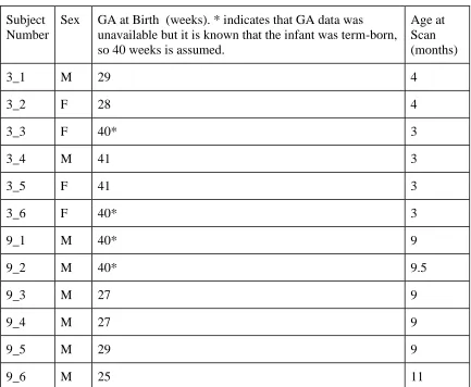
Table 1. Subject demographics. ... 69
Table 2. Subject neurodiagnosis and assessment of white and grey matter integrity. ... 70
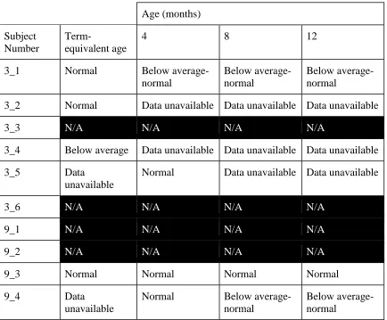
Table 3. Rating of motor ability at four time points. ... 71
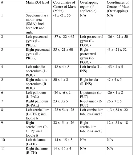
Table 4. Results of preliminary ROI (regions of interest) search. ... 72
Table 5. Final ROIs selected to utilize in analysis. ... 74
vii
List of Figures
Figure 1. Chronologic progression of gross motor development in infants from zero to six
months of age. ... 2
Figure 2. Infants will resist a pull on their visible arm. ... 3
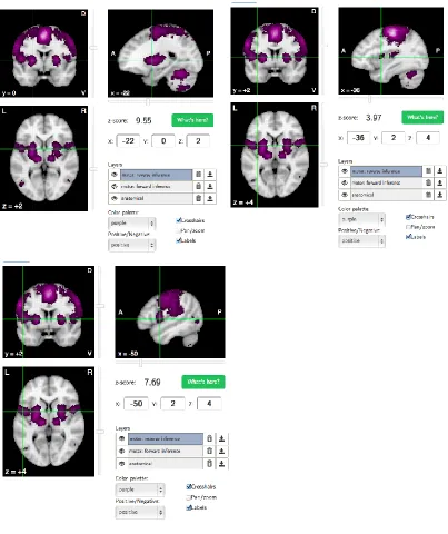
Figure 3. Results of neurosynth.org meta-analysis. ... 9
Figure 4. fMRI task-based activation response to finger movement. ... 18
Figure 5. A visual depiction of functional connectivity MRI. ... 19
Figure 6. BOLD signal time courses... 20
Figure 7. Results of initial reverse inference search on neurosynth.org. ... 75
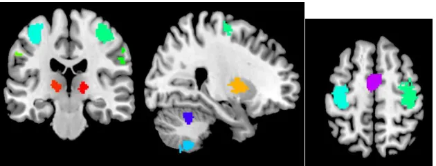
Figure 8. A slice of the brain illustrating the final selected ROIs. ... 76
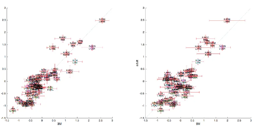
Figure 9. A three-dimensional rendering of the connectivity between each pair of brain regions, for each of the three age groups. ... 76
Figure 10. A matrix representation of the pairwise connectivities shown in Fig. 1. ... 77
Figure 11. For region pair of brain regions, a comparison of the strength of connectivity in the infants and adults. ... 77
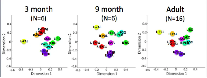
Figure 12. The higher-order structure in the connectivity, as revealed with hierarchical clustering, for each of the groups. ... 78
Chapter 1
1
Introduction and Literature Review
“The passage from the limited motor repertoire of the newborn to the complex locomotor
and manipulatory skills of the toddler stands among the most visible and dramatic
transformations in the human life cycle.”
1.1
Motor Behaviour Changes Dramatically Throughout
Infancy
Motor behaviour changes from birth through childhood and beyond as new skills are
learned and mastered. However, the most rapid and some of the most impressive changes
occur during in the first year of life (Figure 1). Infants enter the world virtually helpless
and – in the span of just a year – become active players in their own lives and the lives of
others.
Using cartoon infants, this image illustrates the monthly milestones that many infants reach. The image has been removed due to copyright restrictions.To view this image, please see Figure 2 in the following manuscript: Johnson, C., & Blasco, P. (1997). Infant growth and development. Pediatrics in review / American Academy of Pediatrics, 18 (7), 224-242. Adapted from Piper, M. C. (1994). Motor assessment of the developing infant. WB Saunders Company. Illustrations by Marcia Smith.
Though vastly limited in their motor abilities, most neonates (infants under four weeks of
age) consistently exhibit a number of actions often referred to as reflexes. Although
originally thought to be simplistic, stereotyped behaviours, reflexes may be more
complex than originally believed. For example, when an infant’s cheek is touched, she
will move her mouth there in search of milk – this is called rooting. However, when she
touches her own cheek, or if she is not hungry, she won’t display this behaviour,
suggesting that she has some voluntary control over this action (Von Hofsten, 2004). An
especially interesting example is the Asymmetric Tonic Neck Reflex; this reflex is
observed when an infant lies on his back with his head turned to one side and he extends
his arm on the side that his head is turned. van der Meer and colleagues hypothesized that Figure 1. Chronologic progression of gross motor development in infants from zero
this “reflex” was actually an attempt by the infant to see his arm, so they placed infants
into an apparatus which could pull both arms down using strings tied to the infant’s
wrists (van der Meer, van der Weel, & Lee, 1995). When both arms were pulled down,
the infant attempted to maintain extension on the side that his head was turned (the
ipsilateral arm) but did not resist the pull on the arm that he couldn't see (the contralateral
arm) (Von Hofsten, 2004) (van der Meer, van der Weel, & Lee, 1995). The researchers
then occluded the infant’s view of both his arms, but allowed him to see the contralateral
arm in a mirror; when both arms were pulled down, he resisted the pull on his
contralateral arm (Figure 2). Finally, when both arms were occluded from the infant’s
view, he did not resist the pull on either arm (van der Meer, van der Weel, & Lee, 1995)
(Von Hofsten, 2004).
This image is a black-and-white sketch of the infant in the arm-pulling apparatus. In this particular sketch, the infant’s head is turned to the right and they are looking into a mirror. In the mirror, they can see their left hand. The apparatus easily pulls down the infant’s right hand; however, since the left hand is visible in the mirror, the infant resists the pull. The image has been removed due to copyright restrictions. To view this image, please see Figure 1 in the following manuscript: Von Hofsten, C. (2004). An action perspective on motor development. Trends in Cognitive Sciences, 8(6), 266-272.
This research suggests there may be hidden complexity in infant motor behaviour as early
as just a few weeks of age (Von Hofsten, 2004) (van der Meer, van der Weel, & Lee,
1995), which marks the first steps in a childhood of increasingly complex motor
behaviour.
Though an infant spends the first few months of life either lying down or fully supported
understanding of their surroundings beyond the womb. Muscle tone in the newborn is
poor, and an infant can only lift her head for a few seconds at a time; however, by 8
weeks of age, she is able to lift her head while prone and maintain this position at will for
a number of minutes by using her arms and torso for support. (Law, Lee, Hulse, &
Tomassetti, 2011). Observing others’ actions plays a crucial role in future motor control
and is likely one of the most important motor behaviours in the first months. One of the
first motor behaviours infants display in their first month are saccades, rapid eye
movements that change the point of eye fixation (Law, Lee, Hulse, & Tomassetti, 2011)
(Purves, et al., 2001). Infants make increasingly more saccades over the first two months
as they learn to focus their attention on objects and visually track moving stimuli (Law,
Lee, Hulse, & Tomassetti, 2011). Gross motor skills develop, beginning with
uncoordinated, jerky arm and leg movements and progressing to prereaching – i.e.
imprecise swiping motions in the direction of stimuli (von Hofsten, 1984) (Law, Lee,
Hulse, & Tomassetti, 2011). By the third month, the infant is able to use the head and
eyes in tandem to track moving objects (Law, Lee, Hulse, & Tomassetti, 2011). Infants
have also developed the fine motor skills required to grasp objects that have been placed
in their hands, but are not yet able to reach for and successfully grasp an object
themselves (Law, Lee, Hulse, & Tomassetti, 2011).
About a third of the way into their first year, infants gain more opportunities to observe
and interact with the world as they can now sit up, though they still need support (White,
Castle, & Held, 1964). Infants also have greater control of their arms and become much
more skilled at reaching and intercepting objects smoothly (White, Castle, & Held, 1964).
Successful coordination of these goal-directed movements depend in part on smooth
pursuit, the ability to accurately track moving objects with the eyes. Infants with the skills
necessary for smooth pursuit also often show the ability to visually predict external
events: when watching an object moving on a path, where the object is at one point
obstructed from view, infants will shift their gaze to the object’s final location before the
to front, pull themselves to sitting, and move their head to look for an object (Gerber,
Wilks, & Erdie-Lalena, 2010) (White, Castle, & Held, 1964). Impressive fine motor
developments also emerge by this age, such as successful reaching and grasping while
seated, transferring objects between hands, and one-hand grasping (Law, Lee, Hulse, &
Tomassetti, 2011). Many infants have also developed sufficiently adequate balance to
allow for reaching up for dangling objects, and down for dropped objects while seated
(Law, Lee, Hulse, & Tomassetti, 2011).
The transition from six months to one year is especially striking, as developments in
motor behaviour are frequent and rapid. Atun-Einy and colleagues studied 27 infants at
two time points within the first year: seven months and 12 months of age (Atun-Einy,
Berger, & Scher, 2013). At the beginning of the study, none of the seven month old
infants could crawl; however, at the conclusion of the study, all infants could crawl, pull
themselves to standing, and walk with support, and a quarter of the infants had begun to
walk unsupported (Atun-Einy, Berger, & Scher, 2013). Though at six months of age most
infants still need some pelvic support to stay seated, many seven month olds can sit
unsupported steadily and have even learned to put their arms out for balance (Gerber,
Wilks, & Erdie-Lalena, 2010). Infants at this age also have sufficient muscle tone and
control in their leg muscles to bounce while held (Gerber, Wilks, & Erdie-Lalena, 2010).
By eight and nine months of age, infants are remarkably autonomous. The infant can
successfully move around on her own accord for the first time by crawling on her
stomach (Gerber, Wilks, & Erdie-Lalena, 2010) (Law, Lee, Hulse, & Tomassetti, 2011).
Infants can also pull themselves to standing and use their arms and legs to get into a
sitting position (Gerber, Wilks, & Erdie-Lalena, 2010) (Law, Lee, Hulse, & Tomassetti,
2011). Infants have now developed the fine motor skills to pincer-grasp objects, take an
object out of a container, hold a bottle on their own, and feed themselves small items
(Gerber, Wilks, & Erdie-Lalena, 2010) (Law, Lee, Hulse, & Tomassetti, 2011). By ten
months, many infants have become experts at creeping (crawling with hands, knees, and
and legs straight) (Gerber, Wilks, & Erdie-Lalena, 2010) (Law, Lee, Hulse, &
Tomassetti, 2011). Infants are also able to walk with both hands holding onto furniture
and stand using only one hand (Gerber, Wilks, & Erdie-Lalena, 2010) (Law, Lee, Hulse,
& Tomassetti, 2011). At 11 and 12 months, fine motor skills have developed
tremendously. Infants begin to use a pincer grip to hold objects using the thumb and
index finger, throw objects, hold a crayon, attempt to stack two cubes, and begin to show
handedness (Gerber, Wilks, & Erdie-Lalena, 2010) (Law, Lee, Hulse, & Tomassetti,
2011). Gross motor development is equally notable as many infants are able to cruise
with only one hand holding onto furniture and most can crawl confidently (Gerber,
Wilks, & Erdie-Lalena, 2010) (Law, Lee, Hulse, & Tomassetti, 2011). Some are able to
stand only for a few seconds; others can stand up confidently for longer by holding their
arms out for balance; and a few may be able to take their first steps independently
(Gerber, Wilks, & Erdie-Lalena, 2010) (Law, Lee, Hulse, & Tomassetti, 2011).
Although there are time points at which many infants reach a milestone, there are of
course innumerable individual differences between infants. For example, many infants
achieve the ability to sit independently, pull themselves up to standing, and crawl at
around eight months of age. However, some infants learn to do so as early as six months
of age or as late as 11 (Atun-Einy, Berger, & Scher, 2013). Rates of growth and
milestone achievement are not stable even within individual infants; that is, an infant that
has reached all major milestones at three months of age may be late to develop at six
months. Darrah and colleagues followed 45 infants from two weeks until they could walk
independently and assessed their gross motor skills monthly using the Alberta Infant
Motor Scale (AIMS) (Darrah, Redfern, Maguire, Beaulne, & Watt, 1998). A
paediatrician assessed these infants at 18 months and all were typically developing;
however, one third of the infants, at some point in their assessment, received a score
below the 10th percentile (Darrah, Redfern, Maguire, Beaulne, & Watt, 1998). An
individual may be precocious in fine motor skills but take 15 months to walk; another
note that both may still grow up to be typically-developing. In fact, (Hadders-Algra,
2002) argues that variability indicates adaptability and is in fact a feature of a healthy
nervous system.
An infant’s motivation to move can also influence their acquisition of motor milestones;
as expected, infants who were more motivated to move reached their motor milestones
earlier (Atun-Einy, Berger, & Scher, 2013). In addition, infants’ motivation increased
shortly, but not immediately, after gaining a new skill (Atun-Einy, Berger, & Scher,
2013). Researchers interpreted that infants became more motivated partly because of
feedback based on experiences gained via their newly acquired skill, highlighting the
interplay between motor behaviour, social interaction, and other sensory and cognitive
abilities (Atun-Einy, Berger, & Scher, 2013).
It’s clear that an infant’s ability to perform complex motor actions, as well as her
confidence, motivation, and skill, change remarkably during the first year of life. It is
therefore logical to infer that major changes in the structure, connectivity, and efficiency
1.2
The Adult Motor System: A Look into the Brain
Before discussing the infant motor network, I will survey the location, function and
connectivity of the areas in the adult brain that are typically related to motor functioning
and/or motor activity. Using neurosynth.org, an open-source database used for large-scale
meta-analysis of functional magnetic resonance imaging data, I identified brain regions
that are activated more often in studies that use the keyword “motor” in their abstracts
(Yarkoni, 2011). A meta-analysis of 2081 studies displays a map with all activated brain
regions (all regions with a positive z-score) in coronal, sagittal, and axial planes
(Yarkoni, 2011) (Figure 3). Scrolling through the slices in each plane, one can see
activation in a number of motor-related regions; however, for brevity, only the major
regions, as selected in Chapter Two, will be described. The regions activated include the
supplementary motor area and the left and right precentral gyrus, rolandic operculum,
thalamus, pallidum, and cerebellum. In addition to these regions, a few other notable
regions will be discussed: the primary motor cortex, which is located on the precentral
gyrus, and the rolandic operculum, which is posterior to the precentral gyrus and is part
of the premotor cortex. Though this list is not exhaustive, it represents a diverse group of
regions that are activated during motor activity. These regions will be discussed briefly to
provide some context for when they are mentioned in the experimental chapter (Chapter
Figure 3. Results of neurosynth.org meta-analysis.
The above figure illustrates the results of an automated meta-analysis of 2081 studies which have the keyword “motor” in their abstract and show increased activation in motor areas.
Frontal brain regions – such as the SMA, precentral gyrus, and rolandic operculum – are
thought to be involved in coordinating voluntary movements. The supplementary motor
area (SMA) has been shown to activate in response to both planning (Roland, Larsen,
Lassen, & Skinhøj, 1980) and executing (Roland, Larsen, Lassen, & Skinhøj, 1980)
(Shibasaki, et al., 1993) (Orgogozo & Larsen, 1979) complex voluntary movements, such
as touching the thumb to each finger in rapid succession in a particular learned order. In
addition, stimulation of the SMA results in a number of diverse motor behaviours, from
vocalizations and head movements to finger, hand, and leg movements (Penfield &
Welch, 1949). Both premotor and primary motor cortices were also activated during a
finger movement task (Baraldi, et al., 1999); additionally, researchers have located an
area of the precentral gyrus that appears to be related to hand movement (Jasper &
Penfield, 1949). Observing or imagining motor activity (Szameitat, McNamara, Shen, &
Sterr, 2012) can also activate the premotor cortex; this is thought to represent the mirror
neuron system within the brain (Culham, 2015). Finally, the rolandic operculum is
activated in response to mouth, tongue, and larynx coordination – i.e. during language
While frontal regions control voluntary movements, more so-called primordial brain
regions – the thalamus, cerebellum and pons, and subcortical structures such as the basal
ganglia (which includes the pallidum, putamen, and striatum) – regulate the involuntary
or automatic components of motor control. Doyon and colleagues studied the basal
ganglia and cerebellum, from early skill learning to automaticity, to tease apart their roles
in the learning process (Doyon, Penhune, & Ungerleider, 2003). They found the
cerebellum to be most active during early skill development, its likely role being
interpreting sensory input in order to moderate motor output (Doyon, Penhune, &
Ungerleider, 2003). In contrast, basal ganglia structures are more active later in learning,
when movement is more automatic and routine (Doyon, Penhune, & Ungerleider, 2003)
(Graybiel, 2005). However, the cerebellum and basal ganglia play many more roles in
complex motor skill development. For example, the cortico-striatal system in the basal
ganglia, including the SMA and premotor area (Roland, Larsen, Lassen, & Skinhøj,
1980), also influences action planning just prior to performance (Doyon, et al., 2009);
part of this involves deciding upon the desired activity and inhibiting undesired motion
(Mink, 1996). Additionally, extensive studies implicate the cerebellum in the encoding,
consolidation, and long-term memory storage of routine motor activity (Doyon, et al.,
2009). The cerebellum, basal ganglia, and cortex connect to the thalamus via a variety of
projections, and the thalamus plays a major role in the circuits that control the
development and execution of motor activity (Haber & Calzavara, 2009). The thalamus
serves as a relay centre that receives and sends out projections and integrates information
from many other brain regions (Haber & Calzavara, 2009).
These subcortical regions connect to areas in the cortex via circuits, such as the
cortico-basal ganglia-thalamocortical loop, that allow them to work together to control movement
(Middleton & Strick, 2000) (Parent & Hazrati, 1994). One such loop is the closed motor
circuit described by Joel and Weiner, where the circuit originates in motor and premotor
cortical areas, leads to the basal ganglia and thalamus, and returns to the original cortical
involved in learning complex motor skills (Doyon, Penhune, & Ungerleider, 2003) as
well as performing simple actions – e.g. basal ganglia- and cerebello-thalamocortical
projections innervate the motor and premotor cortices to support hand and arm
movements (Nakano, 2000).
Together, these regions provide adult humans with the ability to perform a variety of
complex motor behaviours with ease. However, as discussed in Section A, infants’ motor
abilities are much less impressive. A variety of techniques have been used to study fetal
and infant brains to gain a better understanding of motor development at birth and
throughout the first year of life. The remainder of Section B will introduce and examine
structural and microstructural brain development in infants as well as insights from
functional imaging. Finally, disrupted development, and its important contributions to our
1.3
Functional MRI in Brief, and its Challenges and
Opportunities for Infant Neuroimaging
1.3.1
Background
Since its first use in 1991, functional MRI (fMRI) has made a tremendous contribution to
neuroscience research (Bandettini, 2012). Functional MRI records changes in the BOLD
(blood oxygen level-dependent) signal within the brain (Uludag, Dubowitz, & Buxton,
2005) using T2* weighted MRI. Haemoglobin, a group of proteins present in red blood
cells, transports oxygen within the body (National Institutes of Health - National Cancer
Institute, n.d.). In particular, deoxygenated haemoglobin molecules (those that are not
carrying oxygen) have magnetic properties that change the local MR signal; since these
molecules are paramagnetic, they cause a decrease in the MR signal (Uludag, Dubowitz,
& Buxton, 2005). When one is performing a task or using cognitive resources in some
way, blood flow increases to the active areas of the brain in order to provide more oxygen
(Uludag, Dubowitz, & Buxton, 2005). As a consequence of an oversupply in oxygenated
blood and resulting increased oxygenation, the local MR signal will tend to increase
(Uludag, Dubowitz, & Buxton, 2005). It is important to clarify that the BOLD signal does
not directly measure blood flow; only oxygenation (or, more accurately, lack of
deoxygenation) (Buxton, Uludag, & Dubowitz, 2004). The fMRI signal during a task is
compared to baseline activity during rest (in the absence of an overt task) (Uludag,
Dubowitz, & Buxton, 2005). This signal change is statistically evaluated and mapped
onto a structural image of the brain to show which areas have significant activation
1.3.2
MRI Acquisition
To conduct MRI scanning, the scanning protocol for both structural and functional scans
must be decided. Most functional MRI is collected using an echo-planar imaging (EPI)
pulse sequence (Uludag, Dubowitz, & Buxton, 2005). There are typical or standard
parameters for different types of research (e.g. one may choose to use parameters exactly
as described in a previous study); however, sometimes a particular population or type of
analysis requires parameter changes. For example, studying infant populations poses
challenges that will be discussed later in this section.
Researchers must also decide how, and in which order, they would like to collect slices of
the brain; these slices will then be pieced together to provide functional data. Many
researchers are starting to use multiband EPI acquisition, a relatively new method that
allows multiple slices to be collected at once, as it results in a higher sampling rate, and
reduced sensitivity to the effects of motion (Preibisch, Castrillón G., Bührer, & Riedl,
2015) (Feinberg, et al., 2010) (Xu, et al., 2013) (Cusack, Ball, Smyser, &
Dehaene-Lambertz, 2016). Minimizing the amount of time the infant has to spend in the scanner
increases the chance of the infant sleeping through the entire scan, greatly increasing the
1.3.3
Data Analysis
After image acquisition, the data is pre-processed and analyzed. One can pre-process data
manually using an analysis program such as Statistical Parametric Mapping, or SPM (The
FIL Methods group, 1991, 1994-2016); however, sophisticated analysis pipelines have
been programmed to perform standard processing on data automatically (Cusack, et al.,
2014). Pre-processing is necessary in order to “clean up” the data prior to functional
connectivity analysis (Huettel, Song, & McCarthy, 2014). Standard pre-processing
usually includes a number of well-established steps; a few are discussed below.
Though researchers try to minimize head motion as much as possible during the scan,
motion correction is still a crucial part of analysis. Rigid-body realignment, to correct for
motion, involves rotating and translating each slice so they align with one another
(Huettel, Song, & McCarthy, 2014). High-pass filtering (for example, at a threshold of
120 seconds) removes signals slower than a cut-off frequency to reduce the effect of
low-frequency noise; this includes scanner drift as well as physiological noise (such as the
basal metabolic rate) (Huettel, Song, & McCarthy, 2014). Functional images are then
co-registered to (spatially aligned with) the T1-weighted structural image and the structural
images are warped so that they fit a brain template (Cusack, et al., 2014). Templates
representing the average brain are available for adults [e.g. MNI (NeuroImaging and
Surgical Technologies Lab (NIST), 2016)] and infants [e.g. the UNC Neonate Atlas (Shi,
et al., 2011)]. Templates are important when comparing subjects as structural brain
images first have to be normalized in relation to one another. When comparing adults to
infants, the adults will first be normalized to the MNI template and infants to an infant
template; then, the adult images are scaled to fit the infant images. After scaling and
warping structural images, similar distortion is applied to the functional images until they
fit the normalized structural images. Finally, images are smoothed [e.g. with an 8mm
Gaussian kernel (Wylie, et al., 2014)] to increase signal-to-noise and make noise
important step in infant studies because infant data tends to be noisier and of
1.3.4
Opportunities and Challenges of Infant fMRI
The field of infant functional MRI has seen a remarkable increase in published articles
over the last decade, with 24 studies published since 2007. This new field, though still in
its infancy, is proving to be an exciting one that is providing important insights on infant
development. Impressively, over 2/3 of these articles have been published in the past 5
years, demonstrating just how quickly the field is changing. Despite this success,
scanning even one infant is not an insignificant feat. Infant functional imaging (and, of
course, infant research in general) is complicated and poses a number of difficulties
(Almli, Rivkin, & McKinstry, 2007) (Raschle, 2012) (Dean, et al.) (Cusack, Ball,
Smyser, & Dehaene-Lambertz, 2016).
One of the greatest challenges in infant imaging is the reduction of movement, and
researchers have established and refined a number of methods to keep infants as still as
possible (Cusack, Ball, Smyser, & Dehaene-Lambertz, 2016). In clinical settings, infants
sometimes receive sedation to ensure efficient data collection. However, sedation has two
disadvantages. First, although sedatives are thought to be safe, not all of their effects on
the developing brain are currently understood (Dean, et al.). Second, sedatives are likely
to have an effect on brain function, although exactly what is poorly understood. Thus, in
many research settings, infants are not sedated prior to scans; instead, they are most often
scanned while sleeping. The sights and sounds of a new environment can be distracting
and exciting to an infant; it is therefore important to provide a comfortable, calm, and
dimly lit environment to ensure that parents or caregivers are able to feed the infant and
soothe them to sleep prior to scanning. Although infants tend to move less while sleeping
than awake, motion still needs to be addressed and minimized. This is easier in younger
infants as they can be wrapped in a vacuum immobilization bag
(http://cfimedical.com/medvac/), which swaddles and soothes the infants and greatly
reduces movement (Cusack, Ball, Smyser, & Dehaene-Lambertz, 2016). Older infants
the scanner room, so researchers should make efforts to ensure older infants are sleeping
relatively deeply before the scan. Finally, an MRI scanner is noisy and infants must be
protected with proper ear protection; infants are most commonly fitted with earplugs,
Natus® mini muffs (Natus Medical Incorporated, 2016), and ear defenders, which in
combination reduces the exposure to scanner noise by at least 30 decibels.
Additionally, ideal scanning parameters for infants are different than those for adults. For
example, a longer TE is used in infants than in adults because infants have a higher T2*
than adults (in other words, signal strength takes longer to relax after excitation) due to
the increased water content of their brains (Rivkin, et al., 2004).
These changes in relaxation parameters change the contrast between tissues in structural
MRI. Commonly in infant studies, both T1- and T2-weighted structural scans are
acquired for functional imaging analysis because they provide different contrast between
cortex, white matter, sulci and gyri, and so on, allowing researchers to better analyze
structural differences (Kwon, Vasung, Ment, & Huppi, 2014). This is especially useful
for infant scans because, as mentioned previously, infant data tends to be noisier and
better contrast is quite useful in the analysis stages.
Finally, the BOLD signal hemodynamic response in infants is much different than that
observed in adults and must be accounted for in both study design and in post-scan
analysis (Cusack, Ball, Smyser, & Dehaene-Lambertz, 2016) (Cusack, Wild, Linke,
Arichi, Lee, & Han, 2015).
Despite the challenges inherent in conducting developmental functional imaging research
in infants, advances in methodology and technology continue to develop and the field
continues to grow. Of all the innovations in functional imaging, especially promising is
1.4
Resting-State Functional Connectivity as a Tool to
Examine the Infant Brain
In 1995, Bharat Biswal and colleagues discovered what they called “functional connectivity” between the left and right motor cortex (Biswal, Yetkin, Haughton, &
Hyde, 1995). The experiment began with a rest period (resting-state fMRI), where
participants were instructed to close their eyes, rest, and think about nothing in particular.
Then, during the task phase of Biswal’s experiment, participants were instructed to
perform finger tapping between each index finger and thumb; each 20-second period of
finger tapping was followed by a 20-second period of rest (Biswal, Yetkin, Haughton, &
Hyde, 1995). As expected, the motor cortex responded to the finger-tapping task (Figure
4).
This image shows brain activation in the left and right motor cortex, as well as the supplementary motor area, superimposed onto an anatomical image. The activation is a result of bilateral left and right finger movement. The image has been removed due to copyright restrictions. To view this image, please see Figure 3a in the following manuscript: Biswal, B., Yetkin, F., Haughton, V., & Hyde, J. (1995). Functional connectivity in the motor cortex of resting human brain using echo-planar MRI. Magn Reson Med, 34(9), 537-541.
Task-based activation using functional MRI was not novel; however, Biswal and
colleagues were the first to discover spontaneous BOLD fluctuations in participants at
rest (Biswal, Yetkin, Haughton, & Hyde, 1995). Specifically, separate brain regions
within the motor cortex exhibited similar fluctuations, suggesting functional connectivity
between those regions (Van Dijk, et al., 2010) (Figure 5). The analysis method used to
follows. Two regions of the brain are chosen and their fluctuation patterns through time
are correlated and given an r-value (-1.0 < r < 1.0). An r-value of zero indicates zero
correlation and one indicates perfect correlation. A significant correlation suggests that
these two regions may be functionally connected (Figure 5).
In this case, the top graph shows two regions whose BOLD signal patterns are highly correlated (the left motor cortex and right motor cortex). The bottom graph illustrates the connectivity patterns of two regions with low similarity (the left motor cortex and left visual cortex). The image has been removed due to copyright restrictions. To view this image, please see Figure 1 in the following manuscript: Van Dijk, K. R., Hedden, T., Venkataraman, A., Evans, K. C., Lazar, S. W., & Buckner, R. L. (2010). Intrinsic
functional connectivity as a tool for human connectomics: theory, properties, and optimization. Journal of neurophysiology, 103(1), 297-321.
Biswal’s development of functional connectivity MRI (fcMRI) has been influential in the
field of developmental neuroscience. Resting-state fcMRI is ideal for researchers
studying infants and other nonverbal populations, as successful data collection does not
depend on understanding a task, an overt action, or response to stimuli (Smyser, Snyder,
& Neil, 2011) (Seghier & Hüppi, 2010). Resting-state fc-MRI is still quite novel to the
field of infant research, having been successfully executed in infants only during the past
decade; Peter Fransson and colleagues at the Karolinska Institute in Sweden were the first
to identify functional connectivity resting-state networks in the infant brain (Figure 6)
(Fransson, et al., 2007).
This photo illustratesBOLD signal intensity time courses showing coherent spontaneous oscillations in a preterm infant during rest across the hemispheres in the left (green) and right (red) sensorimotor cortex (temporal correlation coefficient, 0.73). The image has been removed due to copyright restrictions. To view this image, please see Figure 2 in the following manuscript: Fransson, P., Skiöld, B., Horsch, S., Nordell, A., Blennow, M., Lagercrantz, H., & Aden, U. (2007). Resting-state networks in the infant brain.
Proceedings of the National Academy of Sciences of the United States of America, 104(39), 15531-15536.
A number of research groups since then have taken on this challenge and produced
exciting results; notably: Wei Gao and colleagues at the Cedars-Sinai Medical Centre in
California, USA (previously at the University of North Carolina); A. David Edwards,
Serena Counsell, Gareth Ball, and colleagues at Imperial and King’s College in London,
UK; and Christopher Smyser, Terrie Inder, and colleagues at Washington University in
Missouri, USA.
To begin, it should be noted that there are a few ways to analyze resting-state functional
connectivity data, and all have been represented in the developmental fcMRI research.
Independent component analysis is a data-driven connectivity analysis. This means that
researchers can analyze the data without first coming up with a hypothesis on which
regions should be connected or not. The algorithm groups together regions with
correlated fluctuations into networks, and separates regions with independent
fluctuations. Researchers can then suggest that these groups of brain regions are part of a
functional network. See: (Fransson, et al., 2007); (Liu, Flax, & Benasich, 2008);
(Fransson, et al., 2009); (Gao W. , Alcauter, Smith, Gilmore, & Lin, 2014); (Doria, et al.,
2010).
Another method, originally introduced by Biswal (1995), is seed-based or ROI-based
functional connectivity, which requires researchers to first have an idea of which brain
– for example, the left and right sensorimotor cortex (Lin, et al., 2008) – after researchers
determine the precise location of these regions (e.g. via an online structural brain
template). Temporal BOLD fluctuations in each of these brain regions are then
determined and these fluctuations can be correlated with the other seeded brain regions.
Researchers can then determine whether or not the correlation between regions is strong
enough to suggest functional connectivity. See: Lin et al., 2008; Weinstein et al., 2016;
1.4.1
Age groups
Most of the current research has been conducted on full-term neonates scanned soon after
birth or preterm infants scanned at term-equivalent age (TEA) or prior to TEA (Fransson,
et al., 2007) (Fransson, et al., 2009) (Doria, et al., 2010) (Smyser, et al., 2010) (Fransson,
Aden, Blennow, & Lagercrantz, 2011) (Smyser C. D., et al., 2013) (Lee, Morgan, Shroff,
Sled, & Taylor, 2013) (van den Heuvel, 2014). Three research groups in the United States
have scanned infants past term. Damaraju and colleagues studied four- and
nine-month-old infants longitudinally (Damaraju, et al., 2014); Liu and colleagues studied infants just
over 12 months of age (Liu, Flax, & Benasich, 2008); and Gao and colleagues conducted
an impressive longitudinal/cross-sectional study of over 100 infants at term, 12 months,
and 24 months of age. For the purposes of this thesis, only results pertaining to infants
1.4.2
Overview of networks identified
Researchers have confirmed, in infants at term, four months, nine months, and 12
months, the existence of a bilateral, functionally- and interhemispherically-connected,
predominantly unlateralized network in sensorimotor, somatomotor, and somatosensory
cortices; typically, this network was mainly comprised of primary motor and sensory
cortices, including the supplementary motor area and left and right M1 (Fransson, et al.,
Resting-state networks in the infant brain., 2007) (Lin, et al., 2008) (Fransson, et al.,
2009) (Doria, et al., 2010) (Smyser, et al., 2010) (Fransson, Aden, Blennow, &
Lagercrantz, 2011) (Gao, Shen, Zhu, & Lin, 2011) (Lee, Morgan, Shroff, Sled, & Taylor,
2013) (van den Heuvel, 2014) (Wylie, et al., 2014) (Alcauter, et al., 2014) (Damaraju, et
al., 2014) (Gao W. , et al., 2014) (Gao W. , Alcauter, Smith, Gilmore, & Lin, 2014)
(Smyser C. D., et al., 2014) (Arichi, et al., 2014) (Gao W. , Alcauter, Smith, Gilmore, &
Lin, 2015) (Weinstein, et al., 2016). Two unilateral, intrahemispheric functional
connectivity networks in the sensorimotor cortices have also been reported (Liu, Flax, &
Benasich, 2008), though these results may be influenced by an inadequate sample size
and differences in analysis (Fransson, et al., 2009). Some research groups have also
identified regions outside of the primary sensorimotor cortex. Using independent
component analysis, researchers identified networks within the following brain regions:
basal ganglia (bilateral) (Fransson, et al., 2009); caudate (bilateral) (Gao, Shen, Zhu, &
Lin, 2011); cerebellum (Doria, et al., 2010) (Smyser, et al., 2010) (Smyser C. D., et al.,
2013); thalamus (Smyser, et al., 2010) (Smyser C. D., et al., 2013) (Alcauter, et al.,
2014). Researchers also identified multi-region networks; notably, networks comprised
of: the basal ganglia, peri-rolandic area, insula, operculum, thalamus, and SMA (Arichi,
et al., 2014); the brainstem and thalamus (Doria, et al., 2010); and a subcortical network
1.4.3
fcMRI of motor networks
fcMRI has greatly contributed to our understanding of the complexity of the infant motor
system just after birth. Though infants were scanned at term, and as young as 29 weeks
PMA, every study reported the existence of at least one motor network. This subsection
will detail the pertinent findings from the 19 studies cited above.
In 2008, Lin and colleagues (Lin, et al., 2008) cross-sectionally studied neonates and
one-year-olds. They chose regions of interest (ROI) in the left and right sensorimotor cortex
and found that the areas that temporally correlated most strongly with their ROI were in
the primary sensorimotor cortex. They also found that the strength of the functional
connectivity between regions, the area of connectivity, and the volume of activation
increased with age.
Using ICA, Liu and colleagues (Liu, Flax, & Benasich, 2008) compared one-year-old
infants to adults. They found that the connectivity within their two unilateral
sensorimotor networks was not adult-like at one year of age (except in two infants);
however, when both unilateral networks were combined, the resulting network was
adult-like.
Doria and colleagues (Doria, et al., 2010) studied infants at four time points: very preterm
infants at birth (29 – 32 weeks PMA); late* preterm infants (33-36 weeks PMA, *though
technically late preterm infants are born past 34 weeks); preterm infants at
term-equivalent age; and a control group of healthy, term-born infants scanned at term. It is
worthwhile to note that infants were sedated, which has the potential to affect the signal.
These researchers selected ROI in the left motor cortex and in the left ventrolateral
nucleus of the thalamus. Interestingly, researchers found that these two ROI were
groups. This sheds some light on the maturation of the motor network in utero and, in the
case of preterm infants, prior to term equivalent age.
In a study of neonates, Smyser and colleagues (Smyser, et al., 2010) found the
sensorimotor network to be better developed than the others; while most networks only
exhibited within-network connectivity between homotopic regions, the sensorimotor
network showed intrahemispheric connections to other ipsilateral supplementary motor
regions. Preterm infants were scanned at between 26 and 40 weeks and compared with
term-born controls scanned at term. Length and strength of sensorimotor network
connectivity increased with age (particularly interhemispheric, rather than localized
intrahemispheric, connections increased) and the preterm group, in general, had fewer
and weaker connections. In addition, connections between sensorimotor cortex and
thalamus were observed; however, they were limited in the preterm infant group when
compared with the term-born controls.
Gao and colleagues (Gao, Shen, Zhu, & Lin, 2011) compared neonates to one-year-olds
using graph theory and found that connectivity between primary sensorimotor cortex and
caudate decreased with age. For example, path length between two regions tended to
decrease. The researchers hypothesized that the infant brain decreases connectivity
between simpler brain areas (such as primary motor cortex), thereby de-emphasizing the
motor network, to better develop higher-order networks (such as the default mode
network).
Smyser and colleagues (Smyser C. D., et al., 2013) studied preterm infants with white
matter injury at term equivalent age. Specifically, they examined the right and left
components of motor cortex, thalamus, and cerebellum. Researchers found that,
compared with term-born controls, connectivity between homologous pairs (e.g.
connectivity between left and right motor cortex) was significantly lower in the preterm
Additionally, the closer the motor cortex and thalamus were to the injury, the more
apparent the decrease in connectivity.
In 2014, Wylie and colleagues (Wylie, et al., 2014) compared infants and adults and
found that, in primary motor areas, adults had greater connectivity than infants; however,
infants showed greater connectivity in areas outside of primary motor areas. The authors
hypothesized that adults’ connectivity was more restricted within primary motor areas
because the adult motor network was generally much more specialized.
Damaraju and colleagues (Damaraju, et al., 2013) studied infants longitudinally at four
and nine months of age and found that connectivity within local networks was generally
strong but decreased with age while between-network connectivity increased with age
(e.g. sensorimotor to frontal areas). The authors also mentioned that the infant networks
showed some similarity to adult networks.
In a 2014 study, Gao and colleagues (Gao W. , et al., 2014) showed that the strength of
sensorimotor network connectivity was lower in one-year-olds when compared with
neonates.
In preterm infants scanned at term-equivalent age and term-born infants, Smyser and
colleagues (Smyser C. D., et al., 2014) showed that connections within the motor cortex
were high compared with other regions (e.g., frontal), providing support for the idea that
the motor network is one of the earliest to develop. The sensorimotor network also
correlated strongly with the thalamus and partially with the cerebellum.
Using a graph theory approach called betweenness centrality, Arichi and colleagues
(Arichi, et al., 2014) studied six preterm infants (three of which had brain injury) and
regions; for example, the basal ganglia appeared to exert more control than the other
brain regions.
Gao and colleagues (Gao W. , Alcauter, Smith, Gilmore, & Lin, 2015) studied healthy
paediatric subjects at term and one year of age and found that sensorimotor networks
were quite adult-like in neonates; in addition, while other networks changed dramatically
during the first year, connectivity within the sensorimotor network remained largely the
same (and in some cases decreased). Interestingly, an infant that later had poor motor
outcomes was found to have weak intrahemispheric connectivity between the insula,
potentially indicating a relationship between motor ability and functional connectivity.
Finally, Weinstein and colleagues (Weinstein, et al., 2016) found that, in preterm infants
scanned at term equivalent age, connectivity was strong between homologous primary
sensory brain regions; additionally, researchers noted a relationship between this
connectivity strength and integrity of the corpus callosum (as measured by
1.5
Interactive Specialization Theory and Gaps in
Literature
In 2001, Mark Johnson postulated a developmental theory referred to as “interactive
specialization”. The basic premise of this theory is that developments in behaviour are
due to changes in connectivity within brain regions. He argues that despite the presence
of structural connections between regions, and even some activity, development of new
actions or skills are ultimately due to changing communication between these regions.
The structural neuroimaging research discussed above suggests that, in many ways, the
infant motor system is quite mature at birth. A variety of structural connections within the
brain are well established, especially within the motor cortex; even resting-state
networks, representative of spontaneous activity fluctuations within motor regions of the
brain, are present at birth and among the first to mature (Smyser & Neil, Use of
resting-state functional MRI to study brain development and injury in neonates, 2015). At first
glance, this evidence appears to be at odds with Johnson’s theory: if the brain is so
incredibly mature at birth, why then is behaviour so immature? However, perhaps the
theory and evidence are not so contradictory: although the motor network is present, with
many of its structural connections formed early in development, it is possible that it is not
yet fully mature. Doria and colleagues provided support for this idea; as discussed above,
connections between the thalamus and motor cortex were only seen in infants at term or
term equivalent age, and not in preterm infants scanned prior to term equivalent age.
Perhaps other changes in connectivity during the first year of life account for the
maturation of behaviour.
While previous research on infant functional connectivity has been informative, our study
is unique in that we compared the infant motor network to that of adults by studying the
regions most activated in adult fcMRI motor studies. Additionally, while the majority of
three and nine months of age (two time-points between which motor behaviour has
developed enormously). With regards to this unique framework, two aspects of
connectivity in the motor network have not been measured. First, in none of the previous
studies has connectivity across the full range of cortical, cerebellar and thalamic regions
in the adult motor system been examined. Second, there has been no quantitative
examination of the relative strengths of connectivity within the motor network. For
example, perhaps the balance of connectivity between cortical regions, versus between
the cortex and thalamus, changes through the first year. I hypothesize that set of regions,
1.6
Thesis Outline
In relation to the Interactive Specialization Theory and the present gaps in infant
functional connectivity MRI literature, the following questions are asked:
When do all of the brain regions that are part of the adult motor network become
connected?
When does the relative strength of connectivity in the motor network become
mature?
Chapter two of this thesis contains the experimental chapter, which details the work done
over the past two years in order to answer the above questions. Finally, chapter three
concludes this thesis and discusses future directions for infant resting-state functional
1.7
References
Alcauter, S., Lin, W., Smith, J. K., Short, S. J., Goldman, B. D., Reznick, J. S., . . . Gao,
W. (2014). Development of thalamocortical connectivity during infancy and its cognitive
correlations. The Journal of neuroscience : the official journal of the Society for
Neuroscience, 34(27), 9067-75.
Alexander, A. L., Lee, J. E., Lazar, M., & Field, A. S. (2007). Diffusion tensor imaging
of the brain. Neurotherapeutics : the journal of the American Society for Experimental
NeuroTherapeutics, 4(3), 316-329.
Almli, C., Rivkin, M., & McKinstry, R. (2007). The NIH MRI study of normal brain
development (Objective-2): newborns, infants, toddlers, and preschoolers. NeuroImage,
35(1), 308-325.
Alonso-Ortiz, E., Levesque, I. R., & Pike, G. B. (2015). MRI-based myelin water
imaging: A technical review. Magnetic Resonance in Medicine, 73(1), 70-81.
Arichi, T., Counsell, S., Allievi, A., Chew, A., Martinez-Biarge, M., Mondi, V., . . .
Edwards, A. (2014). The effects of hemorrhagic parenchymal infarction on the
establishment of sensori-motor structural and functional connectivity in early infancy.
Neuroradiology, 56(11), 985-994.
Atun-Einy, O., Berger, S. E., & Scher, A. (2013). Assessing motivation to move and its
relationship to motor development in infancy. Infant Behavior and Development, 36(3),
Ball, G., Boardman, J., Rueckert, D., Aljabar, P., Arichi, T., Merchant, N., . . . Counsell,
S. (2012). The Effect of Preterm Birth on Thalamic and Cortical Development. Cerebral
Cortex, 22, 1016-1024.
Ball, G., Srinivasan, L., Aljabar, P., Counsell, S. J., Durighel, G., Hajnal, J. V., . . .
Edwards, A. D. (2013). Development of cortical microstructure in the preterm human
brain. Proceedings of the National Academy of Sciences of the United States of America,
110(23), 9541-9546.
Bandettini, P. A. (2012). Twenty years of functional MRI: The science and the stories.
NeuroImage, 62(2), 575-588.
Baraldi, P., Porro, C. A., Serafini, M., Pagnoni, G., Murari, C., Corazza, R., & Nichelli,
P. (1999). Bilateral representation of sequential finger movements in human cortical
areas. Neuroscience Letters, 269(2), 95-98.
Basser, P. J., & Pierpaoli, C. (1996). Microstructural and physiological features of tissues
elucidated by quantitative-diffusion-tensor MRI. Journal of magnetic resonance, 213(2),
560-570.
Belliveau, J., Kennedy, D., McKinstry, R., Buchbinder, B., Weisskoff, R., Cohen, M., . . .
Rosen, B. (1991). Functional mapping of the human visual-cortex by
magnetic-resonance-imaging. Science, 254, 716–719.
Berman, J. I., Mukhaerjee, P., Partridge, S. C., Miller, S. P., Ferriero, D. M., Barkovich,
A. J., . . . Henry, R. G. (2005). Quantitative diffusion tensor MRI fiber tractography of
sensorimotor white matter development in premature infants. NeuroImage, 27(4),
Biswal, B., Yetkin, F., Haughton, V., & Hyde, J. (1995). Functional connectivity in the
motor cortex of resting human brain using echo-planar MRI. Magn Reson Med, 34(9),
537-541.
Brant-Zawadzki, M., Gillian, G., & Nitz, W. (1992). MP RAGE: a three-dimensional,
T1-weighted, gradient-echo sequence--initial experience in the brain. . Radiology, 182(3),
769-775.
Brown, S., Ngan, E., & Liotti, M. (2008). A Larynx Area in the Human Motor Cortex.
Cerebral Cortex, 18(4), 837-845.
Buxton, R., Uludag, K., & Dubowitz, D. (2004). Investigating the Physiology of Brain
Activation with MRI. SPIE Proceedings, 5369, 207-218.
Chugani, H. (1998). A critical period of brain development: studies of cerebral glucose
utilization with PET. Preventive medicine, 27(2), 184-188.
Chugani, H., Phelps, M., & Mazziotta, J. (1987). Positron emission tomography study of
human brain functional development. Annals of neurology, 22(4), 487-497.
Culham, J. (2015). Cortical Areas Engaged in Movement: Neuroimaging Methods. In J.
Wright (Ed.), International Encyclopedia of the Social & Behavioral Sciences (Second
Edition ed.). Elsevier Ltd.
Cusack, R., Ball, G., Smyser, C. D., & Dehaene-Lambertz, G. (2016). A Neural Window
on the Emergence of Cognition. Annals Of The New York Academy Of Sciences, 1369(1),
Cusack, R., Vicente-Grabovetsky, A., Mitchell, D. J., Wild, C. J., Auer, T., Linke, A. C.,
& Peelle, J. E. (2014). Automatic analysis (aa): efficient neuroimaging workflows and
parallel processing using Matlab and XML. Frontiers in neuroinformatics, 8, 90.
Cusack, R., Wild, C., Linke, A., Arichi, T., Lee, D., & Han, V. (2015). Optimizing
stimulation and analysis protocols for neonatal fMRI. PLoS One, 10, e0120202.
Damaraju, E., Caprihan, A., Lowe, J., Allen, E., Calhoun, V., & Phillips, J. (2013).
Functional connectivity in the developing brain: a longitudinal study from 4 to 9months
of age. NeuroImage, 84, 169-80.
Damaraju, E., Caprihan, A., Lowe, J., Allen, E., Calhoun, V., & Phillips, J. (2014).
Functional connectivity in the developing brain: a longitudinal study from 4 to 9months
of age. NeuroImage, 84, 169-80.
Darrah, J., Redfern, L., Maguire, T. O., Beaulne, A. P., & Watt, J. (1998).
Intra-individual stability of rate of gross motor development in full-term infants. Early Human
Development, 52(2), 169-179.
Davidson, R. J., Jackson, D. C., & Larson, C. L. (2000). Chapter Two: Human
Electroencephalography. In J. T. Cacioppo, L. G. Tassinary, & G. G. Berntson (Eds.),
Handbook of Psychophysiology (2nd Edition ed., pp. 27-52). Cambridge University
Press.
De Vries, L., Groenendaal, F., van Haastert, I., Eken, P., Rademaker, K., & Meiners, L.
(1999). Asymmetrical myelination of the posterior limb of the internal capsule in infants
with periventricular haemorrhagic infarction: an early predictor of hemiplegia.
Dean, D., Dirks, H., O'Muircheartaigh, J. W., Jerskey, B., Lehman, K., Han, M., . . .
Deoni, S. (n.d.).
Deoni, S. C., Dean, D. C., O'Muircheartaigh, J., Dirks, H., & Jerskey, B. A. (2012).
Investigating white matter development in infancy and early childhood using myelin
water faction and relaxation time mapping. NeuroImage, 63(3), 1038-1053.
Doria, V., Beckmann, C. F., Arichi, T., Merchant, N., Groppo, M., Turkheimer, F. E., . . .
Edwards, A. D. (2010). Emergence of resting state networks in the preterm human brain.
Proceedings of the National Academy of Sciences of the United States of America,
107(46), 20015-20.
Doyon, J., Bellec, P., Amsel, R., Penhune, V., Monchi, O., Carrier, J., . . . Benaly, H.
(2009). Contributions of the basal ganglia and functionally related brain structures to
motor learning. Behavioural Brain Research, 199, 61-75.
Doyon, J., Penhune, V., & Ungerleider, L. (2003). Distinct contribution of the
cortico-striatal and cortico-cerebellar systems to motor skill learning. Neuropsychologia, 41,
252-262.
Drobyshevsky, A., Bregman, J., Storey, P., Meyer, J., & Prasad, P. V. (2007). Serial
diffusion tensor imaging detects white matter changes that correlate with motor outcome
in premature infants. Developmental neuroscience, 29(4-5), 289-301.
Eyre, J., Miller, S., Clowry, G., Conway, E., & Watts, C. (2000). Functional corticospinal
projections are established prenatally in the human foetus permitting involvement in the
Feinberg, D. A., Moeller, S., Smith, S. M., Auerbach, E., Ramanna, S., Gunther, M., &
… Yacoub, E. (2010). Multiplexed echo planar imaging for sub-second whole brain
FMRI and fast diffusion imaging. PloS One, 5(12), e15710.
Fransson, P., Aden, U., Blennow, M., & Lagercrantz, H. (2011). The functional
architecture of the infant brain as revealed by resting-state fMRI. Cerebral cortex, 21,
145-54.
Fransson, P., Skiöld, B., Engström, M., Hallberg, B., Mosskin, M., Aden, U., . . .
Blennow, M. (2009). Spontaneous brain activity in the newborn brain during natural
sleep--an fMRI study in infants born at full term. Pediatric research, 66(3), 301-5.
Fransson, P., Skiöld, B., Horsch, S., Nordell, A., Blennow, M., Lagercrantz, H., & Aden,
U. (2007). Resting-state networks in the infant brain. Proceedings of the National
Academy of Sciences of the United States of America, 104(39), 15531-15536.
Fransson, P., Skiöld, B., Horsch, S., Nordell, A., Blennow, M., Lagercrantz, H., & Aden,
U. (2007). Resting-state networks in the infant brain. Proceedings of the National
Academy of Sciences of the United States of America, 104(39), 15531-6.
Gao, W. G., Shen, D., Zhu, H., & Lin, W. (2011). Temporal and spatial evolution of
brain network topology during the first two years of life. PLoS ONE, 6(9), e25278.
Gao, W., Alcauter, S., Elton, A., Hernandez-Castillo, C. R., Smith, J. K., Ramirez, J., &
Lin, W. (2014). Functional Network Development During the First Year: Relative
Sequence and Socioeconomic Correlations. Cerebral cortex (New York, N.Y. : 1991),
Gao, W., Alcauter, S., Smith, J. K., Gilmore, J. H., & Lin, W. (2014). Development of
human brain cortical network architecture during infancy. Brain structure & function.
Gao, W., Alcauter, S., Smith, J., Gilmore, J., & Lin, W. (2015). Development of human
brain cortical network architecture during infancy. Brain Structure & Function, 220,
1173-1186.
Gerber, R. J., Wilks, T., & Erdie-Lalena, C. (2010). Developmental milestones: motor
development. Pediatrics in review / American Academy of Pediatrics, 31(7), 267-276.
Graybiel, A. M. (2005). The basal ganglia: Learning new tricks and loving it. Current
Opinion in Neurobiology, 15, 638-644.
Grieve, S., Williams, L., Paul, R., Clark, C., & Gordon, E. (2007). Cognitive Aging,
Executive Function, and Fractional Anisotropy: A Diffusion Tensor MR Imaging Study.
American Journal of Neuroradiology, 28, 226-235.
Haber, S., & Calzavara, R. (2009). The cortico-basal ganglia integrative network: The
role of the thalamus. Brain Research Bulletin, 78, 69-74.
Hadders-Algra, M. (2002). Variability in infant motor behavior: A hallmark of the
healthy nervous system. Infant Behavior and Development, 25(4), 433-451.
Hermoye, L., Saint-Martin, C., Cosnard, G., Lee, S.-K., Kim, J., Nassogne, M.-C., . . .
Mori, S. (2006). Pediatric diffusion tensor imaging: normal database and observation of
the white matter maturation in early childhood. NeuroImage, 29(2), 493-504.
Hirai, M., & Hiraki, K. (2005). An event-related potentials study of biological motion
Hoff, G. A.-J., Van den Heuvel, M. P., Benders, M. J., Kersbergen, K. J., & De Vries, L.
(2013). On development of functional brain connectivity in the young brain. Frontiers in
human neuroscience, 7, 650.
Huang, H., Jeon, T., Sedmak, G., Pletikos, M., Vasung, L., Xu, X., . . . Mori, S. (2013).
Coupling Diffusion Imaging with Histological and Gene Expression Analysis to Examine
the Dynamics of Cortical Areas across the Fetal Period of Human Brain Development.
Cerebral Cortex, 23(11), 2620-2631.
Huettel, S. A., Song, A. W., & McCarthy, G. (2014). Functional Magnetic Resonance
Imaging. Sinauer Associates, Inc.
Hüppi, P. S., & Dubois, J. (2006). Diffusion tensor imaging of brain development.
Seminars in fetal & neonatal medicine, 11(6), 489-497.
Hüppi, P. S., Maier, S. E., Peled, S., Zientara, G. P., Barnes, P. D., Jolesz, F. A., &
Volpe, J. J. (1998). Microstructural Development of Human Newborn Cerebral White
Matter Assessed in Vivo by Diffusion Tensor Magnetic Resonance Imaging. Pediatric
Research, 44, 584-590.
Hüppi, P., Murphy, B., Maier, S., Zientara, G., Inder, T., Barnes, P., . . . Volpe, J. (2001).
Microstructural Brain Development After Perinatal Cerebral White Matter Injury
Assessed by Diffusion Tensor Magnetic Resonance Imaging. Pediatrics, 107(3),
455-460.
Huttenlocher, P. R., & Dabholkar, A. S. (1997). Regional differences in synaptogenesis
Inder, T., Warfield, S., Wang, H., Hüppi, P., & Volpe, J. (2005). Abnormal Cerebral
Structure Is Present at Term in Premature Infants. Pediatrics, 115(2), 286-294.
Jasper, H., & Penfield, W. (1949). Electrocorticograms in man: Effect of voluntary
movement upon the electrical activity of the precentral gyrus. European Archives of
Psychiatry and Clinical Neuroscience, 163-174.
Joel, D., & Weiner, I. (1994). The organization of the basal ganglia-thalamocortical
circuits: Open interconnected rather than closed segregated. Neuroscience, 63(2),
363-379.
Johnson, C., & Blasco, P. (1997). Infant growth and development. Pediatrics in review /
American Academy of Pediatrics, 18(7), 224-242.
Kwon, S. H., Vasung, L., Ment, L. R., & Huppi, P. S. (2014). The Role of Neuroimaging
in Predicting Neurodevelopmental Outcomes of Preterm Neonates. Clinics in
Perinatology, 41(1), 257-283.
Law, J., Lee, M., Hulse, M., & Tomassetti, A. (2011). The infant development timeline
and its application to robot shaping. Adaptive Behavior, 19(5), 335-358.
Lee, W., Morgan, B. R., Shroff, M. M., Sled, J. G., & Taylor, M. J. (2013). The
development of regional functional connectivity in preterm infants into early childhood.
Neuroradiology, 55, 105-11.
Lin, W., Zhu, Q., Gao, W., Chen, Y., Toh, C.-H., Styner, M., . . . Gilmore, J. (2008).
Functional connectivity MR imaging reveals cortical functional connectivity in the