Electronic Thesis and Dissertation Repository
10-23-2017 6:00 PM
Effect of Carbon Monoxide-Releasing Molecule-3 on the Severity
Effect of Carbon Monoxide-Releasing Molecule-3 on the Severity
of Endothelial Dysfunction Due to Elevation of Hydrostatic
of Endothelial Dysfunction Due to Elevation of Hydrostatic
Pressure in an In Vitro Model of Compartment Syndrome
Pressure in an In Vitro Model of Compartment Syndrome
Michel A. Taylor
The University of Western Ontario Supervisor
Dr. AbdelRahmann Lawendy The University of Western Ontario
Graduate Program in Surgery
A thesis submitted in partial fulfillment of the requirements for the degree in Master of Science © Michel A. Taylor 2017
Follow this and additional works at: https://ir.lib.uwo.ca/etd Part of the Orthopedics Commons
Recommended Citation Recommended Citation
Taylor, Michel A., "Effect of Carbon Monoxide-Releasing Molecule-3 on the Severity of Endothelial Dysfunction Due to Elevation of Hydrostatic Pressure in an In Vitro Model of Compartment Syndrome" (2017). Electronic Thesis and Dissertation Repository. 5007.
https://ir.lib.uwo.ca/etd/5007
This Dissertation/Thesis is brought to you for free and open access by Scholarship@Western. It has been accepted for inclusion in Electronic Thesis and Dissertation Repository by an authorized administrator of
ii
Compartment syndrome (CS) is a surgical emergency caused by elevated pressure within a closed osseofascial compartment. It leads to microvascular dysfunction, limiting oxygen and nutrient delivery, gas exchange, resulting in cellular anoxia, muscle necrosis and cell death.
Currently, the only effective treatment is surgical fasciotomy. Recently, carbon monoxide (CO) delivered via carbon monoxide releasing molecule-3 (CORM-3) has been shown to improve microvascular perfusion and convey anti-inflammatory benefits in animal models of CS.
The contribution of elevated hydrostatic pressure (EHP) to the pathophysiology of CS was examined in an in vitro model of CS. We found that EHP led to increased oxidative stress, apoptosis and structural changes within the human vascular endothelial cells; application of CORM-3 diminished the magnitude of these detrimental responses. The data suggest that CORM-3 provides beneficial effects by preventing endothelial activation while preserving endothelial integrity, making CORM-3 an excellent potential adjunct pharmacological therapeutic in CS.
Keywords: compartment syndrome, elevated hydrostatic pressure, human vascular
iii
While each of the co-authors listed below made important contributions to this work, I am the principal author who designed the projects, performed the experimental data acquisition, collection, analysis and manuscript writing.
Abdel-Rahman Lawendy, MD, PhD, FRCSC, in his role as the supervisor, critically reviewed this work and provided leadership and guidance throughout the entire process. His clinical and basic sciences knowledge underlying the pathophysiology, clinical diagnosis and treatment of compartment syndrome is second to none.
Aurelia Bihari, PhD, was the driving force behind this study. She assisted in every facet of this project, from the conception, technical support, data collection, analysis, interpretation and manuscript editing. Her knowledge, effort and persistence are only surpassed by her patience.
Akira Chung, MD, tirelessly worked in the laboratory and dedicated his time to pushing this project along, contributing to the conception, experimentation and data gathering.
iv
To my parents, Bryce and Marie-France, who gave me the opportunity to pursue a career in which I am truly happy. I hope one day to provide my family with as much love, devotion and understanding as they have been able to show me.
To my siblings, Marie-Noelle, Alexandra (Simon) and Jean-Francois, for their humour, understanding and patience, and who at times struggled to put up with a brother who could be, for lack of a better word, a jerk.
To Taylor Smith, although she may never read this, who has had more of a positive influence on me than she will ever know. The most unique person I have ever met; her humour, generosity and patience are only surpassed by her kindness.
To Daniel Peluso, David & Rebecca Gurau, Michael Secter, Michael Czerwinski, Kayvan Nateghi and Danny & Jessica Mendelsohn, for being great friends and raising the bar by simply being significantly more intelligent than me.
v
This work could not have been possible without the help and perseverance of multiple people. I cannot begin to repay them for what they have done for me, but I hope they know that I am truly thankful for everything they have done.
I acknowledge my attendings: Drs. Abdel-Rahman Lawendy, David Sanders and Mark MacLeod, for being three tremendous surgeons, patient teachers, dedicated mentors, part time jokers and good friends. They made the long hours, late nights and weekends truly enjoyable.
I acknowledge Dr. Relka Bihari, whom I simply cannot thank enough. She pulled me through this project with remarkable patience (which I am sure I tested at times). Her understanding of compartment syndrome, ischemia-reperfusion and basically everything else is second to none, and I am truly appreciative of her efforts.
vi
Page
ABSTRACT ... ii
CO-AUTHORSHIP ... iii
DEDICATION ... iv
ACKNOWLEDGEMENTS ...v
TABLE OF CONTENTS ... vi
LIST OF TABLES ... xi
LIST OF FIGURES ... xii
LIST OF APPENDICES ... xiv
LIST OF ABBREVIATIONS ...xv
CHAPTER 1. INTRODUCTION AND HISTORICAL REVIEW ...1
1.1 COMPARTMENT SYNDROME ... 2
1.1.1 Brief Historical Review of CS ... 2
1.1.2 Diagnosis of CS ... 4
1.1.2.1 Clinical ... 5
1.1.2.2 Physical ... 5
1.1.3 Objective ICP Monitoring ... 6
1.1.4 Consequences of Missed CS ... 7
1.2 THERAPEUTIC APPROACHES TO CS ... 9
1.2.1 Fasciotomy ... 9
1.2.1.1 Threshold for Decompression ... 10
vii
1.2.1.2.2 Fasciotomy in the Forearm ... 13
1.2.1.2.3 Fasciotomy in the Hand ... 14
1.2.1.3 Complications of Fasciotomy ... 14
1.2.2 Non-Surgical Interventions ... 16
1.2.2.1 Mannitol ... 16
1.2.2.2 Hyperbaric Oxygen ... 17
1.2.2.3 Tissue Ultrafiltration ... 18
1.2.2.4 Anti-Inflammatories ... 19
1.2.2.5 Anti-Oxidants ... 20
1.3 PATHOPHYSIOLOGY OF CS ... 23
1.3.1 Ischemia ... 25
1.3.1.1 Microvascular Dysfunction ... 25
1.3.1.2 Low Flow Ischemia ... 26
1.3.2 Reperfusion and Inflammation ... 28
1.3.2.1 Reactive Oxygen Species ... 30
1.3.2.2 Endothelial Activation ... 31
1.3.2.3 Cytokines and Chemokines ... 32
1.3.2.3.1 TNF-a ... 33
1.3.2.3.2 IL-1b ... 34
1.3.2.4 Leukocyte Activation ... 35
1.3.2.5 Complement ... 36
viii
1.4.2 Biological Effects of CO ... 42
1.4.2.1 Cellular Signalling ... 42
1.4.2.2 Vasodilation ... 43
1.4.2.3 Anti-Inflammatory Effects ... 44
1.4.2.4 Anti-Apoptotic Effects ... 45
1.4.3 Carbon Monoxide Releasing Molecules (CO-RMs) ... 45
1.4.4 CORM-3 ... 46
1.4.4.1 CORM-3 and Human CS ... 48
1.5 AIM OF THIS THESIS ... 49
1.6 REFERENCES ... 51
CHAPTER 2. ELEVATED HYDROSTATIC PRESSURE ALTERS ENDOTHELIAL CELLS IN AN IN VITRO MODEL OF COMPARTMENT SYNDROME ... 69
2.1 INTRODUCTION ... 69
2.2 MATERIALS AND METHODS ... 70
2.2.1 Reagents ... 70
2.2.2 Cells ... 71
2.2.2.1 HUVECs ... 71
2.2.2.2 Neutrophils ... 71
2.2.3 In Vitro Pressure Model of CS ... 72
2.2.4 Cellular Morphology ... 72
ix
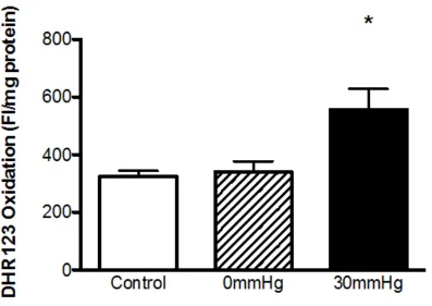
2.2.5 Reactive Oxygen Species (ROS) Production ... 75
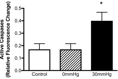
2.2.6 Quantification of Apoptosis ... 75
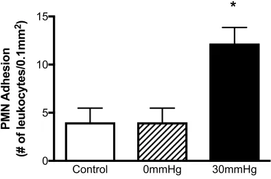
2.2.7 Leukocyte (PMN) Activation - Adhesion ... 76
2.2.8 Statistical Analysis ... 76
2.3 RESULTS ... 77
2.3.1 Structural Changes to Endothelium Due to EHP ... 77
2.3.2 ROS Production ... 77
2.3.3 Apoptosis ... 77
2.3.4 PMN Adhesion ... 81
2.4 DISCUSSION ... 81
2.5 REFERENCES ... 88
CHAPTER 3. CARBON MONOXIDE-RELEASING MOLECULE-3 (CORM-3) IMPROVES ENDOTHELIAL CELL DYSFUNCTION IN AN IN VITRO MODEL OF COMPARTMENT SYNDROME ... 95
3.1 INTRODUCTION ... 95
3.2 MATERIALS AND METHODS ... 97
3.2.1 Reagents ... 97
3.2.2 Cells ... 98
3.2.3 In vitro Models of CS ... 99
3.2.3.1 Elevation of Hydrostatic Pressure ... 99
3.2.3.2 Cytokine Cocktail Stimulation ... 99
x
3.2.6 Quantification of Apoptosis ... 102
3.2.7 PMN Rolling/Adhesion Assay ... 102
3.2.8 Statistical Analysis ... 103
3.3 RESULTS ... 103
3.3.1 ROS Production ... 103
3.3.2 Transendothelial Electrical Resistance (TEER) ... 105
3.3.3 Apoptosis ... 105
3.3.4 Leukocyte Activation ... 108
3.4 DISCUSSION ... 108
3.5 REFERENCES ... 116
CHAPTER 4. GENERAL DISCUSSION ... 123
4.1 OVERVIEW OF RESULTS ... 123
4.1.1 EHP as an in vitro Model of CS ... 124
4.1.2 Effect of CORM-3 ... 125
4.2 LIMITATIONS AND FUTURE DIRECTIONS ... 126
4.3 CONCLUSIONS ... 128
4.4 REFERENCES ... 128
APPENDICES ... 130
APPENDIX I. PERMISSION TO USE COPYRIGHTED MATERIAL ... 131
I.1 Operative Techniques: Orthopaedic Trauma Surgery 2010 ... 131
I.2 Ethics Approval ... 133
xi
Table Page
3.1. Serum levels of cytokines/chemokines detected
xii
Figure Description Page
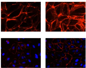
1.1 Anatomy and compartments of the lower leg ... 12 1.2 Leukocyte activation cascade ... 37 1.3 Heme degradation pathway ... 40 2.1 The experimental setup for elevation of hydrostatic pressure (EHP)
in the endothelial cells as an in vitro model of CS ... 74 2.2 The effect of EHP on the expression of F-actin and VE-cadherin
in an in vitro model of CS ... 78 2.3 The effect of EHP on the level of oxidative stress within the endothelial
cells in an in vitro model of CS ... 79 2.4 The effect of EHP on level of apoptosis within the endothelial cells
in an in vitro model of CS ... 80 2.5 The effect of EHP on the endothelial cell activation
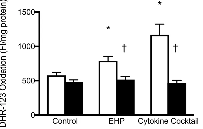
in an in vitro model of CS ... 82 3.1 The effect of CORM-3 on oxidative stress response in human vascular
endothelial cells in an in vitro model of CS ... 104 3.2 The effect of CORM-3 on the integrity of human vascular endothelial
cell monolayer in an in vitro model of CS ... 106 3.3 The effect of CORM-3 on the level of apoptosis in human vascular
xiii
in response to EHP or cytokine cocktail stimulation of human
xiv
Appendix Page
xv AP-1, activator protein-1
ARF, acute renal failure BB, bisbenzimide BR, bilirubin BV, biliverdin
BVR, biliverdin reductase
cGMP, cyclic guanosine monophosphate CO, carbon monoxide
COHb, carboxyhemoglobin
CO-RMs, carbon monoxide-releasing molecules CORM-3, carbon monoxide-releasing molecule-3 COX, cyclooxygenase
COX-1, cyclooxygenase-1 COX-2, cyclooxygenase-2 CS, compartment syndrome DMSO, dimethyl sulfoxide EB, ethidium bromide
EDL, extensor digitorum longus EHP, elevated hydrostatic pressure
xvi HO, heme oxygenase
HO-1, heme oxygenase-1 HO-2, heme oxygenase-2 HO-3, heme oxygenase-3
HUVECs, human vascular endothelial cells ICP, intra-compartmental pressure
ICAM-1, intracellular adhesion molecule-1 Ig, immunoglobulin
IL-1β, interleukin-1 beta IL-6, interleukin-6 IL-8, interleukin-8
IP-10, interferon gamma-induced protein 10 I/R, ischemia-reperfusion
IVVM, intravital video microscopy KC, keratinocyte chemoattractant
LFA-1, lymphocyte function-associated antigen-1 LPS, lipopolysaccharide
xvii NADPH, nicotinamide adenine dinucleotide phosphate NFκB, nuclear factor kappa B
NO, nitric oxide
NOS, nitric oxide synthase NPC, non-perfused capillaries
NSAIDs, non-steroidal anti-inflammatory drugs PAF, platelet activating factor
PECAM-1, platelet-associated cell adhesion molecule-1 PI, propidium iodide
PI3K, phosphatidylinositol 3-kinase PMN, polymorphonuclear leukocytes PSGL-1, P-selectin glycoprotein ligand-1 RFU, relative fluorescence units
RLU, relative luminescence units ROS, reactive oxygen species sGC, soluble guanylate cyclase
TEER, trans-endothelial electrical resistance TNF-α, tumor necrosis factor alpha
TUF, tissue ultrafiltration
TUNEL, terminal deoxynucleotidyl transferase dUTP nick end labelling VCAM-1, vascular cell adhesion molecule-1
CHAPTER 1. INTRODUCTION AND HISTORICAL REVIEW
1.1 COMPARTMENT SYNDROME
Compartment syndrome (CS) is a true medical and surgical emergency, with
potential devastating consequences, caused by an elevated pressure within a closed
osseofascial compartment (Mubarak, Owen et al. 1978, Rorabeck and Clarke 1978,
Matsen, Winquist et al. 1980, Hartsock, O'Farrell et al. 1998). The intercompartmental
fascia is unyielding and as such, individual compartments have limited ability to expand;
this makes them vulnerable to small increases in intracompartmental pressure (ICP) or
decreases in volume.
The increase in pressure within the compartment leads to microvascular
dysfunction and compromise, thereby creating an ischemic environment within the
compartment. This then limits oxygen and nutrient delivery, as well as gas exchange,
resulting in cellular anoxia, muscle necrosis and eventual cell death (Sheridan and
Matsen 1975, Whitesides, Haney et al. 1975, Mubarak, Owen et al. 1978, Rorabeck and
Clarke 1978, Matsen, Winquist et al. 1980). Interestingly, the ischemic environment
occurs in the presence of patent vasculature. Acute CS can result in severe functional
impairment, permanent pain, disability, limb loss, and even death. CS may occur acutely,
following both high- and low-energy trauma, but can also present as a chronic
intermittent condition, such as exertional compartment syndrome, which is most
Various types of injuries and medical conditions have been associated with the
development of acute CS such as fractures, contusions, burns, tight casts & dressings,
blast injuries, gunshot wounds, crush injuries, diabetes, bleeding disorders (Hope and
McQueen 2004), statin medications (Chautems, Irmay et al. 1997, Jose, Viswanathan et
al. 2004), various infections (Schnall, Holtom et al. 1994) and placing patients in
prolonged lithotomy positions for surgical procedures (Goldsmith and McCallum 1996,
Mathews, Perry et al. 2001). CS has been described in the arm, forearm, hand, buttock,
thigh, lower leg, foot, abdomen, thorax and even the orbit (Greene and Louis 1983,
Bonutti and Bell 1986, Brumback 1990, Kym and Worsing 1990, Frink, Hildebrand et al.
2010).
1.1.1 Brief Historical Review of CS
In 1881, the German surgeon Richard von Volkmann first described the clinical
sequelae of CS following traumatic supracondylar distal humerus fractures. He attributed
the devastating clinical outcome to the interruption of arterial blood supply but did not
specify the cause (von Volkmann 1881). This observation by von Volkmann was further
substantiated by Leser in 1884 who, by applying a tight bandage to the limbs of animals,
noted time dependent necrotic changes in the muscle, as well as venous congestion and
swelling (Leser 1884). In 1906, Hildebrand drew attention to the role of nerve
involvement in the pathophysiology of ischemic contractures after replicating the
experimental design of Leser, and coined the term ‘Volkmann’s contracture’ to refer to
the clinical sequelae following supracondylar distal humerus fractures (Hildebrand 1906).
nerve involvement as underlying causes of ischemic contractures, he drew attention to
venous obstruction as the driving force behind CS. Although we now know that venous
obstruction is not a major underlying contributor of CS, Murphy importantly drew
attention to the idea that 1. elevated ICP was a main driving force in the pathophysiology,
that 2. arterial pulses were maintained during the process and finally that 3. by splitting
the underlying deep fascia, the “obstruction” could be relieved (Murphy 1914). Until this
point, treatments had been aimed towards the complications of CS and ischemia, such as
fibrosis and contractures (Rowlands and Lond 1905).
In 1926, through a series of elegant ischemia-reperfusion experiments using the
limbs of dogs, Jepson noted that elapsed time as well as increased pressure was a direct
causal factor in the pathogenesis of ischemic contractures. More importantly, he also
showed that by surgically decompressing the involved compartment, the function of the
limb could be restored (Jepson 1926).
The next significant contribution was likely from the work of Griffiths (1940),
and although he mistakenly argued (for the better part of two decades) that arterial spasm
was the root cause of the resulting ischemic contracture, his research contributed
significantly to our understanding and recognition of early clinical signs and symptoms of
CS, such as pain out of proportion, pain with passive extension and ‘puffiness’, which are
still widely taught to this day (Griffiths 1940).
The next main contribution to our understanding of CS came with the bombing
raids known as the London Blitz in the early 1940s. Patients with crushed extremities
would be taken to hospital, and a relatively stable clinical condition would quickly
condition became known as “crush syndrome” (Bywaters, Delory et al. 1941). This
highlighted the importance of ischemia-reperfusion in the pathogenesis of CS, rather than
strictly speaking of venous congestion and elevated ICP.
In 1975, Matsen delivered his unified theory of CS by combining all the relevant
data available to that point. The important aspects of his theory stated that CS was not
restricted only to the upper extremity, that elevated pressure was a critical feature of the
condition and finally that relieving the ICP via surgical fasciotomy was critical to avoid
the devastating sequelae (Sheridan and Matsen 1975). The importance of Matsen’s
contribution cannot be overstated, as he shifted the discourse from understanding the
underlying pathophysiology to better ways of diagnosing and treating acute CS.
1.1.2 Diagnosis of CS
The early identification, diagnosis and treatment of CS are critical in order to
relieve ICP, prevent ongoing tissue anoxia, necrosis and optimize patient outcome, as
well as prevent long-term disability. The diagnosis of CS is primarily a clinical one,
which, in certain circumstances may be supplemented by direct ICP measurements.
Understanding patient risk factors and the early identification of patient clinical signs and
symptoms are paramount in the diagnosis and appropriate management of CS. Risk
factors include male gender, age under 35, tibia fracture, high energy forearm fractures,
1.1.2.1 Clinical Diagnosis
There are several signs and symptoms that have traditionally been associated with
acute CS. They appear in a stepwise fashion, although the timing can vary significantly
from patient to patient and injury to injury (Myers 2000, Elliott and Johnstone 2003,
Olson and Glasgow 2005, Shadgan, Menon et al. 2008). For this reason, the importance
of serial and thorough clinical evaluations of all patients at risk of developing CS cannot
be overstated; currently, this is considered the standard of care in the management of CS.
The presence of symptoms should not only alert clinicians to the diagnosis of an acute
CS, but also, unfortunately, likely suggests an advanced stage of disease.
The first signs and symptoms associated with CS are usually pain out of
proportion to the apparent injury, and pain with passive stretch of the involved muscle
compartment (Whitesides and Heckman 1996). The sensitivity and specificity of these
clinical findings has been found to be between 13-19% and 97% respectively (Whitesides
and Heckman 1996, Ulmer 2002). Other symptoms associated with CS include tense and
painful muscle compartments, a persistent deep ache or burning pain, paresthesia or
increasing analgesia requirement, with the last of these being an especially important
finding in the pre-verbal pediatric population (Bae, Kadiyala et al. 2001).
1.1.2.2 Physical Examination
The first description of the clinical criteria for the diagnosis of CS was provided
by Griffiths in 1940. Griffiths established the original “four Ps”: pain out of proportion
and pain on passive stretch, paraesthesia, paralysis and ‘puffiness’ (Griffiths 1940).
Wilckens et al. 2005). Unfortunately, some of these physical exam findings, such as
paresthesia and paralysis, are considered late findings of established CS (McQueen,
Christie et al. 1996), and often signify that irreversible vascular, muscular and
neurological injury have likely already occurred (Matsen and Clawson 1975, Ulmer
2002). Although pulselessness was traditionally taught as one of the “5 Ps” in the clinical
diagnosis of CS, the absence of a pulse is no longer considered a feature of CS (Manjoo,
Sanders et al. 2010) and the presence of a pulse certainly does not rule out a diagnosis of
CS. Failing to identify CS and obtain a timely diagnosis is the greatest cause of adverse
clinical outcomes (Matsen and Clawson 1975, Rorabeck 1984, McQueen, Christie et al.
1996, Mars and Hadley 1998). In addition, missed CS is one of the most frequently
argued cases in the field of medico-legal litigation, and is associated with frequent and
significant judgements in the favour of the plaintiff (Bhattacharyya and Vrahas 2004).
1.1.2.3 Objective Compartment Pressure Monitoring
The direct measurement of ICP when attempting to diagnose CS, provided the
proper technique is used, is a valuable tool in the clinician’s armamentarium (Hargens
and Ballard 1995). Various measurement techniques have been described, such as needle
manometer, wick catheter, slit catheter and electronic transducer-tipped catheters
(Hargens and Ballard 1995). In order to capture the peak ICP value, measurements
should be taken at the level of the fracture, as well as additional sites up to 5 cm proximal
and distal to the injury (Heckman, Whitesides et al. 1994). In addition, pressures should
also be measured in the other compartments of the affected limb, to ensure that a CS is
accurate, as they do not rely on limb position or the height of the transducer. All direct
compartment pressure measurement devices have their own specific technical steps, are
user dependent, have their own advantages and disadvantages, and are not immune to
false negatives (Lawendy and Sanders 2010). Although some authors have argued that
all patients (especially young men) presenting with tibial diaphyseal fractures, high
energy fractures of the tibial metaphysis, soft tissue injuries or in patients with an
accompanied bleeding diathesis should undergo objective compartment pressure
monitoring (McQueen, Gaston et al. 2000), this is currently not the usual practice. There
are, however, several indications for using ICP monitors. These include unconscious
patients, pediatric patients, pre-verbal and non-verbal patients, patients with equivocal
signs and symptoms, patients with associated neurological injuries, or in a polytrauma
scenario (Whitesides, Haney et al. 1975, Gelberman, Garfin et al. 1981, Hargens,
Schmidt et al. 1981, Hargens, Akeson et al. 1989). In these cases, continuous
compartment pressure monitoring may help confirm clinical findings, decrease the delay
to fasciotomy and may, as a result, decrease the long-term complications of the disorder
(McQueen, Christie et al. 1996).
1.1.3 Consequences of Missed CS
The early identification and diagnosis of acute CS is critical to its successful
management, and will maximize the chances of a positive clinical outcome. The inability
to obtain a timely diagnosis is the most common cause of adverse clinical outcomes
(Matsen and Clawson 1975, Rorabeck 1984, McQueen, Christie et al. 1996, Mars and
such as muscle infarction, muscle and joint contractures, secondary joint and soft tissue
deformities, limb weakness and neurologic dysfunction (Whitesides and Heckman 1996).
Less common, but nonetheless important complications of missed CS include infection,
gram-negative sepsis, amputation and end-organ involvement (Whitesides and Heckman
1996). The end result of missed CS is often irreversible myoneural ischemia leading to
various degrees of permanent neuromuscular deficits and dysfunction.
The severity of the clinical outcome and dysfunction depends on the amount of
tissue affected, and can range from mild weakness and sensory changes to severe
ischemic contractures and limb dysfunction. When a sufficiently large amount of muscle
tissue is involved (often combined with a weakened and compromised immune system),
CS can lead to severe systemic complications such as crush syndrome, rhabdomyolysis,
renal failure (secondary to myoglobinuria) and systemic shock (Sanghavi, Aneman et al.
2006, West 2007).
A missed or late diagnosis can be the result of clinical inexperience, a lack of
suspicion, or a confusing clinical presentation (McQueen, Christie et al. 1996). These
situations can occur when patients present with altered pain perception, altered level of
consciousness, regional anesthesia, patient-controlled analgesia and nerve injury; all are
known risk factors for late diagnosis (Mubarak and Wilton 1997, Harrington, Bunola et
1.2 THERAPEUTIC APPROACHES TO CS
The therapeutic goals of treating acute CS are to minimize chronic, long lasting
injury and dysfunction of the involved limb, by accurately and efficiently diagnosing the
condition and restoring the compartments microcirculatory environment. This is done in
order to avoid the devastating consequences of myonecrosis, ischemic contracture and
limb dysfunction. Non-operative techniques and adjuncts have been studied in various
animal models and human case series with limited success. These nonoperative
treatments remain unproven, making surgical decompression through fasciotomy of all
involved compartments the only gold standard therapy for acute established CS, provided
it is carried out within 6-8 hours of CS onset (Eaton and Green 1972, Matsen, Winquist et
al. 1980, Rorabeck 1984, McQueen, Hajducka et al. 1996, Lawendy and Sanders 2010).
1.2.1 Fasciotomy
Fasciotomy, as a technique for the surgical treatment of patients with impeding
Volkmann’s contracture, was first described in 1911 by Bardenheuer (Bardenheuer 1911)
although at the time, he used the term ‘aponeurectomy’. Eventually, Murphy in 1914
suggested early surgical fasciotomy for the treatment of increased pressure within a
fascial-enclosed space due to hemorrhage and edema in order to prevent paralysis and
contractures (Murphy 1914). The concept of surgical fasciotomy, as described by
Murphy, is the mainstay for the treatment of CS today. The first detailed record of the
actual operative technique was provided by Benjamin (Benjamin 1957), describing the
surgical approach to the forearm. Fasciotomy is urgently performed to normalize
halting the inflammatory process and ultimately preventing the devastating clinical
sequelae. After 6-8 hours, the risk of permanent tissue damage increases exponentially.
Once muscle and tissue necrosis has occurred, surgical fasciotomy is contraindicated as it
increases the risk of infection significantly. Adding to the complexity of this clinical
presentation, one often does not know the exact time of injury or the subsequent events
that have led to the patient’s current state, making decision making difficult. Therefore,
frequent serial clinical examinations and reassessment are extremely important.
Fasciotomy releases the involved compartment(s), allowing soft tissues to swell
and expand, thus allowing for an increased compartmental volume while decreasing the
ICP. Following decompression, surgical wounds are usually left open for 48-72 hours
prior to skin closure, which is often accompanied by split thickness skin grafting
(Lawendy and Sanders 2010).
1.2.1.1 Threshold for Decompression
Considering the significant cost of missing a CS, some authors have expanded the
indications of using ICP monitoring to include all traumas and fractures with a high risk
of CS (McQueen, Gaston et al. 2000). To complicate matters further, various ICP
thresholds have been proposed, at which fasciotomy should be performed, although there
is currently no clear consensus. Protocols have included absolute values of 30 mmHg, 40
mmHg and 45 mmHg (Mubarak, Owen et al. 1978, Matsen, Winquist et al. 1980,
Schwartz, Brumback et al. 1989) while others, rather than considering the absolute ICP of
a compartment, have used the difference between a compartment’s ICP and the patient’s
fasciotomy (Whitesides, Haney et al. 1975). Most trauma surgeons prefer using this ∆P as
a cut-off measure to perform fasciotomy rather than using an absolute ICP threshold, as
this becomes more useful in hypotensive trauma patients leading to a lower overall
fasciotomy rate when compared to an absolute pressure threshold (Matsen, Winquist et
al. 1980, McQueen, Hajducka et al. 1996). A recent study by Whitney et al (2014) looked
at false positive rates of CS diagnosis based on one-time ICP measurements alone. When
using a ∆P threshold of 30 and 20 mmHg they reported a false positive rate of 35% and
24% respectively (Whitney, O'Toole et al. 2014).
1.2.1.2 Fasciotomy Techniques
Surgical fasciotomy techniques have been well described for the upper and lower
extremities as well as the trunk (McQueen, Gaston et al. 2000). Fasciotomies for CS of
the lower leg (80% of all cases), forearm and hand are among the most commonly
performed.
1.2.1.2.1 Fasciotomy in the Lower Leg
The lower leg is divided into 4 osseofascial compartments: anterior, lateral,
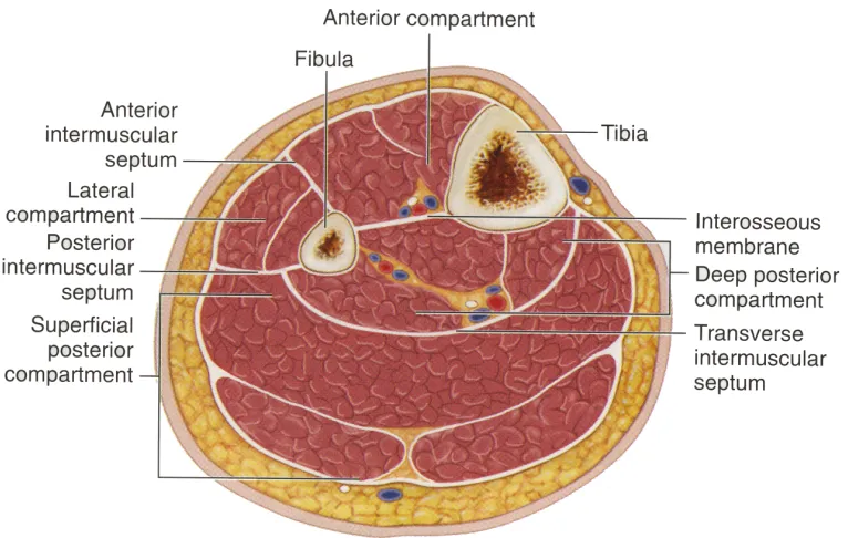
posterior superficial and posterior deep (Gray 2000) (Figure 1.1). The anterior
compartment is the most commonly affected in CS (Rorabeck and Macnab 1976). There
are two commonly described techniques for the surgical decompression of the lower leg:
Figure 1.1. Anatomy and compartments of the lower leg. The lower leg is
comprised of the tibia and fibula, with interconnecting fascial
planes separating the various muscles into anterior, lateral,
superficial posterior and deep posterior compartments.
Some surgeons will routinely employ a two-incision (medial and anterolateral)
technique, while others perform a single anterolateral approach in patients with CS in
order to decompress both the anterior and lateral compartments, and then reassess the
remaining compartments with ICP monitors before releasing the posterior superficial and
posterior deep compartments (Tornetta, Puskas et al. 2016). Better visualization of tissue
planes, neurovascular structures and ability to assess the conditions of soft tissues have
been described as reasons to preferentially perform a two-incision approach (Lawendy
and Sanders 2010). However, both single-incision and two-incision approaches have been
shown to adequately decompress all 4 lower leg compartments (Neal, Henebry et al.
2016).
1.2.1.2.2 Fasciotomy in the Forearm
The forearm is made up of the volar compartment, the extensor compartment and
the mobile wad. The flexor compartment is divided into superficial, middle and deep
muscle layers. The dorsal extensor compartment contains superficial and deep layers
(Gray 2000).
A curvilinear incision is made, extending from proximal and medial aspect to the
elbow flexion crease, which then crosses the flexor surface of the elbow at an oblique
angle. This is then followed by moving lateral to the midline, allowing for an extensile
approach and the ability to release the carpal tunnel if needed. Medial and lateral flaps are
elevated; the lateral antebrachial cutaneous nerve is found and protected. The lacertus
fibrosis, the most proximal tether, is also released. The fascia overlying the superficial
between the flexor carpi ulnaris and the flexor digitorum superficialis is exploited to
reveal the deep volar compartment which can then also be released (Gray 2000).
1.2.1.2.3 Fasciotomy in the Hand
The hand is divided into ten compartments; these include the thenar, hypothenar,
adductor pollicis, four dorsal interossei and three volar interossei compartments (Gray
2000).
Two longitudinal incisions are centered over the index and ring fingers on the
dorsum of the hand. Soft tissues are bluntly dissected on either side of the metacarpals,
incising through the dorsal interossei muscle fascia. If the thenar and hypothenar
compartments need to be released, two separate incisions are made on the volar radial
aspect of the thumb and the volar ulnar aspect of the 5th digit, respectively. The carpal
tunnel can be released through a 4cm longitudinal incision, in line with the ring finger,
with the proximal extent being the flexor crease of the wrist. The transverse carpal
ligament is then released under direct visualization (Kalyani, Fisher et al. 2011).
1.2.1.3 Complications of Fasciotomy
Surgical fasciotomy, although being the gold standard treatment of CS, it is not
without its risks and complications. A high percentage of patients report postoperative
neurologic symptoms and chronic pain associated with their surgical wounds (Fitzgerald,
Gaston et al. 2000). Other complications include dry skin, pruritus, wound discolouration,
swelling, tendon tethering, hypertrophic scarring, ulceration and muscle herniation
infection is also not insignificant, and can create potentially devastating complications;
this is directly related to the timing of the surgical intervention.
Fasciotomies which are delayed for greater than 12 hours have a 28% rate of
infection, while those performed early have an infection rate of 7.3% (Williams, Luchette
et al. 1997). In a retrospective study looking at a trauma patient population, Dover et al
(2011) found an early post-operative complication rate of 20%. Of these, 80%
experienced clinical symptoms which they rated as severe. On long-term follow-up, 70%
of patients experienced persistent symptoms, which severely limited them from either an
occupational or social point of view (Dover, Marafi et al. 2011, Dover, Memon et al.
2012).
Fitzgerald et al (2000) retrospectively assessed complications of fasciotomy in
both upper and lower extremities over an 8-year period (Fitzgerald, Gaston et al. 2000).
They found that one in every ten patients had chronic pain associated with their
fasciotomy wounds and more than 20% of patients covered their scars due to the aesthetic
appearance of the wound. They also found complications to be detrimental both socially
and occupationally, with 28% of patients changing their hobbies and 12% having to
change their occupation, secondary to the complications of their fasciotomy (Fitzgerald,
Gaston et al. 2000). Another post-operative complication of surgical fasciotomies is CS,
which has been found to occur in 3 to 20% of cases (Barr 2008), and is believed to be
caused by excessive post-operative scar tissue formation and/or inadequate release of
1.2.2 Non-Surgical Interventions
Currently, non-operative treatment modalities for CS are utilized in cases where
surgical fasciotomy is contraindicated: when the affected limb is nonviable due to severe
ischemia, or missed CS (Schmidt 2007). Before 1911, non-operative treatment options
mainly consisted of limb mobilization and muscle stretching in order to prevent or treat
ischemic contractures. Today, the most common non operative treatment is the removal
of a cast or occlusive splints in a patient who presents with symptoms suggestive of CS.
In these cases, if symptoms persist, fasciotomy is indicated.
The consequences of a missed CS or of delaying fasciotomy are significant, as a
result, non-operative treatments have been limited to an adjunctive role to fasciotomy. It
would be beneficial to develop non-surgical modalities that could prolong the treatment
window between the onset of CS and the time where irreversible neurological, vascular
or muscular changes occur. Potential medical treatments have been described in both
animal models and human case series. These include mannitol (Better, Zinman et al.
1991), hyperbaric oxygen (Wattel, Mathieu et al. 1998), tissue ultrafiltration (Odland,
Schmidt et al. 2005), anti-inflammatories (Manjoo, Sanders et al. 2010) and anti-oxidants
(Kearns, Daly et al. 2004).
1.2.2.1 Mannitol
Mannitol is an osmotic diuretic, volume expander and free radical scavenger. It is
commonly used to acutely reduce intracranial pressure, prevent or treat acute kidney
injuries associated with CS can lead to rhabdomyolysis, acidosis, acute renal failure
(ARF) and even death (Bywaters, Delory et al. 1941, Better and Stein 1990).
The severity of rhabdomyolysis can be confirmed and clinically followed by
measuring serum creatine kinase (CK) levels. One of the most severe complications of
rhabdomyolysis is ARF, which has a mortality rate of 3-50% (Slater and Mullins 1998,
Malinoski, Slater et al. 2004). One of the tenets in the management of the crush syndrome
and rhabdomyolysis is aggressive fluid resuscitation, in an attempt to prevent both
systemic and renal complications (Odeh 1991, Malinoski, Slater et al. 2004).
Mannitol has been shown to decrease extracellular fluid volume by promoting
water and sodium excretion. It has been shown to reduce ICP in a canine model of CS
(Better, Zinman et al. 1991). Daniels et al. (1998) described the case of a healthy 19–
years old male presenting with heat stroke, who subsequently developed a lower leg CS,
and who was treated only with mannitol. The patient was discharged 10 days after his
admission to the hospital, with only “mild residual weakness” in the involved leg
(Daniels, Reichman et al. 1998).
1.2.2.2 Hyperbaric Oxygen Therapy
Hyperbaric oxygen therapy involves the medical use of oxygen at levels higher
than the atmospheric content of 21%. Hyperbaric oxygen therapy creates a 3-fold
increase in the oxygen diffusion into the tissues (Wattel, Mathieu et al. 1998). This
allows continued delivery of oxygen even in the presence of ischemia. Hyperbaric
oxygen has been described as either the main treatment, or as an adjunct for various
inhalation, severe carbon monoxide (CO) poisoning, osteoradionecrosis, skin flap
healing, clostridial myonecrosis and CS (Leach, Rees et al. 1998).
With respect to CS, hyperbaric oxygen treatment is believed to exert its beneficial
effects on intracompartmental bleeding, swelling and edema by causing oxygen-induced
vasoconstriction and allowing oxygen perfusion at lower perfusion pressures (as are seen
in situations of CS) (Nylander, Nordstrom et al. 1987). As the interstitial edema is
decreased, flow through the microcirculation is restored, or at least improved. The benefit
of hyperbaric oxygen therapy has been reported in several ischemia-related clinical
scenarios including traumatic ischemic lesions, ulcerations, infections and open fractures
(Smith, Stevens et al. 1961, Hanson, Slack et al. 1966, Szekely, Szanto et al. 1973).
Published case studies have reported success in averting fasciotomy in patients presenting
with CS (Strauss, Hargens et al. 1983, Wattel, Mathieu et al. 1998, Gold, Barish et al.
2003); a recent case report by Karam et al. (2010) described the case of an NCAA
football player with acute paraspinal CS following weight-lifting: he was successfully
treated with forced diuresis and hyperbaric oxygen chamber treatment (Karam,
Amendola et al. 2010).
However, due to the lack of definitive evidence and the need for costly and
specialized equipment, hyperbaric oxygen is infrequently used and currently seen only as
an adjunct to, and not a substitute for, surgical fasciotomy.
1.2.2.3 Tissue Ultrafiltration
Tissue ultrafiltration (TUF) was first described as a method of analyzing the
insertion of small-diameter semi-permeable hollow fibers into the tissue compartment of
interest. The catheter is connected to suction, in order to filter interstitial fluid. This
enables researchers to not only decompress the tissues, but also analyze the extracted
fluid for biomarkers (Odland, Schmidt et al. 2005). The use of ultrafiltration in CS has
been shown to lower the intramuscular pressure while maintaining perfusion pressure
(Odland, Schmidt et al. 2005). In their porcine model of CS, using bovine serum
albumin-enriched saline infusion into the anterior compartment of the hind limb, Odland
et al. (2005) measured serum and filtrate creatinine kinase (CK) and lactate
dehydrogenase (LDH) levels over a 10 hour period. They found that the biomarker levels
were 80 times higher in the ultrafiltrate compared to the serum (Odland, Schmidt et al.
2005). Significantly lower pressures were recorded in experimental limbs connected to
negative pressure, coupled with a markedly lessened cellular injury. The authors
undertook a small human clinical trial, to test the safety and efficacy of ultrafiltration.
They examined ten patients with tibial fractures treated with intramedullary nailing with
and without tissue ultrafiltration, and found no difference in ICP between the two groups;
however, 2 patients in the control group developed CS, while none in the ultrafiltration
treatment group (Odland and Schmidt 2011).
1.2.2.4 Anti-Inflammatories
There is a significant body of evidence describing an increase in ICP as the
underlying cause of microcirculatory dysfunction. However, the significant impact of
inflammation and leukocyte activation in the pathophysiology of CS is increasingly being
leukocytes directly impair perfusion, increase intravascular protein leakage, thus
contributing to tissue edema, as well as causing direct parenchymal injury (Kurose,
Anderson et al. 1994, Forbes, Carson et al. 1995, Forbes, Harris et al. 1996, Harris and
Skalak 1996).
Non-steroidal anti-inflammatory drugs (NSAIDs) are a class of medication that
interfere with arachidonic acid metabolism, via inhibition of the cyclooxygenase (COX)
enzyme. Two isoforms have been identified: the constitutively expressed COX-1, and
inducible COX-2. COX-2 expression can be upregulated in response to inflammatory
stimuli and pro-inflammatory cytokines (Jan and Lowry 2009). Manjoo (2010), looked at
the effects of indomethacin, a selective COX-2 inhibitor, on capillary perfusion, cell
damage and inflammatory activation in a rat model of CS. They found that indomethacin
improved tissue perfusion and viability, decreased the number of non-perfused capillaries
and significantly lowered tissue injury, lending support to the suggestion that
anti-inflammatory treatments have the potential to reduce the damage in the presence of
elevated ICP (Manjoo, Sanders et al. 2010).
1.2.2.5 Anti-Oxidants
Ischemia-reperfusion is known to lead to a significant release of reactive oxygen
species (ROS) – extremely damaging free radicals, both locally within the tissue, as well
as from the release of activated neutrophils. Thus, the resulting tissue injury is not only
seen at the local level in skeletal muscle, but also in distant organ systems, such as the
lungs and kidneys (Xiao, Eppihimer et al. 1997, Kearns, Kelly et al. 1999). In a study by
appeared to be the free radical-mediated reperfusion injury: an increase in free radicals
(such as H2O2) causes direct injury to the endothelium. Furthermore, by interacting with
lymphocytes, ROS further stimulate a pro-inflammatory state by increasing cytokines
levels (e.g. TNF-ɑ and IL-8) (Perler, Tohmeh et al. 1990). These, in turn, lead to the
activation of neutrophils and these activated neutrophils then release ROS, which further
contributes to the endothelial injury.
Due to expanding knowledge regarding the contribution of oxidative damage in
CS and ischemia-reperfusion injury, various anti-oxidant therapies have been attempted,
to prevent both the local and systemic injuries. These include N-acetylcysteine (NAC),
taurine and vitamin C. NAC is a free radical scavenger that also restores the host cellular
anti-oxidant defenses by upregulating glutathione levels in the cell (Sjodin, Nilsson et al.
1989). The primary clinical use of NAC is in acetaminophen overdose, to reduce injury to
hepatocytes (Flanagan and Meredith 1991). It is also used for its nephroprotective effects
in patients with kidney failure prior to administering IV contrast (Tepel, van der Giet et
al. 2000), as well as to protect against oxidative injury in lung parenchyma (Bernard
1991). A study by Kearns et al. (1999) examining the effects of NAC in a rat model of
CS found that CS led to decreased muscle contractility and increased tissue
myeloperoxidase activity and treatment with NAC attenuated neutrophil activation and
preserved muscle contractility (Kearns, Kelly et al. 1999).
Taurine (2-aminoethane sulfonic acid) is a sulphur-containing amino acid, derived
from the metabolism of methionine. The major source of taurine is from a person’s diet.
Taurine has been implicated in the inhibition of lipid peroxidation, cell membrane
al. 2004). Studies have found that exogenous administration of taurine can have
protective effects against ischemia-reperfusion tissue injury in the kidney, heart, liver and
skeletal muscle (Oz, Erbas et al. 1999, Wettstein and Haussinger 2000, Michalk,
Hoffmann et al. 2003, Wang, Li et al. 2005). Wang et al. (2005) found that the
administration of taurine reduced anterior compartment pressure, muscle edema, lactate
dehydrogenase and lipid peroxidation products in a rabbit model of CS (Wang, Li et al.
2005).
Vitamin C (ascorbate) is an antioxidant that has been shown to decrease or
prevent reperfusion injury in the lung and skeletal muscle, as well as to reduce oxidant
production in neutrophils (Herbaczynska-Cedro, Wartanowicz et al. 1994, Lehr, Frei et
al. 1995, Kearns, Kelly et al. 1999, Armour, Tyml et al. 2001, Kearns, Moneley et al.
2001). It has scavenging effects on hydrogen peroxide, which is an important component
for neutrophils recruitment and adhesion (Armour, Tyml et al. 2001). Vitamin C also
targets circulating neutrophils and lymphocytes (Levine, Daruwala et al. 1998). It is
believed to exert its beneficial effects by reducing neutrophil recruitment and activation,
as well as their extravasation into the tissues by altering the expression of adhesion
molecules (e.g. ICAM-1). Vitamin C has also shown promising results in
prophylactically treating complex regional pain syndrome (CRPS) or reflex sympathetic
dystrophy (Zollinger, Tuinebreijer et al. 1999). While CRPS and CS are distinct
pathological entities, they do share certain underlying physiological processes such as an
exaggerated inflammatory response, peripheral nervous system dysfunction and an
increase in circulating free radicals causing lipid membrane oxidation (Van der Laan
that pre-treatment with Vitamin C reduced intercellular adhesion molecule-1 (ICAM-1)
expression and myeloperoxidase (MPO) activity as well as muscle swelling, while
preserving muscle contractile function (Kearns, Daly et al. 2004). Although there is a
concern that vitamin C may have pro-oxidant properties when administered at high doses
for a prolonged period of time (Podmore, Griffiths et al. 1998), it has been shown to have
a potent antioxidant effect without associated toxicity at doses less than 500mg per day
(Bendich and Langseth 1995).
1. 3 PATHOPHYSIOLOGY OF CS
The pathophysiology underlying the onset, progression and muscle necrosis
associated with CS is only partially understood. A bony or soft tissue insult, combined
with an inherently rigid and unyielding fascia which prevents volume expansion leads to
increased ICP, which, in turn, leads to microcirculatory dysfunction. This is followed by
the activation of an inflammatory cascade and tissue edema, eventually leading to
impaired gas exchange, restricted oxygen and nutrient delivery. The final common
pathway is cellular anoxia, cell death and myonecrosis.
In 1881, Volkmann was the first to suggest that limb paralysis secondary to CS
was due to the interruption in arterial blood supply, causing ischemia (von Volkmann
1881). He described the devastating hand deformity seen in the paediatric population
following a supracondylar fracture, complicated by CS. The deformity still bears his
name today. However, Volkmann was unable to describe the cause of the ischemia. Leser
experiments, that muscle necrosis was a crucial part of the condition (Leser 1884).
Hildebrand in 1906 demonstrated that nerve involvement also occurred, in addition to
muscle necrosis (Hildebrand 1906). Over the last century, various theories have been
offered and expanded upon to explain the pathophysiological basis of CS. These have
included neurological injury (Thomas 1909), arterial injury and spasm (Griffiths 1940),
venous obstruction (Murphy 1914, Brooks 1922), increased ICP and pressure-induced
ischemia (Jepson 1926).
In 1940, while trying to expand on Volkmann’s findings, Griffiths suggested that
the paralysis and contractures seen were due to an underlying arterial injury with
reflexive spasms (Griffiths 1940). The idea of arterial injury and spasm as a cause of
ischemia and contractures was further supported by Watson-Jones in 1952 (Watson-Jones
1952). Foisie in 1942 believed that autonomic dysfunction mediated the arterial spasm.
As a result, he suggested that autonomic sympathetic blockade could treat CS and prevent
the complications (Foisie 1942). We now know that this was incorrect, on both a
pathophysiological and clinical basis.
The connection between ICP and subsequent ischemia was first made by Hughes
in 1948 (Hughes 1948) and in 1975, while considering the link between pressure,
ischemia, muscle injury and the importance of compartmental decompression through
fasciotomy, Matsen combined these relevant concepts into one unified theory of CS
(Matsen 1975). Through his description, Matsen confirmed that CS could occur in any
anatomical location, and was not a condition exclusive to the upper extremity. In
addition, he suggested that the increase in tissue intracompartmental pressure was a
fasciotomy was the only effective treatment (Matsen 1975). Whitesides (1975) then
helped to define a methodology for directly measuring ICP (Whitesides, Haney et al.
1975). Initially, a threshold ICP was believed to exist above which irreversible changes
and injury would occur (Heckman, Whitesides et al. 1993). Subsequently, rather than an
absolute pressure threshold, others suggested that it is the difference between ICP and
diastolic blood pressure that was relevant, and should be considered in the assessment of
patients (Har-Shai, Silbermann et al. 1992, Heckman, Whitesides et al. 1994, Bernot,
Gupta et al. 1996).
1.3.1 Ischemia
1.3.1.1 Microvascular Dysfunction
Three theories have attempted to describe the microcirculatory dysfunction and
ischemia associated with increased tissue pressure seen in CS: microvascular occlusion
theory, critical closing pressure theory and arterio-venous gradient theory.
The microvascular occlusion theory states that CS results from capillary occlusion
caused by increased ICP. The theory postulates that increased ICP above capillary
pressure leads to a reduction in the patency of capillaries and thus subsequent blood flow.
This then creates an ischemic state, impairing gas exchange and nutrient delivery, leading
to cellular anoxia and cell death. However, a study by Hartsock et al (1998) found that
while compartment pressures could be experimentally raised well beyond the level to
cause complete cessation of capillary blood flow, collapse of capillary vessels was not
seen, essentially discrediting the microvascular occlusion theory (Hartsock, O'Farrell et
The critical closing pressure theory describes an absolute ICP above which
arteriole closure occurs, caused by an elevated differential between tissue pressure and
intravascular pressure (Burton and Yamada 1951). This would then lead to arteriolar
collapse and tissue ischemia. The validity of this theory however was put into question by
an experiment by Vollmar et al (1999) who assessed the response of arterioles, capillaries
and venules to pressure elevation and found no signs of arteriolar spasm or collapse
(Vollmar, Westermann et al. 1999).
Finally, the arterio-venous gradient theory states that CS is caused by increased
tissue pressure, which reduces the pressure gradient from the high pressure seen in the
arterial system to the low pressure on the venous side. As ICP rises, the gradient is
reduced and blood flow decreases, causing cellular anoxia and tissue injury (Matsen,
Winquist et al. 1980). This phenomenon also leads to pooling of venous blood, fluid
extravasation, interstitial edema and swelling and causes a further rise in ICP (Matsen
and Krugmire 1978). Although all three theories attempt to explain the link between
raised ICP and microcirculatory dysfunction, the AV gradient theory provides the closest
link, and is most easily reconciled with our current understanding of CS and
microcirculatory dysfunction vis-à-vis pressure gradient changes.
1.3.1.2 Low Flow Ischemia
We now know that, rather than being due to a state of complete occlusive vascular
ischemia and spasm, CS creates a microcirculatory “low flow” environment, occurring in
the presence of patent arterial vessels. Under normal conditions, microvascular perfusion
there occurs a shift in perfusion toward intermittently perfused capillaries (IPC), and
nonperfused capillaries (NPC) (Lawendy, Sanders et al. 2011, Lawendy, Bihari et al.
2015). Lawendy (2011) used intravital video microscopy (IVVM) to directly observe the
microvascular perfusion changes seen in early CS. After artificially raising ICP in a rat
model of CS, a decrease in the number of CPC (representing healthy perfusion) and an
increase in intermittent and non-perfused capillaries was found. These changes in
microvascular perfusion were accompanied by significant leukocyte activation, as well as
parenchymal injury (Lawendy, Sanders et al. 2011).
Despite the presence of microvascular dysfunction, some degree of perfusion
remains during CS, creating a “low flow” ischemic state, where CPCs are present in the
same capillary bed as IPC and NPC. NPCs have no ability for nutrient or gas exchange,
and represent a state of ischemia, unable to meet the metabolic demands of the tissue
(Lawendy, Sanders et al. 2011). This creates a partial ischemic state, which triggers an
early and significant inflammatory response (Gute, Ishida et al. 1998, Lum and Roebuck
2001, Schlag, Harris et al. 2001).
Heppenstall (1986) considered the ischemic process in CS in a canine model.
They found that the low flow ischemic state (specifically associated with CS) caused
tissue injury that was significantly greater than what was seen in a state of complete
ischemia. This finding was believed to be due to the intense inflammatory reaction
(Heppenstall, Scott et al. 1986). The association between partial ischemia and intense
inflammatory response has been substantiated by Conrad (2005), who compared partial
ischemic state caused a significant early increase in pro-inflammatory mediators when
compared to complete ischemia (Conrad, Stone et al. 2005).
There are important distinctions which must be drawn between CS and complete
ischemia-reperfusion (I/R) injury: CS causes tissue injury and necrosis despite a patent
macrocirculatory system in the face of a palpable distal pulse (Seddon 1966). In addition,
the injury occurring as a result of CS is of greater magnitude compared to a complete
ischemic insult of the same duration (Heppenstall, Scott et al. 1986). While our
understanding of the pathophysiological basis underlying CS is not complete,
microcirculatory dysfunction caused by an ongoing ischemia-reperfusion type injury,
early leukocyte activation and a pro-inflammatory state appear to be the driving forces
behind the generation of CS, and its potentially devastating sequelae.
1.3.2 Reperfusion and Inflammation
The greatest paradigm shift with respect to our understanding of the
pathophysiology underlying CS came in 1941 during World War II. Researchers noted
the systemic clinical collapse which occurred in otherwise stable patients following the
revascularization of injured limbs. They noted a decrease in urine output, clinical
deterioration and multi-organ failure, followed by death in certain cases. Interestingly,
this occurred even when the injured limbs had been amputated (Bywaters and Beall 1941,
Bywaters, Delory et al. 1941). This led to the concept of ‘crush syndrome’, defining the
clinical entity associated with what we know today as ‘reperfusion injury’.
Reperfusion injury occurs when tissues are perfused after a period of ischemia.
effects. The return of oxygen during reperfusion causes the formation of reactive oxygen
species (ROS), which, along with activated neutrophils, cause the local and systemic
injury seen following reperfusion.
It has been well documented, in complete ischemia-reperfusion (I/R), that
increasing ischemia time leads to an increasing accumulation of activated leukocytes
(particularly neutrophils) in the post-capillary venules. Neutrophils contain intracellular
granules made up of various proteases and myeloperoxidase, which are very damaging to
cellular and extracellular targets. Upon activation, these granules are released into the
affected tissues. Thus, leukocyte activation leads to increased vascular permeability to
plasma protein leakage, tissue edema, and increased interstitial pressure. An increase in
interstitial pressure is believed to physically compress capillaries altering the arterial
venous gradient, leading to further failure of capillaries to reperfuse upon restoration of
blood flow. Correlation has been noted between the number of leukocytes in the
capillaries of post-ischemic tissue and the percentage of capillaries exhibiting no-reflow
(Engler, Dahlgren et al. 1986, Barroso-Aranda, Schmid-Schonbein et al. 1988, Gute,
Ishida et al. 1998).
The I/R process also leads to the expression of cell surface ischemic antigens; this
leads to complement activation cascade, which eventually results in the formation of the
membrane attack complex (MAC). In addition, cytokines are also released, providing
signals between the responding immunological cells, leading to adhesion, migration and
1.3.2.1 Reactive Oxygen Species (ROS)
ROS are small chemically reactive substances containing oxygen. These include
peroxides, superoxide and hydroxyl radical. ROS are formed as a normal byproduct of
the mitochondrial electron transport chain, peroxisomal fatty acid metabolism and
oxygen metabolism, and play an important role in cellular signalling as well as
maintenance of homeostasis (Toyokuni 1999).
During periods of ischemia, xanthine oxidase (XO) (an enzyme located in
microvascular endothelial cells of skeletal muscle) is converted from its oxidized
nicotinamide-adenone dinucleotide (NAD+)-dependent dehydrogenase (XDH) state into
XO (Korthuis, Granger et al. 1985, Korthuis, Grisham et al. 1988, Carden, Smith et al.
1990, Carden, Smith et al. 1991). Upon reperfusion (ie: the re-introduction of oxygen),
molecular oxygen now acts as the substrate which XO converts to ROS, such as
superoxide and hydroxyl radicals. The newly formed ROS will cause further tissue
damage by attacking cell membrane lipids, proteins and glycosaminoglycans.
Furthermore, the process will further stimulate the pro-inflammatory state, bringing
leukocytes to the affected tissues.
Under normal conditions, host cells are protected from the damaging effects of
ROS by endogenous anti-oxidants, such as superoxide dismutase, catalase and
glutathione peroxidase. During oxidative stress, when ROS overwhelm the anti-oxidant
defense of the host, ROS will damage cellular membranes, and as a result, severe
1.3.2.2 Endothelial Activation
Under normal circumstances, resting endothelial cells do not interact with
leukocytes; on the contrary, they actually play a role in maintaining leukocyte quiescence
(Ley, Laudanna et al. 2007). Leukocyte quiescence is due, in part, to adhesion molecules
not being expressed (like E-selectin or VCAM-1), expressed at very low levels (like
ICAM-1), or sequestered internally (like P-selectin). In response to reperfusion injuries,
the activation of endothelial cells consists of three time-dependent stages: immediate
(within minutes), acute (within hours) and chronic (within days) (Ley and Reutershan
2006). The immediate activation of endothelial cells is triggered by inflammatory
chemokines, which leads to endothelial degranulation and contraction (Maier and Bulger
1996). P-selectin, which is normally stored within the cytoplasmic Weibel-Palade bodies,
is delivered to the cell surface and functions to facilitate leukocyte recruitment (Weibel
and Palade 1964), by interacting with the P-selectin glycoprotein ligand-1 (PSGL-1)
found on leukocytes.
The acute endothelial activation is triggered by the release of pro-inflammatory
cytokines, such as tumor necrosis factor-alpha (TNF-α) and interleukin-1 beta (IL-1β);
this leads to an upregulation of gene transcription and production of E-selectin, as well as
ICAM-1 (Kurose, Anderson et al. 1994, Gute, Ishida et al. 1998, Ley, Laudanna et al.
2007). The process appears to be reversible once the source of inflammation is resolved
1.3.2.3 Cytokines/Chemokines
Cytokines and chemokines are a family of cell-derived secreted polypeptides that
act as communication messengers between cells (Feghali and Wright 1997). They can
communicate through autocrine, paracrine and/or endocrine mechanisms. Chemokines
are a subset of cytokines possessing chemotactic properties. These messengers are
responsible for cellular activation, communication, feedback loops and the initiation of
the systemic response to inflammation. The majority of cytokines are multifunctional
and, through their binding to cell surface receptors, can initiate a series of intracellular
signal transduction pathways (Feghali and Wright 1997).
Cytokines can alter the expression of various transcription factors and, therefore,
regulate gene transcription, further altering and modifying the production of cytokines
and cell surface receptors. Their effects are varied, and include synergistic and
antagonistic action, as well as exerting both negative and positive feedback regulatory
loops. They provide signals between leukocytes and endothelial cells eventually leading
to adhesion and transmigration of leukocytes (Gillani, Cao et al. 2012).
Acute inflammatory reactions, such as those seen in I/R injury and CS, are
mediated by a number of pro-inflammatory cytokines, most notably IL-1β, TNF-ɑ, IL-6,
IL-8, thromboxane A2; these are produced in the acute phase of inflammatory response.
Their upregulation stimulates downstream leukocyte activation and recruitment to the
involved tissues. The end result is the effects on leukocyte activation, increased reactive
oxygen species, the production and upregulation of adhesion molecules, phagocytosis and