1556-6811/09/$08.00⫹0 doi:10.1128/CVI.00368-08
Copyright © 2009, American Society for Microbiology. All Rights Reserved.
Qualification of the Hemagglutination Inhibition Assay in Support
of Pandemic Influenza Vaccine Licensure
䌤
Diana L. Noah,
1* Heather Hill,
2David Hines,
3E. Lucile White,
1and Mark C. Wolff
2Southern Research Institute, 2000 9th Avenue South, Birmingham, Alabama1; EMMES Corporation, 401 North Washington St.,
Suite 700, Rockville, Maryland 208502; and CBR International Corporation, 2905 Wilderness Place,
Suite 202, Boulder, Colorado 803013
Received 9 October 2008/Returned for modification 3 November 2008/Accepted 6 February 2009
Continued outbreaks of highly pathogenic avian influenza over the past decade have spurred global efforts to develop antivirals and vaccines. As part of vaccine development, standard methods are needed for deter-mining serum antibody titers in response to vaccination. Hemagglutination inhibition (HAI) assays are appropriate for assessing the immunogenicity of pandemic influenza vaccines in support of license approval. We demonstrate that a rigorous qualification of the HAI assay for H5N1 influenza virus, evaluating for precision, intermediate precision, linearity, range, specificity, and robustness, satisfies the intent of regulatory guidance for assay validation despite the lack of availability of specific reference standard antigens and antisera.
Repeated outbreaks of highly pathogenic avian influenza viruses within animal and human populations over the past decade have resulted in global efforts to develop antivirals and vaccines which would serve as important control measures for a pandemic situation (6). As part of vaccine development, the international community has recognized the need to standard-ize the assays used for determining serum antibody responses to vaccination, thereby allowing all vaccines to be compared under equivalent conditions. There are several candidate as-says, including neutralization and hemagglutination inhibition (HAI) (3, 5, 7). HAI is a relatively inexpensive and simple method for measuring hemagglutinin (HA)-specific antibodies in the serum of influenza virus-vaccinated (or -infected) indi-viduals, and this antibody titer has been correlated with pro-tective immunity in humans in some studies (2). In addition, the HAI assay is used extensively in epidemiological studies of influenza virus infection. However, no globally accepted stan-dard has been developed for HAI, primarily because of the myriad influenza virus subtypes and strains observed and evolv-ing in nature to infect humans.
To demonstrate the suitability of an HAI assay for reliably detecting and quantifying human antibody responses to influ-enza A virus H5N1 vaccines and to provide confidence in HAI antibody titrations performed according to a standard operat-ing procedure (SOP) in the absence of an international stan-dard of reference, Southern Research Institute (SR) developed and executed a rigorous HAI method qualification protocol. We provide herein a description of the qualification procedures and the results generated by SR. This qualification serves as a standard for operation that will facilitate global implementa-tion of this critical assay for vaccine development and influenza surveillance.
MATERIALS AND METHODS
Virus.H5N1 reassortant virus rgH5VN (rg A Vietnam/1203/2004⫻A/PR/8/34 6:2 modified reassortant FP 70, 155; select agent exempt virus strain) was grown in 10-day-old embryonated chicken eggs and allantoic fluid was harvested as previously described (7). For these studies, three individual lots of virus were used: one lot from the initial amplification in chicken eggs (E1) and two lots amplified from E1 (E2).
Horse red blood cells.Horse red blood cells (HRBCs) in acid citrate dextrose (Lampire Biologicals, Pipersville, PA) were used for assays performed up to 6 days post-bleed date. For robustness studies, HRBCs from three different horses bled on the same day and collected individually were tested on days 1 and 6 after bleeding.
Receptor-destroying enzyme.Serum samples were incubated with receptor-destroying enzyme II (RDE;Vibrio choleraeneuraminidase; catalog number YCC-340; Accurate Chemical and Scientific, Westbury NY) at a 1:3 ratio (vol/ vol) at 37⫾2°C in a water bath for 18 to 20 h (7). RDE was subsequently inactivated by heating in a 56⫾2°C water bath for 30 to 60 min. Postinactivation, 6 volumes of phosphate-buffered saline (PBS) were added to the treated serum resulting in a dilution of serum of 1:10 before testing. RDE-treated and diluted serum was stored at 2 to 8°C for up to 1 week or at⫺20⫾5°C for long-term storage. For robustness testing, three separate lots of RDE were evaluated on a single day.
Positive and negative control sera.A commercially obtained pool of normal human serum (Cambrex Biosciences), tested and shown to be negative for antibody to H5 rg A/Vietnam/1203/2004 in the HAI assay, was used as the negative control serum. The positive control serum was prepared by diluting hyperimmune sheep serum with the negative control serum. The sheep serum was from an animal immunized first with bromelain-cleaved, purified H5HA from A/Turkey/Wisconsin/68 and finally with bromelain-cleaved, purified H5HA from rg A/Vietnam/1203/2004.
Clinical serum samples.Clinical samples were obtained during the conduct of NIH-sponsored clinical trials of an A/Vietnam/1203/04 vaccine(s) in 2005 and 2006. Twenty-seven samples were selected by the data coordinating center from the specimen repository, based on the results obtained during the clinical trials testing. The samples had previously been determined to yield titers in the HAI assay ranging from 10 to 1,280. Three samples selected for specificity analysis were from subjects known by the data center to be H5 positive, and an additional three were H3 positive and H5 negative. Laboratory staff were blinded to the prior results. All clinical samples were collected from subjects who provided written informed consent agreeing to the future use of their sera. Specimens were identified by bar code and contained no personal identifiers.
HAI assay. (i) Procedure.The HAI assay detects serum antibodies to the viral hemagglutinin by measuring the inhibition of virus-mediated agglutination of erythrocytes. Potentially pandemic avian influenza virus strains are tested in the HAI assay using HRBCs, as HRBCs express only (␣2,3Gal)-N-acetyl sialic acid * Corresponding author. Mailing address: Southern Research
Insti-tute, 2000 9th Avenue South, Birmingham, AL 35205. Phone: (205) 581-2586. Fax: (205) 581-2093. E-mail: noah@sri.org.
䌤Published ahead of print on 18 February 2009.
558
on August 17, 2020 by guest
http://cvi.asm.org/
residues and not (␣2,6Gal)-N-acetyl sialic acid residues, which are required by human viruses to agglutinate red blood cells (5). Hence, seasonal influenza virus strains are unable to agglutinate HRBCs. The HAI assay was performed using a previously described method (5) according to the Southern Research SOP. Briefly, HRBCs were washed and resuspended to a final concentration of 1% (assuming that the packed HRBCs were at a concentration of 75%) in 1⫻PBS containing 0.5% bovine serum albumin (from fraction V; Invitrogen). The rgH5VN virus was adjusted to 8 hemagglutination units (HAU) per 50l (4 HAU per 25l) in PBS. In 96-well round-bottom plates, 25l of PBS was added to all wells except A1-11. An additional 25l of PBS was added to wells in column 12, and wells E to H in this column served as the cell controls. RDE-treated serum samples were placed on a VWR DMS-2500 high-speed plate mixer (or equivalent) and mixed for a minimum of 2 min at 1,200 rpm. To row A, columns 1 to 9, 50l of individual clinical test sera was added. To row A, column 10, 50l of negative control serum was added. To row A, column 11, 50l of positive control serum was added. All sera were serially diluted in twofold increments through row H, resulting in dilutions from 1:10 to 1:1,280 (Fig. 1). After serial dilution of the test and control sera, 25l (4 HAU) of the diluted rgH5VN virus was added to each well except for the cell control wells (rows E to H in column 12). Rows A to D in column 12 served as the virus controls. Quality control (QC) plates, included in every assay, were similarly designed to include positive, negative, virus, and cell controls and reconfirmation of the virus amount through viral back-titer dilutions. Plates were then incubated for at least 30 min at room temperature. After incubation, 50l of 1% HRBCs was added to every well on all plates. Plates were tapped to ensure mixing, covered, and incubated for 60 min at room temperature. Plates were then tilted and wells observed for agglutination (Fig. 2). Nonagglutinating cells were defined by a button of cells at the very bottom of the well, which could be seen running or streaking down the side of the well to the bottom edge of the well when the plate was tipped at approximately a 60° angle. Agglutinating cells were defined as a diffuse pattern of settling on the well bottom with no running or streaking to the edge of the well when the plate was tipped. Wells that displayed streaks or lines that ran but did not pool at the bottom edge of the well were also recorded as agglutinating. The HAI titer of the individual serum sample was determined to be the inverse of the last dilution where cells were not agglutinated.
(ii) HAI assay acceptance criteria.Each time the HAI assay is performed, a set of acceptance criteria, based on the performance of the sera and virus controls, must be fulfilled for the results for the test sera to be accepted. These acceptance criteria require the following. (i) Values on individual test plates for the positive control sera must be within 1 dilution of the calculated median value for all plates within that assay. (ii) Values on individual test plates for the negative control sera
should equal 10 or 20. If the value is neither 10 nor 20, the results of the individual plate are not accepted. (iii) Viral back-titers demonstrate the amount of virus used in the assay and are determined as a duplicate titer on a single QC plate for the assay. The individual viral back-titers must have values of 4⫾1 dilution or the assay is not accepted and no test titers are analyzed. If any single value equals 8, the assay positive and negative controls are evaluated to deter-mine the overall acceptance of the assay.
Qualification protocol.A qualification protocol was created to demonstrate precision, intermediate precision, linearity, range, specificity, and robustness of the HAI assay. Test samples for the qualification protocol included the H5 negative control serum, dilutions of the H5 positive control serum, and H5-negative and H5-positive human clinical sera. Consistency of the assay over time was determined from trend analyses of positive and negative control results. The testing for each of the qualification parameters is described below.
(i) Precision testing.Precision testing was designed to determine the variabil-ity, or reproducibilvariabil-ity, within an assay by a single operator on the same day and variability for that same operator over multiple days. A master plate was gener-ated that contained (i) 36 total samples of diluted H5 positive control serum, including 4 samples of each of 9 dilutions spanning the range from 1:10 to 1:2,560; (ii) 9 H5 negative control sera, undiluted; and (iii) 27 human clinical sera that had previously provided titers of 10 to 1,280 in the HAI assay. The order of loading the samples on the plates was coded and not disclosed to the operator during performance of the assay (i.e., the operators were blinded to the sample order for reading the assay endpoints on the plates). Samples were randomized prior to analysis by nonoperators and the actual order of the samples was recorded. The master plate was RDE treated as detailed above. A single oper-ator prepared horse red blood cells and diluted virus on each assay date. The operator established a plate order such that the entire master plate of 72 samples tested in triplicate on a single assay date. The test plate order was established such that all samples were tested once on the first eight assay plates, tested a second time on the next eight assay plates, and tested a third time on the final eight assay plates. Each set of eight assay plates was separated by a single QC plate, which evaluated the positive and negative controls and the virus concen-tration. The above procedure was repeated on three different days by the same operator for assessment of precision and also by an additional operator for assessment of intermediate precision.
(ii) Intermediate precision testing. Intermediate precision testing was de-signed to determine variability between operators. Intermediate precision was assessed by having the precision testing performed independently by a second operator testing the same sera in the same laboratory using the same method and SOP.
FIG. 1. Test plate setup prior to the addition of virus. VC, virus controls; CC, cell controls.
FIG. 2. Agglutination patterns observed with HAI plate tilted at a 60° angle.
on August 17, 2020 by guest
http://cvi.asm.org/
(iii) Specificity testing.Specificity of the HAI assay was tested using human clinical sera positive for HAI antibody for the H5N1 or H3 influenza A virus hemagglutinin subtypes and an international standard rubella virus-specific an-tiserum obtained from the National Institute for Biological Standards and Con-trol (catalog number RUBI-1-94). The rubella standard antiserum was tested at 10, 40, and 100 units. Normal sheep serum was also included in the specificity assay because the positive control serum for the HAI assay is prepared by spiking of hyperimmune sheep serum into normal human serum. All sera were RDE treated and tested by a single operator on a single day. Specificity was tested as described above on two separate occasions. To further demonstrate the speci-ficity of the assay for detecting antibody to H5N1 Vietnam virus, a third test was conducted using antibody-positive hyperimmune sera to H1, H7, H5 Indonesia, and influenza B virus, as well as normal sera from a variety of species predicted to be negative for antibodies to H5N1 Vietnam.
(iv) Linearity testing.Linearity, demonstrating that the test results are directly proportional to the amount of analyte in a sample, was determined using the data from the experiments run for precision and intermediate precision testing. Rep-licate titers obtained by both operators for the dilution series of the positive control sheep serum were evaluated by plotting the log-transformed value of each predicted titer against the corresponding experimentally derived trans-formed geometric mean titer (GMT; or equivalently, the mean of the log-transformed titer). A regression line and the correlation coefficient for the data were calculated and used to assess the linearity of the results for the positive control serum across the dilution spectrum.
(v) Robustness testing.Robustness of the HAI assay was tested by determin-ing the effects of varydetermin-ing critical reagents. For robustness, the assays were per-formed by a single operator using the method described for precision testing, but each assay was performed on only a single day. The critical reagents tested were HRBC lots, RDE lots, and virus lots. HRBCs from three different horses bled on the same day were tested on days 1 and 6 after bleeding. Three separate lots of receptor-destroying enzyme were used to treat separate master plates. Each master plate was tested once. Virus from three separate lots was tested, including one from the initial amplification in chicken eggs (E1 passage level, generated on 2/28/2005) and two lots further amplified from the E1 passage (E2 passage level, generated on 5/20/2005 and 3/23/2006).
RESULTS
Precision.Precision was evaluated from the results gener-ated by a single operator with extensive experience in conduct-ing the assay, predetermined to be Operator 1 before execu-tion of the assay. All assays performed by Operator 1 met the acceptance criteria and no assays were repeated. Prospectively defined criteria for acceptable assay precision included the following: (i) for the nonclinical positive and negative control
sera, at least 80% of the 45 nonclinical sera must have tripli-cate test results no more than twofold (1 dilution) from the GMT of the respective triplicates on each assay day and at least 75% of the individual sample results across all three assay days (n⫽405) must be within twofold (1 dilution) of the GMT for that individual sample across all three days; (ii) for the 27 clinical sera, at least 22 of the 27 clinical sera (81.5%) must have triplicate test results no more than twofold (1 dilution) from the GMT of the respective triplicates on a single day.
TABLE 1. Precision testing for nonclinical and clinical samples by Operator 1
Sample type
No. (%) with indicated result within twofold of GMT on:
Day 1 Day 2 Day 3 Days 1–3a
Negative Positive Negative Positive Negative Positive Negative Positive
Nonclinical 3 (6.7) 42 (93.3) 6 (13.3) 39 (86.7) 5 (11.1) 40 (88.9) 58 (14.3) 347 (85.7) Clinical 3 (11.1) 24 (88.9) 4 (14.8) 23 (85.2) 2 (7.4) 25 (92.6) 46 (18.9) 197 (81.1)
aCombined data for all 3 days.
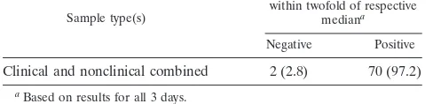
TABLE 2. Assessment of intermediate precision (Operators 1 and 2)
Sample type(s)
No. (%) with indicated result within twofold of respective
mediana
Negative Positive
Clinical and nonclinical combined 2 (2.8) 70 (97.2)
aBased on results for all 3 days.
TABLE 3. HAI specificity test results
Sample Titer(s)
Clinical and control samplesa
H5 positive clinical serum CS 25 ...320, 320 H5 positive clinical serum CS 08 ...320, 320 H5 positive clinical serum CS 28 ...80, 40 H3 positive clinical serum CS 29 ...10,b10
H3 positive clinical serum CS 30 ...10, 10 H3 positive clinical serum CS 31 ...10, 10 Rubella standard antiserum 10 units...10, NDc
Rubella standard antiserum 40 units...10, ND Rubella standard antiserum 100 units...10, 10 Normal sheep serum control ...ND, 10
HP antiserum
H5 Vietnam HP undiluted ...1,280
H5 Vietnam HP 1:5...160
H5 Indo HP undiluted ...640
H5 Indo HP 1:5...80
H7 HP undiluted...10
H7 HP 1:5 ...10
H3 HP undiluted...10
H3 HP 1:5 ...10
H1 HP undiluted...10
H1 HP 1:5 ...10
B/HK (Victoria lineage) HP undiluted...10
B/HK (Victoria lineage) HP 1:5 ...10
B/Yamagata lineage(2008) HP undiluted ...10
B/Yamagata lineage(2008) HP 1:5 ...10
B/Yamagata HP undiluted...10
B/Yamagata HP 1:5 ...10
B/Sh Yamagata HP undiluted...10
B/Sh Yamagata HP 1:5 ...10
Normal (unvaccinated) animal serum from various species Normal human, undiluted...10
Rabbit, undiluted ...10
Ferret, undiluted ...10
Goat, undiluted ...10
Sheep, undiluted ...10
Horse, undiluted ...10
aTiters from two replicates are reported for these samples. bA titer of 10 is negative for H5N1 HAI.
cND, not determined.
on August 17, 2020 by guest
http://cvi.asm.org/
Table 1 shows the results obtained by Operator 1 for each of the 3 days of testing for the nonclinical positive and negative control samples. This analysis was based on 45 samples, tested in triplicate, and all three members of the triplicate set were evaluated to determine if they were within twofold of the triplicate’s GMT. This test was conducted for each day, inde-pendent of the other days. Also in Table 1, the combined results for all 3 days for the testing of the nonclinical samples by Operator 1 are presented. This analysis was based on 405 results (45 samples per day for 3 days with 3 replicates per sample). Each result was tested, independently of the others, to determine that it was within twofold of the GMT of the nine results for the sample across all 3 days. For days 1, 2, and 3, greater than 80% of the nonclinical samples (93.3%, 86.7%, and 88.9%, respectively) had three replicate measurements that were within twofold of the GMT value for all replicates of that individual sample (Table 1). For all individual nonclinical test results across the 3 days, 85.7% were within twofold of the
GMT value for the respective nonclinical serum sample (Table 1), thereby meeting the acceptance criteria for precision. Table 1 shows the results obtained by Operator 1 for each of the 3 days of testing for the clinical samples. For days 1, 2 and 3, greater than 85% of the individual clinical samples (88.9%, 85.2%, and 92.6%, respectively) had three replicate measure-ments that were within twofold of the GMT value for all replicates of that individual sample. For all individual clinical test results across the 3 days, 81.1% were within twofold of the GMT value for the respective clinical serum sample (Table 1). Therefore, the prospective acceptance criteria for qualification of the H5N1 HAI assay for precision (reproducibility) were met.
Intermediate precision.Intermediate precision was assessed by having the precision testing performed independently by a second operator (Operator 2) using the same samples in the same laboratory and the same methodology for comparison with results obtained by Operator 1. All assays performed by Operator 2 met the acceptance criteria and no assays were repeated. Intermediate precision acceptance criteria estab-lished in advance required median values for an individual sample computed separately for each operator across all 3 days to be within twofold (1 dilution). This criterion had to be met for 51 of the 72 individual samples (71%).
The data analysis for intermediate precision is presented in Table 2. Seventy of 72 (97.2%) of the median values for indi-vidual clinical and nonclinical samples for the two operators were within twofold across all 3 days. Therefore, the interme-diate precision of the HAI assay was considered acceptable.
Specificity. Specificity was tested three times and all tests showed acceptable specificity of the HAI assay. The results are shown in Table 3. The assay showed specificity for detection of
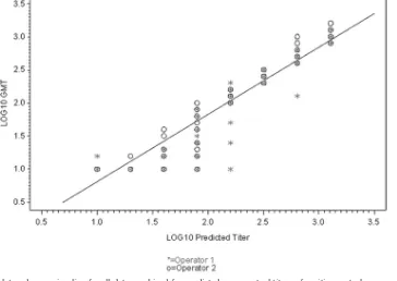
FIG. 3. Scatter plot and regression line for all data combined for predicted versus actual titers of positive control serum using a log10scale.
TABLE 4. Linearity results
Operator(s) Day(s)
Correlation coefficient, expected vs. actual
titer (log10scale)
1 and 2 (combined) 1–6 0.95
1 1 0.88
2 0.97
3 0.98
2 1 0.96
2 0.99
3 0.96
on August 17, 2020 by guest
http://cvi.asm.org/
antibody to H5 hemagglutinins in that positive titers were obtained with the H5 Vietnam and H5 Indonesian (Indo) hyperimmune (HP) antisera as well as the H5N1 human clin-ical sera known to have demonstrable titers to rgH5VN virus. No cross-reaction was seen with H7 antiserum, another avian influenza virus hemagglutinin subtype. The H3, H1, and influ-enza B (both the Yamagata and Victoria lineages) seasonal influenza virus HP antisera, as well as the rubella standard antisera and the normal sheep, rabbit, ferret, goat, horse, and human sera, had no detectable titers to rgH5VN virus. There-fore, the HAI assay was shown to be specific for H5N1 virus and suitable for testing of clinical serum samples from H5N1 vaccine trials.
Linearity. The prospectively defined criterion for demon-stration of linearity of the assay required a correlation
coeffi-cient of the regression line of the log-transformed data for the entire intermediate precision data set to be atⱖ0.9. Linearity was evaluated separately for each operator for each of the 3 days tested as well as for a combined data set including the results from all six assays. All assays met the assay acceptance criteria and no assays were repeated.
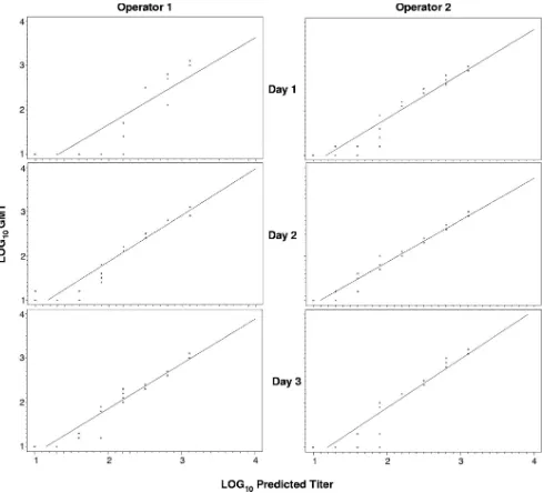
The tabulated results for linearity are presented in Table 4. Scatter plots of the linearity data are presented in Fig. 3 (all days combined) and Fig. 4 (scatter plot of individual testing dates and operators). The correlation coefficient for the com-bined (six-assay) data set was 0.95, which exceeded the accep-tance criterion ofⱖ0.90. In addition, the correlation coefficient for five of the six individual data sets exceeded the acceptance criterion, with the remaining individual data set having a cor-relation coefficient of 0.88, largely due to the decreased
linear-FIG. 4. Scatter plots and regression lines by operator and day for predicted versus actual titers of positive control serum using a log10scale.
on August 17, 2020 by guest
http://cvi.asm.org/
ity for titers ofⱕ80. The H5N1 HAI assay was demonstrated to be linear, with test results directly proportional to the amount of analyte (antibody in serum) in a sample.
Range.The range of the assay, which is the interval between the upper and lower amounts of analyte for which suitable levels of precision, specificity, and linearity have been demon-strated, was inferred from the precision and linearity data and determined to be from a titer of 10 to 1,280. The range of the assay was demonstrated by the range of dilutions over which the results were linear (correlation coefficient ofⱖ0.9), but it should be noted from the scatter plots in Fig. 3 and 4 that predicted titers of 80 (1.9 log10) or below were less frequently
matched by the measured titer than titers predicted to be greater than 80.
Robustness.All assays met the assay acceptance criteria and no repeat analyses were required. For each critical reagent evaluated, the results were considered acceptable if at least 80% of the sample titers were within twofold (one dilution) of the median titer for that sample on the day of assay or across two different assay dates (as was tested for robustness of HRBC storage time). For this comparison, the results were considered acceptable if at least 80% of the sample titers on each assay day were within twofold (1 dilution) of the median titer for that sample across the two assay days. Table 5 shows the primary combined analysis of sample-specific median HAI titers obtained using HRBCs after 1 day or 6 days of storage. The sample titers were within twofold (1 dilution) of the me-dian titer across the two assay days for 86.1% of the samples tested. Table 6 contains analyses for robustness of the horse red blood cells based upon blood lot. Three individual lots of blood from three individual donor horses were analyzed in the same day, on two different dates. Each assay result was com-pared to the median for the day on which it was tested. The analyses from both dates of testing suggested that the assay is robust across multiple donor horses; however, the nonclinical control samples were more reproducible than the clinical samples.
Robustness was demonstrated for RDE lot in that, for both the nonclinical (93.3%) and clinical (85.2%) samples sepa-rately and combined (90.3%), at least 80% of the sample titers
were within twofold (1 dilution) of the median endpoint value (Table 7).
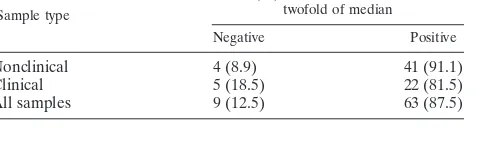
Similarly, robustness was demonstrated for virus lot in that, for both the nonclinical (91.1%) and clinical (81.5%) samples separately and combined (87.5%), at least 80% of the sample titers were within twofold (1 dilution) of the median endpoint value (Table 8).
The H5N1 HAI assay was shown to be robust by the primary analysis for variations in animal source of HRBCs (different horses), age of HRBCs for up to 6 days postcollection, for different lots of RDE of the same source and catalog number, and for virus lots differing between one and two passages in eggs.
Consistency of controls.Southern Research maintains a his-torical database on the performance of the positive and nega-tive controls over time. Figure 5 demonstrates the consistency of the controls over 2 years and more than 6,000 HAI assay plates. The graph denotes two major changes in the assay, with point A delineating the addition of a mixing step to the pro-tocol. This addition stabilized the titers obtained for the pos-itive control. Point B denotes the alteration in the dilution factor used in the assay such that the positive control endpoint would be near the middle of the plate. After implementation of this new dilution, 98% of all plates had titers of 1,600, 3,200, or 6,400 (all within twofold of each other). A new positive control was implemented around the plate with a titer of 4,800; how-ever, this control was pretested and used in the assays at a dilution that provided endpoints in the same region of the plate. These results clearly show high levels of consistency over time and with different operators.
DISCUSSION
Our results demonstrate that the H5N1 HAI assay is robust and linear and that precision and intermediate precision are attainable with appropriately trained staff. These qualification data are critical for evaluating current and future vaccines to potentially pandemic strains of influenza virus, as they estab-lish uniform acceptance criteria that may be applied
interna-TABLE 5. Assessment of robustness for HRBC storage time
Sample
No. (%) with indicated result within twofold of combined median for
days 1 and 6
Negative Positive
HRBCs (all samples) 10 (13.9) 62 (86.1)
TABLE 6. Assessment of robustness for HRBC lot
Sample type
No. (%) with indicated result within twofold of median on:
Day 1 Day 6
Negative Positive Negative Positive
Nonclinical 6 (13.3) 39 (86.7) 5 (11.1) 40 (88.9) Clinical 6 (22.2) 21 (77.8) 6 (22.2) 21 (77.8) All samples 12 (16.7) 60 (83.3) 11 (15.3) 61 (84.7)
TABLE 7. Assessment of robustness for receptor-destroying enzyme lot
Sample type
No. (%) with indicated result within twofold of median
Negative Positive
Nonclinical 3 (6.7) 42 (93.3)
Clinical 4 (14.8) 23 (85.2)
All samples 7 (9.7) 65 (90.3)
TABLE 8. Assessment of robustness for virus lot
Sample type
No. (%) with indicated result within twofold of median
Negative Positive
Nonclinical 4 (8.9) 41 (91.1)
Clinical 5 (18.5) 22 (81.5)
All samples 9 (12.5) 63 (87.5)
on August 17, 2020 by guest
http://cvi.asm.org/
tionally and can be used to support licensure of these vaccines as part of the foundation for pandemic preparedness. The approach outlined here provides a scaffold for laboratories to develop and qualify HAI assays by establishing uniform meth-ods and acceptance criteria that may be applied internationally and enable rapid implementation of international standards for pandemic vaccines once available.
All assays met the assay acceptance criteria as outlined above.
The FDA guidelines for pandemic influenza vaccine evalu-ation state that an HAI titer greater than or equal to 40 (1) is the best immunological parameter currently available to pre-dict protection from natural infection. Therefore, an HAI as-say should optimally be qualified for measurement of antibody titers in this range. While the linear range of the H5N1 HAI assay was determined to include positive titers as low as 20 based on the correlation coefficient of expected versus ob-served antibody titers, we obob-served a trend toward greater
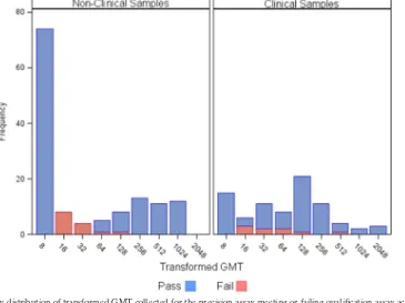
FIG. 6. Frequency distribution of transformed GMT collected for the precision assay meeting or failing qualification assay acceptance criteria across all 3 days.
FIG. 5. Performance of the control sera over time, as tracked by our historical database, indicates that carefully stored controls provide consistent results over a 2-year period that encompassed hundreds of assays and multiple analysts. Arrow A, implementation of a serum mixing step; arrow B, modification of the dilution factor used for the positive control serum.
564
on August 17, 2020 by guest
variability and a modest deviation from linearity at titers of 80 or below (Fig. 3 and 4). Indeed, 55.6% of all clinical samples and 85.7% of nonclinical samples not passing acceptance cri-teria during precision and linearity testing had titers between 12 and 45 (Fig. 6). These values increased to 77.8% and 92.9%, respectively, when this range was extended to titers of 90 or lower. The erythrocyte settling pattern is often more difficult to interpret in low-titer samples due to the presence of high concentrations of serum (4), which may account, in part, for
greater variability at that end of the assay range. In addition, this settling pattern difficulty most commonly resulted in re-corded values below the expected titer, possibly explaining the deviation from linearity at low titers. Any inaccuracies in the estimation of titers below 80 would likely manifest as under-estimates rather than overunder-estimates.
As a result of these observations during qualification of the H5N1 HAI assay, we undertook additional method develop-ment to determine if increased time of incubation for the
FIG. 7. Seven operators analyzed 72 nonclinical samples at 45 min, 90 min, or 135 min. (A) GMT from the data of all operators at each time point was converted to a log10scale and linear regression against predicted titers was performed. (B) GMT from the data of all seven operators at each time point is
graphically displayed for titers of 80 or lower to underscore the increased frequency of detecting these lower titers with an increase in incubation time.
on August 17, 2020 by guest
http://cvi.asm.org/
serum and virus improved the linearity and precision for sera with titers of 80 or lower. We found improvement in correlat-ing predicted and measured titers of control serum by incubat-ing virus and serum for periods longer than those outlined in the current SR SOP (30 to 60 min) and in other manuals (7) (Fig. 7). Indeed, linearity increased across the entire range of the assay from a correlation coefficient of 0.95 at 45 min to 0.97 at 90 and 135 min (Fig. 7A). Furthermore, the ability of the operator to visibly detect titers of 80 or lower dramatically improved with the increase in incubation time (Fig. 7B). There was no significant effect of longer incubation times on mea-sured titers at the upper end of the range (Fig. 7A). This was not an unexpected result, as we provide more time for anti-body-antigen interactions to reach equilibrium, thereby im-proving detection, especially when the antibody is present at a low concentration or is of low avidity.
Our historical database that tracks the performance of the control sera over time indicated that carefully stored controls provided consistent results over a 2-year period that encom-passed hundreds of assays and multiple analysts, as noted above in “Consistency of controls” (Fig. 5). These data suggest that an international standard reference serum, if developed, would likely have a long shelf life.
Our results clearly demonstrate that the HAI assay as adapted for use with potentially pandemic influenza viruses can be precise, consistent, linear, and robust, and the range of the assay can span the entire set of dilutions possible on a 96-well microtiter plate (1:10 to 1:1,280). These results provide a strong framework for subsequent qualification of the HAI assay in other laboratories and will greatly enhance the capa-bilities to examine and compare pandemic vaccines in a rapid and high-throughput fashion.
ACKNOWLEDGMENTS
This project has been funded with federal funds from the National Institute of Allergies and Infectious Diseases, National Institutes of Health, Division of Microbiology and Infectious Disease, Department of Health and Human Services, under contract number N01-AI-30068 to PPD, Inc., Wilmington, NC.
We extend special thanks to Michelle Culp, Roland Levandowski, Emily Kough, and Jean Hu-Primmer at the NIAID Division of Micro-biology and Infectious Disease, Fred Batzold from PPD, Inc., and Catherine McCall and Judy Ruckman from CBR International Corp. for their assistance and critique. Additional thanks go to Crystal Coleman, Valerie Johnson, Lisa Slappey, Melinda I. Sosa, Shuang Feng, Tracy Williams, and Rachel Sayer from SR for their technical assistance.
REFERENCES
1.Food and Drug Administration.2007. Guidance for industry: clinical data needed to support the licensure of seasonal inactivated influenza vaccines. U.S. Food and Drug Administration, Rockville, MD. http://www.fda.gov/cber /gdlns/trifluvac.htm#iii.
2.Hobson, R., R. L. Curry, A. S. Beare, and A. Ward-Gardner.1972. The role of serum haemagglutination-inhibiting antibody in protection against chal-lenge infection with influenza A2 and B viruses. J. Hyg.70:767–777. 3.Rowe, T., R. A. Abernathy, J. Hu-Primmer, W. W. Thompson, X. Lu, W. Lim,
et al.1999. Detection of antibody to avian influenza A (H5N1) virus in human serum by using a combination of serologic assays. J. Clin. Microbiol.37:937– 943.
4.Schild, G. C., and W. R. Dowdle.1975. Influenza virus characterization and diagnostic serology, p. 315–372. InE. D. Kilbourne (ed.), The influenza viruses and influenza. Academic Press, New York, NY.
5.Stephenson, I., J. M. Wood, K. G. Nicholson, A. Charlett, and M. C. Zambon.
2004. Detection of anti-H5 responses in human sera by HAI using horse erythrocytes following MF59-adjuvanted influenza A/Duck/Singapore/97 vac-cine. Virus Res.103:91–95.
6.World Health Organization. 2006. WHO Influenza Task Force meeting. World Health Organization, Geneva, Switzerland.
7.World Health Organization.2002. WHO manual for animal influenza diag-nosis and surveillance. World Health Organization, Geneva, Switzerland.