Evaluation of Two Enzyme-Linked Immunosorbent Assay Kits for
Chikungunya Virus IgM Using Samples from Deceased Organ and
Tissue Donors
Harry E. Prince, Michelle L. Altrich, Marek J. Nowicki Serology Laboratory, Viracor-IBT Laboratories, Los Angeles, California, USA
The identification of nearly 3,500 cases of chikungunya virus (CHIKV) infection in U.S. residents returning in 2014 and 2015 from areas in which it is endemic has raised concerns within the transplant community that, should recently infected individuals become organ and/or tissue donors, CHIKV would be transmitted to transplant recipients. Thus, tests designed to detect recent CHIKV infection among U.S. organ and tissue donors may become necessary in the future. Accordingly, we evaluated 2 enzyme-linked immunosorbent assays (ELISAs) for CHIKV IgM readily available in the United States using 1,000 deidentified serum or plasma specimens collected from donors between November 2014 and March 2015. The Euroimmun indirect ELISA identified 38 reactive specimens; however, all 38 were negative for CHIKV IgG and IgM in immunofluorescence assays (IFAs) conducted at a reference laboratory and, thus, were falsely reactive in the Euroimmun CHIKV IgM assay. The InBios IgM-capture ELISA identi-fied 26 reactive samples, and one was still reactive (index>1.00) when retested using the InBios kit with a background subtrac-tion modificasubtrac-tion to identify false reactivity. This reactive specimen was CHIKV IgM negative but IgG positive by IFAs at two reference laboratories; plaque reduction neutralization testing (PRNT) demonstrated CHIKV-specific reactivity. The IgG and PRNT findings strongly suggest that the InBios CHIKV IgM-reactive result represents true reactivity, even though the IgM IFA result was negative. If testing organ/tissue donors for CHIKV IgM becomes necessary, the limitations of the currently available CHIKV IgM ELISAs and options for their optimization must be understood to avoid organ/tissue wastage due to falsely reactive results.
C
hikungunya virus (CHIKV) is an alphavirus transmitted from person to person via mosquito bites (1). Symptoms include fever, rash, and debilitating arthralgia; 15% to 60% of patients develop chronic arthralgia leading to arthritic joint damage (2). After a large CHIKV outbreak in India and southeast Asia in 2004 through 2006, in which nearly 2 million people became infected (3,4), epidemiologists predicted that CHIKV might move to other geographic areas where the mosquito vectors are found (5). This prediction was realized in December 2013, when local transmis-sion of CHIKV was reported on the Caribbean island of St. Martin (6). CHIKV infection has since spread throughout the Caribbean basin (7) and is now also endemic in Mexico, Central America, and South America and in the Caribbean island nations. In con-junction with this outbreak, 3,490 cases in U.S. residents (from 49 of 50 states) were reported to the CDC during 2014 and 2015; 3,478 cases represented infections acquired during international travel to areas where CHIKV is endemic, whereas 12 cases repre-sented local transmission (8).There is concern within the transplant community that CHIKV could be transmitted from organ and/or tissue donors to recipients. Donor-derived transmission of other mosquito-borne viruses with similar epidemiologic and biologic features, notably dengue virus and West Nile virus, has been documented (9,10). Although no cases of CHIKV transmission by transplantation have yet been reported, studies have shown that CHIKV can be isolated from corneas of acutely infected individuals (11), and atypical manifestations of CHIKV infection were reported in a recipient who became infected 7 years after receiving a liver trans-plant (12). However, the likelihood of CHIKV transmission by transplantation, and what organs/tissues might harbor the virus, remains unknown.
As additional information regarding CHIKV transmission by transplantation becomes available, there might be a future need for tests to identify recent CHIKV infection, particularly among U.S. donors living in geographic areas where many residents travel internationally and have close ethnic ties to areas where CHIKV is endemic (13). There are two accepted methods for identifying recent CHIKV infection, detection of CHIKV IgM and CHIKV RNA. CHIKV RNA is detectable in serum within the first week after symptom onset but then subsides to undetectable levels. CHIKV IgM, in contrast, becomes detectable by day 5 after the onset of symptoms and remains detectable for approximately 4 months. Thus, CHIKV RNA testing would identify organ/tissue donors who were infected within 1 week prior to donation, whereas CHIKV IgM detection would identify donors who were infected 5 days to 4 months prior to donation. From this stand-point, CHIKV IgM appears to be the more robust indicator of recent infection in organ/tissue donors (14,15,16). Thus, we eval-uated the performance of two commercially available CHIKV IgM
Received24 June 2016 Returned for modification2 August 2016
Accepted9 August 2016
Accepted manuscript posted online17 August 2016
CitationPrince HE, Altrich ML, Nowicki MJ. 2016. Evaluation of two enzyme-linked immunosorbent assay kits for chikungunya virus IgM using samples from deceased organ and tissue donors. Clin Vaccine Immunol 23:825–830.
doi:10.1128/CVI.00330-16.
Editor:P. P. Wilkins, CDC
Address correspondence to Harry E. Prince, [email protected]. Copyright © 2016, American Society for Microbiology. All Rights Reserved.
on August 17, 2020 by guest
http://cvi.asm.org/
enzyme-linked immunosorbent assay (ELISA) kits using 1,000 ar-chived serum or plasma samples from organ or tissue donors.
MATERIALS AND METHODS
Donor specimen selection and deidentification.The 1,000 specimens evaluated were collected from organ or tissue donors in the United Net-work for Organ Sharing (UNOS) region 5, composed of several south-western and south-western U.S. states, including California. All six UNOS region 5 organ procurement organizations (OPOs) served by our laboratory pro-vided permission to utilize their specimens for the study. For each month of the 5-month period (November 2014 through March 2015), the first 60 serum or plasma samples from deceased prospective organ (heart-beat-ing) donors and the first 140 serum or plasma samples from deceased prospective tissue (cadaveric) donors submitted to our facility were re-trieved from⫺80° storage. The study panel thus contained 300 organ donor samples and 700 tissue donor samples; this distribution reflects the relative proportions of deceased organ donor versus deceased tissue do-nor specimens submitted to our facility for infectious disease screening. Samples were deidentified as requested by the participating OPOs; a sam-ple within a given group (defined by donor type [organ or tissue] and month of collection) was randomly selected, and 1 ml of the sample was transferred to a randomly selected, numbered plastic vial within a desig-nated numeric range. No key was created to enable tracking of which donor specimen was transferred to which numbered vial. Thus, the only demographic data available for a given specimen were donor type (organ or tissue) and month of collection; no result could be linked to a donor’s protected health information.
Known positive specimens.Five serum samples known to be positive for CHIKV IgM by an immunofluorescence assay (IFA) were obtained from Focus Diagnostics (San Juan Capistrano, CA); titers ranged from 1:40 to 1:640 (reference value,⬍1:10) (16).
Euroimmun assay.Samples were tested for CHIKV IgM using the anti-chikungunya virus ELISA (IgM) kit (designated for research use only) purchased from Euroimmun US (Mountain Lakes, NJ), according to the manufacturer’s instructions (17). Briefly, serum or plasma samples diluted 1:101 in sample diluent containing anti-human IgG were added to microtiter wells coated with a proprietary mixture of recombinant CHIKV antigens. After the wells sat for 1 h at 37°C and were washed, peroxidase-labeled anti-human IgM was added to them. After 30 min at room temperature (RT), the wells were washed again and then reacted with a chromogen-substrate solution (tetramethylbenzidine [TMB] plus hydrogen peroxide) for 15 min at RT; the color reaction was stopped by the addition of 0.5 M sulfuric acid. Optical densities (ODs) were measured at 450 nm and 630 nm (reference wavelength) for each well using a BioTek ELx800 photometric reader (Winooski, VT). Per the manufacturer’s in-structions, the results were expressed as index values, calculated by divid-ing a specimen’s OD by the OD of a kit-supplied calibrator serum in-cluded in the same run. Index values of⬍0.8 were considered negative, values of 0.8 to⬍1.1 were considered equivocal, and values ofⱖ1.1 were deemed positive.
InBios screening assay.Samples were also tested for CHIKV IgM using the CHIKjj Detect IgM-capture ELISA kit (designated for research use only) purchased from InBios International (Seattle, WA) (18). Briefly, samples were diluted 1:100 in dilution buffer and added to microtiter wells coated with polyclonal anti-human IgM (the animal source is con-sidered proprietary). After wells sat for 30 min at 37°C and were washed, they then were incubated for 30 min at 37°C with an antigen solution containing a recombinant CHIKV envelope (E2/E1) glycoprotein pro-duced in C7/10 mosquito cells (19). Wells were again washed and then incubated with peroxidase-labeled monoclonal anti-CHIKV antibody (CHK-175) for 30 min at 37°C. After the wells underwent a final wash, a TMB substrate solution was added, and after 10 min at RT, the color reaction was stopped by the addition of 1 N sulfuric acid. OD at 450 nm (no reference wavelength) was measured for each well using a BioTek ELx800 photometric reader. Per the manufacturer’s instructions, results
were expressed as index values, calculated by dividing a specimen’s OD by the OD of a kit-supplied cutoff serum included in the same run. Index values of⬍1.00 were considered nonreactive, and values ofⱖ1.00 were considered reactive.
InBios background subtraction modification.All samples reactive in the InBios screening assay were retested using the InBios kit modified to identify samples falsely reactive due to IgM antibodies that recognize im-munoglobulins of other animal species, known as heterophilic IgM anti-bodies. These antibodies, like all IgM antibodies, are captured by the im-mobilized anti-IgM antibody attached to the microtiter well. Then, due to their heterophilic activity, they bind the peroxidase-labeled monoclonal anti-CHIKV antibody reagent when added, both in the presence and in the absence of antigen (20). For this modified assay, the diluted kit-sup-plied cutoff control and each diluted donor sample were added to 2 wells, and after the initial incubation and washing, one well received CHIKV antigen and the other well received sample dilution buffer. The assay was then finished per the manufacturer’s instructions, and the OD of each well was measured as described for the screening assay. For each sample, the net OD was calculated by subtracting the OD of the well receiving dilution buffer from the OD of the well receiving antigen (hence the designation of this modification as background subtraction [BS]). These net OD values were then used to calculate the BS index, defined as the net sample OD divided by the net cutoff OD (21); the same interpretive guidelines em-ployed for the screening assay were used for the modified assay. In addi-tion, the value observed from the antigen well OD (Ag OD) was divided by the value observed from the diluent well OD (dil OD) to calculate the Ag OD/dil OD ratio; this value has proven useful for identifying false-reactive samples in the InBios dengue capture and West Nile virus IgM-capture ELISAs (22,23).
Reference laboratory testing.All samples considered reactive (equiv-ocal or positive) in the Euroimmun assay and 2 samples with InBios BS index values of⬎0.80 were sent to Focus Diagnostics, where they were tested for CHIKV IgG and IgM using validated IFAs (16). For both IFAs, samples were screened at a 1:10 dilution. The 5 samples with the highest positive Euroimmun ELISA index values and the 2 samples with InBios BS index values of⬎0.80 were also sent to the California Department of Public Health (CDPH) Viral and Rickettsial Disease Laboratory (Rich-mond, CA), where they were tested for CHIKV IgG and IgM by IFA; for both IFAs, samples were screened at a 1:8 dilution, and the titers of the positive samples were determined by endpoint dilution. Per CDPH pro-tocol, one sample positive for CHIKV IgG was reflexed to alphavirus plaque reduction neutralization testing (PRNT), which assessed the abil-ity of various dilutions of the sample to inhibit the infectivabil-ity of CHIKV and western equine encephalitis virus (WEEV).
Statistics.Differences between proportions were assessed by calculat-ing the chi-square statistic (23), with significance defined as aPvalue of⬍0.01.
RESULTS
Reactivity in screening assays.Table 1presents a summary of the findings observed for organ and tissue donor serum or plasma specimens when tested for CHIKV IgM using the Euroimmun and InBios screening assays. Of 1,000 donor samples, 38 (3.8%) were reactive (equivocal or positive) in the Euroimmun ELISA, with 6.0% of 300 organ donor samples and 2.86% of 700 tissue donor samples reactive. Similarly, 26 of 1,000 (2.6%) samples were reac-tive in the InBios screening assay, with 2.3% of samples from organ donors and 2.7% of samples from tissue donors reactive. Only one specimen was reactive in both the Euroimmun (equiv-ocal) and InBios screening assays. When assessing differences be-tween proportions for various groups, the only difference that reached statistical significance was the proportion of organ donor versus tissue donor samples with an equivocal Euroimmun result (4.3% versus 1.1%,P⫽0.001). The index range of positive donor
on August 17, 2020 by guest
http://cvi.asm.org/
samples in the Euroimmun ELISA was 1.13 to 3.21, with 13/17 (76%) between 1.13 and 2.00; the index range of reactive donor samples in the InBios ELISA was 1.06 to 5.98, with 22/26 (85%) between 1.06 and 2.00. All 5 known CHIKV IgM-positive samples obtained from Focus Diagnostics were positive in the Euroimmun ELISA (index range, 2.07 to 6.91) and reactive in the InBios screening assay (index range, 6.48 to 12.75).
Figure 1presents the proportions of donor samples reactive in
the 2 assays by month of sample submission and donor group. With one exception, reactive results were observed in both assays from both donor groups during each month; the exception was that no InBios-reactive samples were found among the March 2015 organ donor samples. Although the proportion of organ do-nor samples reactive in the Euroimmun assay was higher in those obtained in November 2014 than in those obtained in other months, this difference was not statistically significant. Similarly, the proportion of tissue donor samples reactive in the InBios assay was higher in samples obtained in November 2014 than in those obtained in other months, but this difference was not statistically significant.
Reactivity in the InBios assay with background subtraction. Samples reactive in the InBios CHIKV IgM screening assay were retested using the BS modification of the InBios assay to identify samples falsely reactive due to heterophilic IgM antibodies (20).
Table 2provides examples of various reactivity data patterns
ob-served in the BS assay. Note in particular that the false-reactive sample exhibited a reactive screening index (1.23) but a nonreac-tive BS index (0.14) and an Ag OD/dil OD ratio that was identical to that of the nonreactive sample (1.08). In contrast, the true-reactive sample (from Focus Diagnostics) exhibited both
screen-ing index and BS index values of⬎1.00 and an Ag OD/dil OD ratio that was markedly higher than the ratios of the nonreactive and false-reactive samples.
Figure 2presents the In Bios BS index and Ag OD/dil OD ratio
results for the 26 donor specimens that were reactive in the InBios screening assay. Screen-reactive specimen 5 was clearly distinc-tive, exhibiting a BS index of⬎1.00 and an Ag OD/dil OD ratio that was more than 2.5-fold higher than the ratios observed for the other 25 samples. Screen-reactive specimen 17 also exhibited a BS index that was markedly higher than the indices of all other sam-ples (except specimen 5), but the Ag OD/dil OD ratio of specimen 17 was not increased compared to the ratios of other samples (except specimen 5). Screen-reactive specimen 1 was the sample that exhibited reactive results in both the Euroimmun and InBios screening ELISAs; as shown inFig. 2, this sample yielded a nega-tive result in the InBios BS assay. As expected, all 5 known posinega-tive samples from Focus Diagnostics exhibited increased BS index val-ues (ⱖ7.98) and increased Ag OD/dil OD ratios (ⱖ15.73) (data not shown).
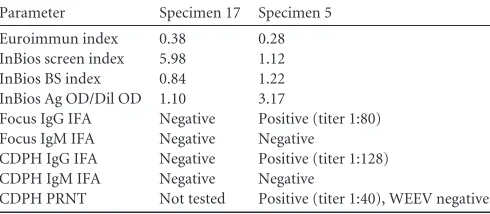
Reference laboratory test results.All 38 samples reactive in the Euroimmun ELISA were negative for CHIKV IgG and CHIKV IgM when tested by IFA at Focus Diagnostics; similarly, the 5 samples with the highest positive index values in the Euroimmun ELISA were also negative for CHIKV IgG and IgM when tested by IFA at CDPH. The 2 samples with a BS index value of⬎0.80 in the InBios BS assay (screen-reactive specimens 5 and 17) were also sent to both Focus Diagnostics and CDPH for CHIKV antibody testing; the results are summarized inTable 3. Reactive specimen 17 was negative for CHIKV IgG and IgM at both reference labo-ratories. In contrast, reactive specimen 5 was negative for CHIKV
TABLE 1Results for deceased-donor specimens tested for CHIKV IgM using Euroimmun and InBios screening ELISAs
Donor group n
Euorimmun assay results (n[%]) InBios assay results (n[%])
Negative Equivocal Positive Nonreactive Reactive
Organ 300 282 13 (4.3) 5 (1.7) 293 7 (2.3)
Tissue 700 680 8 (1.1) 12 (1.7) 681 19 (2.7)
All 1,000 962 21 (2.1) 17 (1.7) 974 26 (2.6)
FIG 1Proportions of samples reactive in screening assays in relation to donor group and month of sample collection.
on August 17, 2020 by guest
http://cvi.asm.org/
IgM but positive for CHIKV IgG at both reference laboratories, with similar CHIKV IgG titers. PRNT results from the CDPH for reactive specimen 5 demonstrated that it had specific neutralizing activity toward CHIKV (titer, 1:40) but not WEEV (titer,⬍1:20). These results are consistent with the interpretation that the InBios CHIKV IgM-reactive result for reactive specimen 5 (collected from an organ donor in January 2015) represents a true-reactive result.
DISCUSSION
A forecasting model recently identified several U.S. urban centers as high-risk areas for travel-associated CHIKV infections (13). Should a CHIKV-infected traveler become an organ/tissue donor within a few months of his or her return to the United States, it is possible that the transplanted organ/tissue will still harbor enough of the virus to transmit the infection to the recipient. Thus, if the number of travel-related CHIKV cases continues to increase, or local mosquito-borne transmission escalates, an assay for identi-fying donors recently infected with CHIKV might be required by UNOS and other transplant organizations. Thus, the goal of our study was to evaluate the suitability of commercially available CHIKV IgM ELISA kits for testing samples from deceased organ and tissue donors, most of whom resided in southern California, an area at high risk for CHIKV transmission. The investigation produced several interesting findings regarding the performance characteristics of the CHIKV IgM ELISA kits evaluated.
On the basis of the small number (n⫽5) of known CHIKV IgM-positive samples evaluated, both the Euroimmun and InBios assays exhibited excellent sensitivity. Specificity, however, was an-other matter. We found that 3.8% of donor samples were reactive (equivocal or positive) in the Euroimmun indirect ELISA, a value nearly 5-fold higher than the reactive rate of 0.8% presented in the package insert for a panel of 498 blood donors (17). Interestingly,
the package insert value was based on a single cutpoint index of 1.0 to define reactivity as negative or positive rather than following the package insert guidelines for defining negative, equivocal, and positive reactivity. If we used this single cutpoint approach, our reactivity value would be 2.4%, which is still 3-fold higher than the value presented by the manufacturer. This difference most likely reflects differences in the populations studied (deceased organ/ tissue donors versus living blood donors). Further, we found that all 38 samples reactive in the Euroimmun ELISA were falsely re-active on the basis of IFA results. The factors responsible for this false reactivity remain unknown; however, the observation that the proportion of samples exhibiting an equivocal Euroimmun result was significantly higher in organ donors than the propor-tion observed in tissue donors suggests that some types of false reactivity may be related to organ donor-specific predonation treatment factors. Further studies are needed to characterize these factors and to identify ways to optimize the assay to reduce or eliminate false reactivity.
Similarly, 2.6% of donor samples were reactive in the InBios IgM-capture ELISA when performed in accordance with the pack-age insert. Although the data supplied in the packpack-age insert did not include expected reactivity levels among blood donors or a similar population (18), this finding was not unexpected on the basis of published findings for similar IgM-capture ELISAs (20,24). As discussed by Levinson and Miller (20), the majority of samples reactive in IgM-capture assays that target pathogens in areas of low endemicity are falsely reactive due to heterophilic antibodies, which recognize immunoglobulins of other animal species. Inher-ent to the IgM-capture format, however, is the ability to distin-guish false reactivity from true reactivity using the BS modifica-tion. When this modified assay is performed, samples falsely reactive in the screening assay exhibit essentially the same signal intensity in the presence and absence of antigen, whereas truly reactive samples exhibit much higher signal intensity in the pres-ence of antigen than in the abspres-ence of antigen (21,25). This signal difference is the basis for calculating the BS index and the Ag OD/dil OD ratio; both have been used to distinguish false from
TABLE 2Examples of BS index and Ag OD/dil OD ratios in the InBios assay with BS modification
Sample Antigen well OD Diluent well OD Net OD (Ag OD⫺dil OD) Screen index BS index Ag OD/dil OD
Calibrator 0.145 0.049 0.096 2.96
Nonreactive 0.060 0.051 0.009 0.41 0.09 1.08 False reactive 0.178 0.165 0.013 1.23 0.14 1.08 True reactive 0.818 0.052 0.766 5.64 7.98 15.73
FIG 2InBios background subtraction (BS) assay results for 26 specimens reactive in the InBios screening assay.
TABLE 3Reference laboratory testing results for 2 samples with InBios BS index values of⬎0.80
Parameter Specimen 17 Specimen 5
Euroimmun index 0.38 0.28 InBios screen index 5.98 1.12 InBios BS index 0.84 1.22 InBios Ag OD/Dil OD 1.10 3.17
Focus IgG IFA Negative Positive (titer 1:80) Focus IgM IFA Negative Negative
CDPH IgG IFA Negative Positive (titer 1:128) CDPH IgM IFA Negative Negative
CDPH PRNT Not tested Positive (titer 1:40), WEEV negative
on August 17, 2020 by guest
http://cvi.asm.org/
true reactivity (21–23,25). Thus, IgM-capture ELISAs offer a ma-jor advantage over indirect IgM ELISAs, since identification of falsely reactive samples by a simple assay modification is not pos-sible for the indirect format (22,23,25).
The most interesting finding in our study was the identification of a specimen apparently truly reactive for CHIKV IgM using the InBios IgM-capture ELISA with BS modification, even though it was CHIKV IgM negative by IFA. This sample exhibited a BS index higher than 1.00 and an Ag OD/dil OD ratio more than 2.5-fold higher than the ratios of 25 falsely reactive samples. As further support for this true-reactive interpretation, the sample was CHIKV IgG positive by IFA at 2 different reference laborato-ries, and this reactivity was specific for CHIKV by PRNT; these observations indicate that the donor was truly exposed to CHIKV, and thus, the InBios IgM reactivity represents true reactivity. The explanation for why the sample yielded a positive signal using the InBios ELISA but not IFA remains unknown; one possibility is that the antigenic epitopes on the recombinant protein used in the ELISA are of sufficient abundance to yield a reactive signal but are too sparsely distributed or insufficiently exposed within infected cells to yield a positive signal in the IFA. Additional studies are needed to determine the source of these discrepant reactivities.
Our study has some limitations. The InBios CHIKV IgM ELISA package insert does not include instructions for perform-ing the BS modification; thus, interpretation of a BS index of⬎1.0 as reactive has not been validated by the kit manufacturer. How-ever, data from the reference laboratories for the two samples with the highest InBios BS index values suggest that a BS index of 1.0 is an appropriate cutpoint for distinguishing positive from negative reactivity. Similarly, there has been no systematic determination of the appropriate Ag OD/dil OD ratio cutpoint for discriminat-ing true reactivity from false reactivity in the InBios CHIKV IgM assay. The InBios dengue IgM and West Nile virus IgM ELISAs both employ BS modification, with the results expressed as Ag OD/dil OD ratios (21,22); however, the cutpoints are different in the 2 assays, indicating that the cutpoint for CHIKV IgM is assay specific. It remains unclear why InBios elected to market the CHIKV IgM assay without BS modification, since other InBios IgM-capture ELISAs employ this format. A limitation from the specimen standpoint is that some of the donor samples evaluated may have been collected after the donors were transfused with blood products, crystalloids, and/or colloids, which might have altered the results via blood volume dilution. Lastly, the sample deidentification process precluded us from accessing demographic information for the donor who supplied the specimen reactive in the InBios BS assay; such in-formation would have enabled a clearer assessment of the likelihood that this donor was truly infected with CHIKV and when and where the exposure occurred.
If CHIKV IgM ELISAs are ever utilized to screen organ/tissue donors for recent CHIKV infection, a complete understanding of the limitations of the currently available assays will be required to prevent organ/tissue wastage due to falsely reactive results. Thus, further studies are needed to optimize currently available assays (or develop new assays) for identifying individuals with recent CHIKV infection.
ACKNOWLEDGMENTS
We thank the following UNOS Region 5 OPOs for permission to include their samples in this study: Donor Network West, Intermountain Donor Services, LifeSharing, Nevada Donor Network, OneLegacy, and Sierra
Donor Services. We also thank Dr. Sharon Messenger and the staff of the CDPH for performing IFA and PRNT.
FUNDING INFORMATION
This work, including the efforts of Harry E. Prince and Michelle L. Altrich, was funded by Mendez National Institute for Transplantation Founda-tion (005).
REFERENCES
1.Powers AM, Logue CH.2007. Changing patterns of chikungunya virus: re-emergence of a zoonotic arbovirus. J Gen Virol88:2363–2377.http://dx .doi.org/10.1099/vir.0.82858-0.
2.Borgherini G, Poubeau P, Jossaume A, Gouix A, Cotte L, Michault A, Arvin-Berod C, Paganin F.2008. Persistent arthralgia associated with chikungunya virus: a study of 88 adult patients on Reunion Island. Clin Infect Dis47:469 – 475.http://dx.doi.org/10.1086/590003.
3.Staikowsky F, Talarmin F, Grivard P, Souab A, Schuffenecker I, Le Roux K, Lecuit M, Michault A.2009. Prospective study of chikungunya virus acute infection in the Island of La Reunion during the 2005–2006 outbreak. PLoS One4:e7603.http://dx.doi.org/10.1371/journal.pone .0007603.
4.Lakshmi V, Neeraja M, Subbalaxmi MVS, Parida MM, Dash PK, Santhosh SR, Rao PV.2008. Clinical features and molecular diagnosis of chikungunya fever from south India. Clin Infect Dis46:1436 –1442.http: //dx.doi.org/10.1086/529444.
5.Burt FJ, Rolph MS, Rulli NE, Mahalingam S, Heise MT.2012. Chikungu-nya: a re-emerging virus. Lancet379:662– 671.http://dx.doi.org/10.1016 /S0140-6736(11)60281-X.
6.World Health Organization.Chikungunya fact sheet no. 327.http://www .who.int/mediacentre/factsheets/fs327/en/. Accessed 23 October 2014. 7.Pan American Health Organization.Number of reported cases of
chi-kungunya fever in the Americas, by country or territory 2015.http://www .paho.org/hq/index.php?option⫽com_topics&view⫽article&id⫽343 &Itemid⫽40931. Accessed 14 December 2015.
8.Centers for Disease Control and Prevention.Chikungunya virus in the United States.http://www.cdc.gov/chikungunya/geo/united-states.html. Accessed 23 June 2016.
9.Saigal S, Choudhary NS, Saraf N, Kataria S, Mohanka R, Soin AS.2013. Transmission of dengue virus from a donor to a recipient after living donor liver transplantation. Liver Transpl19:1413–1414.http://dx.doi .org/10.1002/lt.23755.
10. Winston DJ, Vikram HR, Rabe IB, Dhillon G, Mulligan D, Hong JC, Busuttil RW, Nowicki MJ, Mone T, Civen R, Tecle SA, Trivedi KK, Hocevar SN; West Nile Virus Transplant-Associated Transmission In-vestigation Team.2014. Donor-derived West Nile virus infection in solid organ transplant recipients: report of four additional cases and review of clinical, diagnostics, and therapeutic features. Transplantation97:881– 889.http://dx.doi.org/10.1097/TP.0000000000000024.
11. Couderc T, Gangneux N, Chretien F, Caro V, Luong TL, Ducloux B, Tolou H, Lecuit M, Grandadam M.2012. Chikungunya virus infection of corneal grafts. J Infect Dis206:851– 859.http://dx.doi.org/10.1093/infdis /jis296.
12. Kee ACL, Yan S, Tambyah P.2010. Atypical chikungunya virus infec-tions in immune-compromised patients. Emerg Infect Dis16:1038 –1040. http://dx.doi.org/10.3201/eid1606.091115.
13. Fredericks AC, Fernandez-Sesma A.2014. The burden of dengue and chikungunya worldwide: implications for the southern United States and California. Ann Glob Health 80:466 – 475. http://dx.doi.org/10.1016/j .aogh.2015.02.006.
14. Staples JE, Beiman RF, Powers AM.2009. Chikungunya fever: an epide-miological review of a re-emerging infectious disease. Clin Infect Dis49: 942–948.http://dx.doi.org/10.1086/605496.
15. Renault P, Solet J-L, Sissoko D, Balleydier E, Larrieu S, Filleul L, Lassalle C, Thiria J, Rachou E, de Valk H, Ilef D, Ledrans M, Quatresous I, Quenel P, Pierre V.2007. A major epidemic of chikungunya virus infection on Reunion Island, France, 2005–2006. Am J Trop Med Hyg77:727–731. 16. Prince HE, Seaton BL, Matud JL, Batterman HJ.2015. Chikungunya
virus RNA and antibody testing at a national reference laboratory since the emergence of chikungunya virus in the Americas. Clin Vaccine Immunol 22:291–297.http://dx.doi.org/10.1128/CVI.00720-14.
17. Euroimmun Medizinische Labordiagnostika AG.Anti-chikungunya vi-rus ELISA (IgM) test instruction. EI_293aM-_A_UK_C02.doc, version 15
on August 17, 2020 by guest
http://cvi.asm.org/
September 2014. Euroimmun Medizinische Labordiagnostika AG, Mountain Lakes, NJ.
18. InBios International.CHIKjj Detect IgM ELISA. Insert part no. 900179-03. Effective date: 15 September 2015. InBios International, Seattle, WA. 19. Erasmus JH, Needham J, Raychaudhuri S, Diamond MS, Beasley DWC,
Morkowski S, Salje H, Fernandez Salas I, Kim DY, Frolov I, Nasar F, Weaver SC.2015. Utilization of an Eilat virus-based chimera for serolog-ical detection of chikungunya infection. PLoS Negl Trop Dis9:e0004119. http://dx.doi.org/10.1371/journal.pntd.0004119.
20. Levinson SS, Miller JJ.2002. Towards a better understanding of het-erophile (and the like) antibody interference with modern immunoas-says. Clin Chim Acta 325:1–15. http://dx.doi.org/10.1016/S0009 -8981(02)00275-9.
21. Prince HE, Yeh C, Lapé-Nixon M.2008. Development of a more efficient algorithm for identifying false-positive reactivity results in a dengue virus immunoglobulin M screening assay. Clin Vaccine Immunol15:1304 – 1306.http://dx.doi.org/10.1128/CVI.00157-08.
22. Welch RJ, Anderson BL, Litwin CM.2008. Evaluation of a new commer-cial enzyme immunoassay for the detection of IgM antibodies to West Nile virus using a ratio method to eliminate nonspecific reactivity. J Clin Lab Anal22:362–366.http://dx.doi.org/10.1002/jcla.20271.
23. Welch RJ, Chang GJ, Litwin CM.2014. Comparison of a commercial dengue IgM capture ELISA with dengue antigen focus reduction micro-neutralization test and the Centers for Disease Control dengue IgM cap-ture-ELISA. J Virol Methods 195:247–249. http://dx.doi.org/10.1016/j .jviromet.2013.10.019.
24. Stangroom J.Chi-square calculator.www.socscistatistics.com/tests/chisquare. Accessed 12 September 15.
25. Hogrefe WR, Moore R, Lapé-Nixon M, Wagner M, Prince HE.2004. Performance of immunoglobulin G (IgG) and IgM enzyme-linked immu-nosorbent assays using a West Nile virus recombinant antigen (preM/E) for detection of West Nile virus- and other flavivirus-specific antibodies. J Clin Microbiol42:4641– 4648.http://dx.doi.org/10.1128/JCM.42.10.4641 -4648.2004.