Spatiotemporal pattern of the mouse chondromodulin-I gene
expression and its regulatory role in vascular invasion into
cartilage during endochondral bone formation
CHISA SHUKUNAMI
1, KEN-ICHI IYAMA
2, HIROYUKI INOUE
1and YUJI HIRAKI
1*
1Department of Molecular Interaction and Tissue Engineering, Institute for Frontier Medical Sciences, Kyoto University, Kyoto and 2Department of Surgical Pathology, Kumamoto University School of Medicine, University Hospital, Kumamoto, Japan
ABSTRACT During endochondral bone formation, vascular invasion into cartilage initiates the replacement of cartilage by bone. Chondromodulin-I, a 25 kDa glycoprotein purified from bovine epiphyseal cartilage, was recently identified as a novel endothelial cell growth inhibitor. Here we cloned the mouse chondromodulin-I cDNA from a mouse whole embryo cDNA library. Northern blot analysis revealed that the chondromodulin-I transcripts were expressed in association with the formation of cartilage expressing type II collagen from days 11 to 17 of gestation in mouse embryos, at which time cartilaginous bone rudiments were gradually replaced by bone. Chondromodulin-I mRNA was also detected in the thymus and eyes at a lower level. In situ hybridization revealed significant expression in all cartilaginous tissues in the embryos at days 13.5 and 16 of gestation. However, the expression was completely abolished in the hypertrophic cartilage zone prior to calcification. Upon chondrogenic differentiation of mouse ATDC5 cells in vitro, the expression of chondromodulin-I transcripts was induced concomitantly with the formation of type II expressing chondrocytes. The expression of the transcripts then declined as type X collagen-expressing hypertrophic chondrocytes appeared in the culture. Purified chondromodulin-I protein inhibited the vascular invasion into cartilage ectopically induced by demineralized bone matrix in nude mice, leading to the suppression of bone formation in vivo. These results suggest that chondromodulin-I is involved in the anti-angiogenic property of cartilage, and that the withdrawal of its expression allows the vascular invasion which triggers the replacement of cartilage by bone during endochondral bone development.
KEY WORDS:
endochondral bone formation, chondromodulin-I, angiogenesis inhibitor, vascular invasion,
cartilage
0214-6282/99/$15.00
© UBC Press Printed in Spain
www.lg.ehu.es/ijdb
*Address for reprints: Yuji Hiraki, Department of Molecular Interaction and Tissue Engineering, Institute for Frontier Medical Sciences, Kyoto University, 53 Shogoin-Kawahara-cho, Sakyo-ku, Kyoto 606-8507, Japan. FAX: +81-75-751-3808. e-mail: hiraki@frontier.kyoto-u.ac.jp
Original Article
Abbreviations used in this paper: ALP, alkaline phosphatase; αMEM, alpha
modified essential medium; BMP-2, bone morphogenetic protein-2; BSA, bovine serum albumin; CE, cartilage-extracts; ChM-I, chondromodulin-I; DBM, demineralized bone matrix; DME/F12, Dulbecco’s modified Eagle’s medium and Ham’s F-12 medium; FBS, fetal bovine serum; FGF, fibroblast growth factor; PTH(1-34), parathyroid hormone-(1-34) amide; RACE, rapid amplification of cDNA ends; RT-PCR, reverse transcription polymerase chain reaction; TGF-β, transforming growth factor-β; VEGF, vascular endothelial growth factor.
Introduction
During embryonic development, most bones of the skeleton are formed through a process called endochondral bone formation (Erlebacher et al., 1995). This process is initiated by the conden-sation of mesenchymal cells to form cartilaginous bone rudiments. In the bone rudiments, chondrocytes undergo a series of events that include proliferation, cellular hypertrophy, and calcification of the matrix. Subsequently, calcified cartilage allows vascular inva-sion which recruits bone precursor cells from the neighboring tissues, leading to the replacement of cartilage by bone. Vascular invasion thus coordinates chondrogenesis and the subsequent osteogenesis in endochondral bone development. Cartilage is generally avascular and exhibits resistance to vascular invasion due to an intrinsic angiogenesis inhibitor (Kuettner and Pauli,
A number of angiogenic molecules have been found in cartilage, including fibroblast growth factor (FGF) (Gonzalez et al., 1990; Twal et al., 1994), vascular endothelial growth factor (VEGF) (Harada et al., 1994) and a 120-kDa angio-genic molecule (Alini et al., 1996). Cartilage has also been described as one of the major sources of transforming growth factor-β (TGF-β) (Gelb et al., 1990), which stimu-lates angiogenesis in vivo (Yang and Moses, 1990). Carti-lage is thus potentially “angiogenic” in the absence of the intrinsic inhibitor for angiogenesis.
We recently purified a vascular endothelial cell growth inhibi-tor from fetal bovine cartilage, and found it to be identical to chondromodulin-I (I) (Hiraki et al., 1997a,b). Bovine ChM-I is a glycoprotein with 121 amino acid residues which is secreted from cells after post-translational modification and cleavage from the transmembrane precursor protein (Hiraki et al., 1997a). In the present study, we cloned the mouse ChM-I cDNA from a 17-day mouse whole embryo cDNA library and then characterized the spatiotemporal pattern of the ChM-I gene expression in mouse embryos. Taking advantage of the endochondral bone formation ectopically induced in mice, we demonstrated that ChM-I interfered with vascular invasion and the replacement of cartilage by bone in vivo.
Results
Molecular cloning and analysis of mouse ChM-I precursor cDNA
The mouse chondrogenic cell line ATDC5 can be used to study the multistep differentiation process encompassing the stages from mesenchymal condensation to calcification in vitro (Shukunami et al., 1996, 1997). Initially we attempted to clone mouse ChM-I precursor cDNA from differentiated cultures of ATDC5 cells. Total RNA was extracted from the differentiated
culture of ATDC5 cells at the mature chondrocyte stage on day 21. The adaptor-ligated double strand cDNA was then con-structed by using a Marathon cDNA Amplification Kit (Clontech). The full-length mouse ChM-I precursor cDNA was isolated by the reverse transcription polymerase chain reactions (RT-PCR) and the rapid amplification of cDNA ends (RACE). This strategy yielded three overlapping clones encompassing a total 1433bp region which contained a 1002bp open reading frame. The
Fig. 1.The deduced amino acid sequences of mature ChM-I. In (A), the deduced amino acid sequence of mouse ChM-I precursor cDNA is shown. The putative transmembrane domain is double-underlined, and the portion corresponding to mature ChM-I is boxed. The potential precursor-processing signal (RERR) is underlined. The nucle-otide sequence of mouse ChM-I precursor cDNA was deposited in the GenBank data base under the accession number U43509. In (B), the amino acid sequence of mouse mature ChM-I is compared with those of human and bovine counterparts. The asterisk indicates the putative N-glycosylation site. The sequences of human and bovine ChM-I are available from GenBank under the accession number AB006000 and M65081, respectively.
cDNA clone containing the entire coding region for ChM-I precursor protein was also isolated from a mouse 17-day embryo-derived λgt 11 cDNA library. Fifty positive clones were identified from 5x10⁵ recombinant phage clones.
The primary amino acid sequence of mouse ChM-I precursor protein (334 amino acid residues) was deduced from the nucle-otide sequence of the above-mentioned cDNA (Fig. 1A). The overall amino acid sequence identity was 89%, compared to the human counterpart. Similarly to the human counterpart, mouse mature ChM-I (120 amino acid residues) was encoded as a C-terminal part of the putative transmembrane precursor protein preceded by the processing signal (Arg-Glu-Arg-Arg) (Hiraki et al., 1991). The sequence identity of mature ChM-I was 87%, which was lower than that of the entire precursor. Most amino acid substitutions (15 of the 16 amino acid substitutions) were found in the N-terminal hydrophilic domain of the molecule (Fig. 1B). However, the N-glycosylation site (Asn29) was conserved as indicated by the asterisk in Figure 1B. Bovine ChM-I has a cluster of five Thr residues (Thr8 to Thr12) in the N-terminal domain, one of which (Thr9) was glycosylated (Hiraki et al., 1991). In mouse ChM-I, this cluster was replaced by Pro-Ser-Thr-Thr. In comparison to the human counterpart, the C-termi-nal hydrophobic domain (Phe42 to Val120), which contained all eight Cys residues, was completely conserved except for one amino acid residue (Val107). It is thought that the proteolytic
cleavage of the ChM-I precursor generates the putative trans-membrane protein (termed chondrosurfactant protein, Ch-SP). The amino acid sequence identity of the Ch-SP domain was 91%, compared to the human counterpart. The membrane-spanning domain and all three Cys residues in this domain were completely conserved.
Northern blot analysis of ChM-I mRNA in mice
The tissue distribution of ChM-I transcripts in 4-week-old mice was examined by Northern blotting, and compared to that of the phenotype marker for cartilage, type II collagen (Fig. 2). For comparison, growth-plate chondrocytes were isolated from the ribs of 4-week-old mice and grown for 10 days until they accumulated extracellular matrix in culture. As shown in Figure 2, the growth-plate chondrocytes abundantly expressed the 1.7 kb ChM-I transcripts in the culture. Compatible with the previ-ous observation (Hiraki et al., 1991), the 1.7 kb transcripts for ChM-I gene were readily detectable in rib cartilage. In addition, we found that ChM-I transcripts were expressed in some extra-cartilaginous tissues to a lesser extent, such as the thymus and eye.
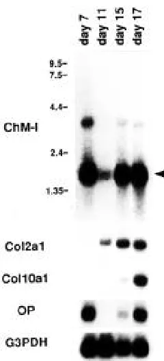
The temporal pattern of ChM-I expression was examined by Northern blot analysis using poly A+ RNA isolated from the whole embryos at days 7, 11, 15, and 17 of gestation (Fig. 3). As indicated by the expression of type II collagen mRNA, cartilaginous bone rudiments were formed around 11 days after gestation in the mouse embryos. The level of type II collagen mRNA was progressively elevated in association with the expansion of skeletal elements in the embryos until day 17 (Lyons et al., 1990), at which time the cartilage was invaded by blood vessels, resulting in the formation of a primary ossifica-tion center in the cartilaginous mold of bone (Kaufman, 1992). Prior to the vascular invasion, the expressions of type X col-lagen and osteopontin mRNAs were induced in association with the formation of hypertrophic and calcified chondrocyte zones (Fig. 3) (Iyama et al., 1991). The lower level of ChM-I transcripts was detected in the mouse embryos at day 11, and elevated in parallel with the expression of type II collagen mRNA (Fig. 3). Interestingly, a highly elevated expression of ChM-I mRNA was detected in the embryos at day 7 prior to organogenesis. In addition to the 1.7 kb transcripts, embryos at this stage ex-pressed the ChM-I transcripts approximately 3.9 kb in size which were only faintly discernible at the later stages of devel-opment (at days 15 and 17) (Fig. 3). The expression of the osteopontin gene was also detected at day 7.
In situ hybridization of ChM-I transcripts during endochon-dral bone formation
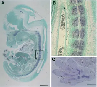
To explore the spatiotemporal pattern of the ChM-I gene expression, we performed in situ hybridization using mouse embryos at various stages of development. The cartilaginous structure first became apparent in the occipital bone rudiment in the sagittal section of mouse embryos at day 11. Evident hybridization signals of ChM-I mRNA were detected exclusively in cartilage in this region, although hybridization signals were also faintly discernible in the intermediate mesoderm flanking notochord (data not shown). The sagittal sections of embryos at day 13.5 revealed the specific expression of ChM-I mRNA in all of the cartilaginous tissues including the nasal septum, tracheal
rings and ribs (Fig. 4). The cartilaginous precursors of the vertebral column were composed of prehypertrophic chondrocytes at this stage of development. As shown in Figure 4B, strong hybridization signals were clearly detected in chondrocytes, whereas no signal was detected in the noto-chord. Cartilage in the limbs also expressed ChM-I mRNA (Fig. 4C).
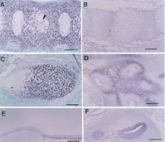
As the embryonic stage progressed, hypertrophic chondrocytes developed in cartilaginous bone rudiments at day 16. The expression of ChM-I markedly declined in the hyper-trophic chondrocyte zone in the vertebral column (Fig. 5A). The specificity of the ChM-I antisense cRNA probe used here was confirmed by the absence of signals using the sense cRNA probe (Fig. 5B). As shown in Figure 5C, no hybridization signal was detected in the hypertrophic chondrocyte zone of Meckel’s cartilage, in contrast to the prehypertrophic chondrocyte zone. There were some additional sites of ChM-I expression to a lesser extent in embryos at day 13.5 in the chondrocytes surrounding the auditory canal of the otic vesicle (Fig. 5D) and immature osteoblasts in the membranous neurocranium (Fig. 5E) as well as chondrocranium. There were positive hybridiza-tion signals in the neural layer of the retina, which was sepa-rated by a hyaloid artery from the lens at this stage (data not shown). The lateral wall of the cochlear duct of the inner ear at day 12 also hybridized with the ChM-I antisense cRNA probe (Fig. 5F). The in situ hybridization using the [35S]-labeled cDNA probe indicated that the epithelial layer of the pancreatic ducts and developing whisker follicles were also minor sites of ChM-I expression on the sections at days 13.5 and 16, respectively (data not shown).
Expression of ChM-I transcripts in the course of chondro-genic differentiation in vitro
Mouse ATDC5 cells reflect the multistep differentiation pro-cess encompassing the stages from mesenchymal condensa-tion to calcificacondensa-tion in vitro (Shukunami et al., 1996, 1997). We monitored the time-course of the expression of ChM-I mRNA during the multistep differentiation of these cells, and compared it with those of type II and type X collagen mRNAs (Fig. 6). As indicated by the expression of type II collagen mRNA, the ATDC5 cells began to express the differentiated phenotype of prehypertrophic chondrocytes by day 6 of culture in the pre-formed condensation area, and pre-formed cartilage nodules (Shukunami et al., 1996). In association with the expression of type X collagen mRNA (Fig. 6), the cells matured to become hypertrophic in the center of cartilage nodules (Shukunami et al., 1997).
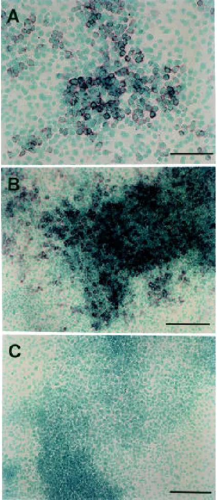
No transcript for the ChM-I gene was detectable in undiffer-entiated ATDC5 cells on day 3, as in mouse osteoblastic MC3T3-E1 cells, or mouse embryonic C3H10T1/2 cells (Fig. 6). However, the ChM-I transcripts were induced to express in ATDC5 cells upon chondrogenic differentiation on day 12, slightly later than the induction of type II collagen mRNA. The ChM-I mRNA level reached a maximal level on day 15, at which time the cells ceased to grow and became mature. As the hypertrophic chondrocytes appeared in cartilage nodules in parallel with the induction of type X collagen expression, the expression of ChM-I transcripts gradually declined (Fig. 6). Figure 7A shows a developing cartilage nodule at an early phase of the nodule formation. The in situ hybridization analysis using the differentiated culture of ATDC5 cells revealed that the
differentiated chondrocytes forming the nodu-lar structure evidently expressed ChM-I tran-scripts (Fig. 7A). Mature chondrocytes piled up in cartilage nodules heavily stained with the ChM-I cRNA probe, but no hybridization signals were detected in the undifferentiated cells filling the inter-nodular space (Fig. 7B). The specificity of the hybridization probe was confirmed by the use of the sense cRNA probe (Fig. 7C).
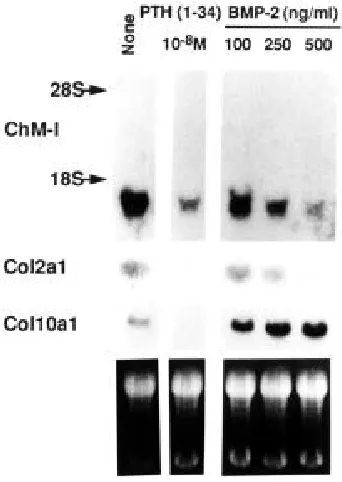
As previously reported (Shukunami et al., 1997), the culture of ATDC5 cells reached the stage of maturation approxi-mately from day 15 to 21, at which time the differentiated cells in cartilage nodules gradually ceased to grow. The progres-sion of differentiation can be perturbed by the treatment of the culture at this stage with recombinant bone morphogenetic pro-tein-2 (BMP-2) or parathyroid hormone (1-34) amide [PTH(1-34)]; BMP-2 facili-tated the progression of differentiation to-ward the type X collagen-expressing hy-pertrophic stage, and PTH(1-34) caused a de-differentiation of the cells to loose both the type II and type X collagen expres-sions (Shukunami et al., 1998). As shown in Figure 8, the culture of ATDC5 cells reached the state at the entry for hyper-trophic stage around day 21, and the cells expressed type II collagen and type X collagen as well as ChM-I mRNA in cul-ture. When the cells were de-differenti-ated by the treatment with 10-8 M PTH(1-34) for 48 h, both the type II and type X collagen gene expressions were markedly down-regulated. Similarly, the expression of ChM-I mRNA declined. On the other hand, when the cells were stimulated to become hypertrophic by the treatment with
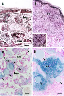
on the left side. When the DBM pellets were recovered 9 days after implantation, we confirmed that only the cartilaginous tissue was induced in the pellets in the presence and absence of CE10-50 kDa. Three weeks after the implantation, the radiologi-cal examination suggested the formation of ectopic bone at the sites of the control pellets in all five animals (Fig. 9A). The histological analysis indicated the formation of trabecular bone and marrow cavities. Hematopoietic cells were found in the marrow cavities developed, indicating that vascular invasion into cartilaginous implants from the surrounding vasculature took place. In three of the five pellets containing CE10-50 kDa, there was no indication of bone formation by soft X-ray analysis (data not shown). The formation of cartilage was confirmed histologically in the implants containing CE10-50 kDa (Fig. 9B). Alcian blue staining indicated the formation of cartilage matrix in the implants (data not shown). In most parts of the implants, a number of chondrocytes became hypertrophic (Fig. 9B, in-set). The implants were surrounded by fibrous and/or adipose tissue as well as capillaries, but there was little vascular
Fig. 5.In situ hybridization for ChM-I transcripts in mouse embryos at various developmental stages. Sagittal sections of mouse embryos were hybridized with the ChM-I antisense cRNA probe. Vertebral columns at day 16 were hybridized with the antisense probe (A) or with the sense probe as a negative control (B). The expression of the ChM-I transcripts declined in the late hypertrophic chondrocytes (arrowhead). In (C), Meckel’s cartilage at day 16 is shown. Positive hybridization signals were detected in the prehypertrophic and early hypertrophic zones, but were completely abolished in the late hypertrophic zone. In (D), the otic vesicle at day 13.5 is shown. Hybridization signals were seen in chondrocytes surrounding the auditory canal. In addition to the chondrocranium, immature osteoblasts in the dermocranium hybridized to the probe at day 13.5 (E). The lateral wall of the cochlear duct of the inner ear at day 12 also hybridized to the probe (F). The sections were counter-stained with methyl green or hematoxylin. Bars, 100
µm in (A), (B), (C), and (F); 200 µm in (D) and (E).
BMP-2 for 48 h, the expression of type X collagen gene was evidently up-regulated in a dose-dependent manner, whereas the expression of type II collagen gene declined (Fig. 8). As the cells expressed a hypertrophic phenotype more, the level of ChM-I mRNA declined in a manner dependent on the dose of BMP-2 (Fig. 8).
Effect of mature ChM-I protein on endochondral bone formation
invasion into the cartilaginous implants. The two other pellets had been absorbed without bone formation within three weeks after implantation.
We next examined whether purified ChM-I can mimic the effect of CE10-50 kDa on bone formation. In contrast to the crude prepa-ration of cartilage matrix CE10-50 kDa, purified ChM-I rapidly dif-fused out from the DBM pellets during implantation because of its higher solubility in aqueous body fluid. In the present study, we used ChM-I that had been bound to heparin-Sepharose beads. The control DBM (15 mg) pellets that had been previously mixed with heparin beads alone resulted in the formation of trabecular bone and marrow in all five animals three weeks after implantation (Fig. 9C). Alcian blue-positive cartilaginous tissue occupied less than 5% of the total area of the section by the histological examination. On the other side of each animal, the DBM pellets mixed with purified ChM-I (2 µg)-bound heparin-Sepharose beads were implanted. After three weeks, bony tissue was found to occupy about 30% of the total area of the section. Cartilaginous tissue persisted in the area surrounded by the ChM-I-containing beads (Fig. 9D). When the DBM pellets were recovered earlier (on day 9 after implantation), only the cartilaginous tissue was in-duced in the heparin bead-containing pellets regardless of the presence of ChM-I.
Discussion
Most bones of the skeleton are first formed as cartilaginous bone rudiments during organogenesis. Following the vascular invasion, cartilaginous tissue is replaced by bone at a later time during embryonic development (Erlebacher et al., 1995). Invad-ing blood vessels are accompanied by bone precursor cells such as osteoblasts and osteoclast precursors (Whitson, 1994). Thus, vascular invasion is a highly regulated process tempo-rally and spatially during endochondral bone development, and it coordinates chondrogenesis and the subsequent osteogen-esis (Iyama et al., 1991). Cartilage is unique among the tissues of mesenchymal origin in that it is avascular and extraordinarily resistant to vascular invasion (Kuettner and Pauli, 1983). Cap-illaries invade the lower hypertrophic and/or calcified cartilage in bone rudiments at the site of contact with the bony collar and periosteum. It is believed that cartilage undergoes phenotypic switching from anti-angiogenic to angiogenic during the late stage of cartilage differentiation.
On the basis of studies on tumor angiogenesis, Hanahan and Folkman (1996) proposed the balance hypothesis that angio-genesis is regulated by a balance of inducers and inhibitors for endothelial cell proliferation. The cumulative levels of inducer and inhibitor signals maintain the endothelial cells in alternative states of quiescence and angiogenesis. A balance of agonistic and antagonistic signals appears to be a fundamental regula-tory motif in the tissue specification and cell-fate determination during embryonic development, as demonstrated by the ac-tions of BMP and Noggin (Reshef et al., 1998). Although a number of angiogenic molecules has been reported in cartilage (Harada et al., 1994; Twal et al., 1994; Alini et al., 1996), the cartilage-specific macromolecules that counteract angiogenic signals have not been identified.
Previous studies highlighted the participation of proteinase inhibitors in the anti-angiogenic properties of cartilage (DiMuzio et al., 1987; Moses et al., 1990, 1992), since angiogenesis involves a local degradation of the basement membrane sur-rounding the endothelium (Folkman and Shing, 1992). Mem-bers of the thrombospondin family are other extracellular matrix components with anti-angiogenic properties (Rastinejad et al., 1989; Taraboletti et al., 1990). These molecules are also found in cartilage, but have a broader distribution pattern in the body (O’Shea and Dixit, 1988; Iruela-Arispe et al., 1993). It is not clear whether these molecules function as components of the angiogenic switch in cartilage. It was reported that an endothe-lial growth inhibiting agent (TNP-470) inhibited the ectopic bone formation induced by BMP-2, in which the agent interfered with the vascular invasion into cartilage (Mori et al., 1995). Thus, we hypothesize that the endogenous endothelial cell growth inhibi-tor participates in the angiogenic switching in cartilage, and that withdrawal of the inhibitor permits vascular invasion into carti-laginous bone rudiments to trigger the subsequent bone forma-tion.
As previously reported (Hiraki et al., 1997b), the fractionated bovine cartilage-extracts CE10-50 kDa exhibit a potent inhibitory action on the growth of endothelial cells in vitro. Crude extracts from fetal bovine cartilage CE10-50 kDa apparently inhibited the replacement of cartilage induced in the DBM pellets by bone (Fig. 9). In the implants of DBM, cartilage once formed was
sisted even after three weeks (Fig. 9B). There was no obvious evidence of vascular invasion in the implants. These results indicated that exogenous CE10-50 kDa inhibited the angiogenic switching of cartilage induced in the implants.
ChM-I was originally identified as a growth stimulating com-ponent in the extracts of fetal bovine cartilage (Hiraki et al., 1991). It also stimulated the proteoglycan synthesis and colony formation of growth plate chondrocytes in culture (Inoue et al., 1997). We purified the endothelial cell growth inhibitor from the cartilage extracts, and found that it was identical to ChM-I (Hiraki et al., 1997a,b). It inhibited the proliferation and tube morphogenesis of vascular endothelial cells in vitro (Hiraki et al., 1997a). We demonstrated that ChM-I was secreted from chondrocytes and localized in the inter-territorial matrix of cartilage where it was bound to some anchoring molecules yet to be identified (Hiraki et al., 1997a). As shown in Figure 9C and D, ChM-I-bound heparin-Sepharose beads mimicked the effect of CE10-50 kDa on ectopic bone formation in vivo. These results indicate that ChM-I is one of the key components in the angio-genic switching in cartilage. In the present study, we isolated mouse ChM-I precursor cDNA and characterized the primary structure of the ChM-I precursor protein (Fig. 1).
The mouse cartilage expressed the ChM-I transcripts at the highest level among the tissues examined (Fig. 2). The thymus and eye also expressed ChM-I mRNA at a lower level. The eye is another example of avascular tissue. In mammals, the devel-oping eyes are vascularized by hyaloid arteries which are degenerated until birth. The expression of ChM-I in the retina of mouse embryo at day 14.5 suggests a role of ChM-I in the development of the eye. In situ hybridization identified some minor sites of ChM-I gene expression (Fig. 5). Osteoblasts did not express ChM-I mRNA (Fig. 6). However, immature osteo-blasts in the neurocranium transiently expressed the gene (Fig. 5E). Since ChM-I stimulates the proliferation of osteoblasts (Mori et al., 1997), there is a possibility that ChM-I also partici-pates in the proliferation of osteoblast precursors as an autocrine factor under certain physio-pathological conditions. In any case, the expression of ChM-I mRNA was associated with the formation of cartilaginous tissue, and was obviously detected in prehypertrophic cartilage (Figs. 4 and 5). Vascular invasion into vertebral column takes place at around day 17 in mouse development (Kaufman, 1992). Prior to this, the expression of ChM-I was abolished in hypertrophic cartilage which appeared at day 16. As shown in Figures 6 and 7, the induction of ChM-I expression occurred in association with chondrogenic induc-tion of ATDC5 cells in vitro. Moreover, the expression of ChM-I mRNA was regulated in a differentiation stage-specific man-ner (Figs. 6 and 8). The perturbation of cartilage differentiation by exogenous signaling molecules profoundly affected the level of ChM-I mRNA (Fig. 8). Although the functional role of ChM-I in thymus is not clear at present, these expression profiles of ChM-I gene were compatible with its regulatory role in angiogenic switching in cartilage and eye.
Cartilage formation is initiated in mouse embryos at around day 11. At this stage, the expression of ChM-I gene was confined almost exclusively to the cartilaginous bone rudi-ments. No ChM-I transcripts were detected in the notochord. However, we found that ChM-I transcripts were also expressed prior to organogenesis at a markedly higher level in mouse
Fig. 7.Localization of ChM-I transcripts in the developing cartilage nodules in the ATDC5 cell cultures in vitro. Undifferentiated ATDC5 cells were plated in a two-well Lab-Tek Chamber Slide at a density of 2x10⁴ cells/well, and cultured for 12 days (A) or 18 days (B and C) in the medium described in the Fig. 6 legend. In situ hybridization was then performed with a digoxigenin-labeled antisense cRNA (A and B) or sense cRNA (C) probe for ChM-I. The nuclei of the cells were stained with methyl green. Bars, 100 µm in (A) and 200 µm in (B) and (C).
per-embryos at day 7 (Fig. 3). In chick per-embryos, ChM-I gene was expressed transiently in the notochord and sclerotome as well as the roof plate and floor plate of the neural tube during the early stages of development (U. Dietz, GSF-Research Center, Munich, Germany, personal communication). The functional role of ChM-I expression will be elucidated by the generation of mice carrying the ChM-I null mutations. Studies along this line are now underway.
Materials and Methods
Materials
Fractionated cartilage-extracts containing 10-50 kDa compo-nents (CE10-50kDa) were prepared from the guanidine extracts of
fetal bovine epiphyseal cartilage, as previously described (Hiraki et al., 1997b). Bovine ChM-I was purified from fetal bovine epiphy-seal cartilage as previously described (Hiraki et al., 1991), and was provided by Dr. J. Kondo (Mitsubishi Chemical Corporation, Yokohama, Japan). Human recombinant BMP-2 was a gift from Dr. J. M. Wozney (Genetics Institute Inc., Cambridge, MA). Bovine parathyroid hormone-(1-34) amide [PTH(1-34)] was purchased from Bachem (Torrance, CA). Reconstituted bovine demineralized bone matrix (DBM) was a generous gift from Dr. Y. Kuboki (Hokkaido University, Sapporo, Japan) which was prepared from the mixture
of 4 M guanidine extract and 4M guanidine-insoluble materials of demineralized bone matrix by dialysis as previously described by Sampath and Reddi, (1981).
Cell culture
Chondrogenic differentiation was induced in mouse ATDC5 cells by culturing the cells in a 1:1 mixture of Dulbecco’s modified Eagle’s medium (DMEM) and Ham’s F-12 (DME/F12) medium (JRH Biosciences, Lenexa, KS) containing 5% fetal bovine serum (FBS, JRH Biosciences), 10 µg/ml human insulin (Boeheringer, Mannheim, Germany), 10 µg/ml human transferrin (Boehringer), and 3x10⁻⁸ M sodium selenite (Sigma, St. Louis, MO) at 37°C under
5% CO2 in air, as previously described (Shukunami et al., 1996).
The inoculum size of the cells was 4x10⁴ cells/well in 24-multiwell
plates or 6x10⁴ cells/well in 6-multiwell plates (Corning, New York).
In some experiments, the cells were allowed to differentiate in the above medium for 21 days, and then incubated for another 48 h in the presence of PTH(1-34) or BMP-2, which modified the differen-tiated state of the cells (Shukunami et al., 1996, 1998). Mouse embryonic C3H10T1/2 cells were plated at a density of 6x10⁴ cells/
well in 6-multiwell plates and grown to confluence in DMEM con-taining 10% FBS for 3 days. Mouse osteoblastic MC3T3-E1 cells were plated at a density of 6x10⁴ cells/well in 6-multiwell plates and grown to confluence in alpha modified essential medium (αMEM, Dainippon Pharmaceutical, Tokyo) containing 5% FBS for 3 days. The culture medium was replaced every other day. For the Northern blot analysis, growth-plate chondrocytes were isolated from the ribs of 4-week-old DDY mice by following the reported method with some modification (Shimomura et al., 1975). Isolated chondrocytes were plated in 6-well multiwell plates (105 cells/well)
and grown in DME/F12 medium containing 10% FBS for 10 days.
Isolation of mouse ChM-I cDNA
Mouse ChM-I cDNA was isolated by the RT-PCR and RACE
methods, using RNA isolated from ATDC5 cells. First-strand cDNA was synthesized by SuperScript II RNase H- Reverse Transcriptase
(GIBCO BRL, Grand Island, NY) with total RNA extracted from the differentiated cultures of ATDC5 cells on day 21. Purified total RNA (2.5 µg) was incubated at 45°C for 60 min with a mixture of 100 units of SuperScript II RNase H- Reverse Transcriptase, 2.5 µM of
oligo-d(T)16 primer (Perkin Elmer, Norwalk, CT), 3 mM MgCl2, 50
mM Tris-HCl (pH 8.3), 75 mM KCl, 5 mM DTT and 1 mM each of dCTP, dTTP, dGTP and dATP in a volume of 10 µl. Aliquots of one-tenth of the cDNA were used to amplify a partial ChM-I cDNA clone. The primer set used was designed on the basis of the nucleotide sequence conserved in human and bovine ChM-I cDNAs (Hiraki et al., 1991): primer 1, 5’-GGGTCAATGGAAATAGACGCTG-3’; primer 2, 5’-ACACCATGCCCAGGATGCGG-3’. 3'- and 5'-RACE were then employed to obtain the full-length mouse ChM-I cDNA. Adaptor-ligated double-stranded cDNAs were prepared using a Marathon cDNA Amplification Kit (Clontech, Palo Alto, CA) according to the manufacture’s instructions. The 5’-end of ChM-I cDNA was ampli-fied using an anchor primer and the nested primer 3 (5’-GCTCTGCTTGGTCACTGTGCC-3’) immediately upstream of primer 4 (5’-GGCATGATCTTGCCTTCCAG-3’). The 3’-end of the cDNA was amplified using an anchor primer and the nested primer 5 (5’-CCAATGTTCAGGACGACGCAG-3’) downstream of primer 6 (5’-GGAAGGCAAGATCATGCCAGT-3’). The amplified PCR prod-ucts were subcloned into pCRII (Invitrogen, San Diego, CA).
Mouse ChM-I cDNA was also isolated from a λgt11 cDNA library constructed from 17-day mouse whole embryos (Clontech). A total of 5x10⁵ independent recombinant phages was initially screened by
hybridization with a partial cDNA clone obtained from the above-mentioned RT-PCR product. The hybridization-positive phage clones were isolated by the repeated plaque purification. The inserts isolated Fig. 8.Effects of PTH(1-34) and BMP-2 on the expression of ChM-I
from the clones were subcloned into the EcoRI site of pBlueScript SK(+) (Stratagene, La Jolla, CA). The nucleotide sequence of the inserts was determined using a Dye Terminator Cycle Sequencing Kit with an ABI 310 Genetic Analyzer (Applied Biosystems, Foster City, CA). Both cDNA strands were sequenced using standard sequencing primers. The DNA sequences obtained were compiled and analyzed by the Gene Works computer software program (IntelliGenetics, Tokyo). Fig. 9.Effect of ChM-I on the replacement of cartilage by bone during ectopic bone formation. Pellets of bovine demineralized bone matrix (DBM) were implanted into the fascia of the back muscle of nude mice. The histological appearances of the implants three weeks after implantation are shown. In (A), trabecular bone (tb) and bone marrow (bm) developed in the DBM pellet (15 mg) alone. In (B), cartilaginous tissue (ca) with little vascular invasion developed in the DBM pellet which was previously mixed with the fractionated bovine cartilage-extracts CE10-50 kDa (2 mg). The inset shows a large number of chondrocytes undergoing hypertrophy. In (C), trabecular bone (tb) and bone marrow (bm) developed in the pellet of DBM (15 mg) mixed with heparin-Sepharose beads alone. In (D), typical cartilage (ca) formed in the pellet of DBM (15 mg) mixed with bovine purified ChM-I (2 µ g)-bound heparin-Sepharose beads. Arrowheads indicate heparin-Sepharose beads in the implanted DBM pellets. The sections were stained with hematoxylin-eosin staining in (A) and (B) or with Alcian blue and Kernechtrot in (C) and (D). Bars, 100 µm in (A) and (B) and 50 µm in (C) and (D).
Northern blot analysis
Total RNA was prepared from the cultures of ATDC5 cells or tissues of 4-week-old DDY mice by a single-step method according to Chomczynski and Sacchi (1987). Total RNA (20 µg) was dena-tured with 6% formaldehyde, fractionated by 1% agarose gel (SeaKem GTG, FMC Bioproducts, Rockland, ME), and transferred onto Nytran membranes (Schleicher and Schuell, Dassel, Ger-many) with a Turboblotter (Schleicher and Schuell) apparatus. A mouse embryo multiple tissue Northern blot (Clontech) filter was also used in some experiments. Hybridization was performed overnight at 42°C with an appropriate α-[32P]-dCTP labeled cDNA
probe (106 cpm/ml) in a solution containing 50% formamide, 6xSSPE
(0.9 M NaCl, 20 mM NaH2PO4, 6 mM EDTA at pH 7.4), 0.2% bovine
serum albumin (BSA), 0.2% Ficoll 400, 0.2% polyvinylpyrolidone, 0.1% SDS, and 200 µg/ml denatured salmon sperm DNA. Hybrid-ization probes were prepared by the random primer method with a BcaBEST labeling kit (Takara, Otsu, Japan) using the appropriate cDNA fragments; a 1.4 kb EcoRI fragment of pKT1180 (Kimura et al., 1989) as a probe for α1(II) collagen; a 0.65 kb HindIII fragment of pSAm10h (Apte et al., 1992) as a probe for α1(X) collagen; a 1.4 kb EcoRI fragment as a probe for osteopontin (Oldberg et al., 1986); a 1.3 kb EcoRI fragment of mouse ChM-I cDNA. A probe for glyceraldehyde-3-phosphste dehydrogenase (GAPDH) was
ampli-fied by RT-PCR with a mouse GAPDH control amplimer set
(Clontech). After hybridization, the filters were washed at 55°C for 30 min in 2xSSPE, 0.1% SDS, then washed at 55°C for 30 min in 0.1xSSPE, 0.1% SDS. The membrane filters were exposed to X-OMAT film (Eastman Kodak, Rochester, NY) at -80°C.
In situ hybridization
Balb/c mouse embryos at days 11, 12.5, 13.5, 14.5 and 16 of gestation were collected and fixed with 4% paraformaldehyde in 0.01 M phosphate buffer saline (pH 7.4) overnight at 4°C. Whole embryos were dehydrated in a graded series of ethanol and em-bedded in paraffin. Sections were cut at 6 µm and then processed for in situ hybridization. In some experiments, ATDC5 cells were plated in Lab-Tek Chamber Slides (Nalgen Nunc International, Naperville, IL) at a density of 2x10⁴ cells/well, and cultured for 12
days in DME/F12 medium containing 5% FBS, 10 µg/ml human insulin, 10 µg/ml human transferrin, and 3x10⁻⁸ M sodium selenite.
The 0.5 kb mouse ChM-I cDNA fragment containing the mature ChM-I region was used as a hybridization probe. Digoxigenin-labeled antisense and sense cRNA probes for mouse ChM-I were prepared with a DIG RNA labeling kit (Boehringer). Hybridization was performed at 50°C for 16 h. After hybridization, the slides were washed under high stringency conditions. Hybridization signals were detected immunohistochemically by an alkaline phosphatase-conjugated antibody using a nucleic acid detection kit (Boehringer). Appropriate controls to exclude false-positive staining due to en-dogenous alkaline phosphatase activity were performed and were negative. The sections were counterstained with methyl green or hematoxylin.
Ectopic bone formation
Pellets of DBM were mixed with cartilage extracts or ChM-I-bound heparin-Sepharose beads as follows: Reconstituted bovine DBM (15 mg) was suspended in 2 ml water in a conical tube and centrifuged at 1500xg for 15 min. The supernatant was discarded, and then CE10-50
kDa (2 mg) was added. The resultant suspension was vigorously mixed
The lyophilized pellets were implanted bilaterally into the fascia of the back muscle of male 6-week-old nude mice (BALB/c:nu/nu, Clea, Osaka, Japan). Three weeks later, the mice were anesthetized and photographed with a soft X-ray apparatus (type C-SM Softex, Tokyo, Japan) for the assessment of bone formation. The implants were then dissected out, cleaned of adherent tissue, and fixed in neutral formalin. The fixed samples were decalcified, dehydrated, embedded in paraffin, and sectioned (6 µm thick) for histological examination by hematoxylin and eosin staining, or alcian blue and kernechtrot staining.
Acknowledgments
We are grateful to Dr. J. Kondo (Mitsubishi Chemical Corporation), Dr. J. M. Wozney (Genetics Institute), and Dr. Y. Kuboki (Hokkaido University) for providing purified bovine ChM-I, recombinant human BMP-2, and bovine DBM, respectively. We also thank Dr. Uwe Dietz and Dr. Rudi Balling (GSF-Research Center in Munich) for their discussion and communication of their data on the pattern of chicken ChM-I expression prior to publication. Chisa Shukunami is a recipient of a Research Fellowship of the Japan Society for the Promotion of Science for Young Scientists. This work was partly supported by Special Coordination Funds of Promoting Science and Technology from the Science and Technology Agency of Japan (to YH) and Grants-in-Aid from the Ministry of Education, Science and Culture of Japan (#08672363 to HI; #09671892 to YH).
References
ALINI, M., MARRIOTT, A., CHEN, T., ABE, S. and POOLE, A.R. (1996). A novel angiogenic molecule produced at the time of chondrocyte hypertrophy during endochodnral bone formation. Dev. Biol. 176: 124-132.
APTE, S.S., SELDIN, M.F., HAYASHI, M. and OLSEN, B.R. (1992). Cloning of the human and mouse type X collagen genes and mapping of the mouse type X collagen gene to chromosome 10. Eur. J. Biochem. 206: 217-224.
CHOMCZYNSKI, P. and SACCHI, N. (1987). Single-step method of RNA isola-tion by acid guanidinium thiocyanate-phenol-chloroform extracisola-tion. Anal. Biochem. 162: 156-159.
DIMUZIO, M.T., TRIPIER, D. and KUETTNER, K.E. (1987). Purification and characterization of an elastase inhibitor derived from hyaline cartilage. J. Rheumatol. 14: 45-48.
ERLEBACHER, A., FILVAROFF, E.H., GITELMAN, S.E. and DERYNCK, R. (1995). Toward a molecular understanding of skeletal development. Cell 80: 371-378.
FOLKMAN, J. and SHING, Y. (1992). Angiogenesis. J. Biol. Chem. 267: 10931-10934.
GELB, D.E., ROSIER, R.N. and PUZAS, J.E. (1990). The production of trans-forming growth factor-beta by chick growth plate chondrocytes in short term monolayer culture. Endocrinology 127: 1941-1947.
GONZALEZ, A.-M., BUSCAGLIA, M., ONG, M. and BAIRD, A. (1990). Distribu-tion of basic fibroblast growth factor in the 18-day rat fetus: LocalizaDistribu-tion in the basement membranes of diverse tissues. J. Cell Biol. 110: 753-765.
HALL, B.K. (1983). Tissue interactions and chondrogenesis. In Cartilage (Ed. B.K. Hall). Academic Press, New York, pp. 209-210.
HANAHAN, D. and FOLKMAN, J. (1996). Patterns and emerging mechanisms of the angiogenic switch during tumorigenesis. Cell 86: 353-364.
HARADA, S.-I., NAGY, J.A., SULLIVAN, K.A., THOMAS, K.A., ENDO, N., RODAN, G.A. and RODAN, S.B. (1994). Induction of vascular endothelial growth factor expression by prostaglandin E2 and E1 in osteoblasts. J. Clin. Invest. 93: 2490-2496.
HIRAKI, Y., INOUE, H., IYAMA, K.-I., KAMIZONO, A., OCHIAI, M., SHUKUNAMI, C., IIJIMA, S., SUZUKI, F. and KONDO, J. (1997a). Identification of chondromodulin-I as a novel endothelial cell growth inhibitor: purification and its localization in the avascular zone of epiphyseal cartilage. J. Biol. Chem. 272: 32419-32426.
HIRAKI, Y., KONO, T., SATO, M., SHUKUNAMI, C. and KONDO, J. (1997b). Inhibition of DNA synthesis and tube morphogenesis of cultured vascular
endothelial cells by chondromodulin-I. FEBS Lett. 415: 321-324.
HIRAKI, Y., TANAKA, H., INOUE, H., KONDO, J., KAMIZONO and SUZUKI, F. (1991). Molecular cloning of a new class of cartilage-specific matrix, chondromodulin-I, which stimulates growth of cultured chondrocytes. Biochem. Biophys. Res. Commun. 175: 971-977.
INOUE, H., KONDO, J., KOIKE, T., SHUKUNAMI, C. and HIRAKI, Y. (1997). Identification of an autocrine chondrocyte colony-stimulating factor; chondromodulin-I stimulates colony formation of growth plate chondrocytes in agarose culture. Biochem. Biophys. Res. Commun. 241: 395-400.
IRUELA-ARISPE, M.L., LISKA, D.J., SAGE, E.H. and BORNSTEIN, P. (1993). Differential expression of thrombospondin 1, 2, and 3 during murine develop-ment. Dev. Dynamics 197: 40-56.
IYAMA, K., NINOMIYA, Y., OLSEN, B.R., LINSENMAYER, T.F., TRELSTAD, R.L. and HAYASHI, M. (1991). Spatiotemporal pattern of type X collagen gene expression and collagen deposition in embryonic chick vertebrae undergoing endochondral ossification. Anat. Rec. 229: 462-472.
KAUFMAN, M.H. (1992). The atlas of mouse development. Academic Press, San Diego, pp. 291-335.
KIMURA, T., MATTEI, M.-G., STEVENS, J.W., GOLDRING, M.B., NINOMIYA, Y. and OLSEN, B.R. (1989). Molecular cloning of rat and human type IX collagen cDNA and localization of the α1(IX) gene on the human chromosome 6. Eur. J. Biochem. 179: 71-78.
KUETTNER, K.E. and PAULI, B.U. (1983). Vascularity of cartilage. In Cartilage (Ed. B.K. Hall). Academic Press, New York, pp. 281-312.
LYONS, K.M., PELTON, R.W. and HOGAN, B.L.M. (1990). Organogenesis and pattern formation in the mouse: RNA distribution patterns suggest a role for Bone Morphogenetic Protein-2A (BMP-2A). Development 109: 833-844.
MORI, S., YOSHIKAWA, H., UEDA, T., HASHIMOTO, J., KATO, M., MIYAZONO, K. and TAKAOKA, K. (1995). Inhibition by anti-angiogenic agent (TNP-470) of ectopic bone formation induced by bone morphogenetic protein-2. Bone 17: 569.
MORI, Y., HIRAKI, Y., SHUKUNAMI, C., KAKUDO, S., SHIOKAWA, M., KAGOSHIMA, M., MANO, H., HAKEDA, Y., KUROKAWA, T., SUZUKI, F. and KUMEGAWA, M. (1997). Stimulation of osteoblast proliferation by the carti-lage-derived growth promoting factors chondromodulin-I and II. FEBS Lett. 406: 310-314.
MOSES, M.A., SUDHALTER, J. and LANGER, R. (1990). Identification of an inhibitor of neovascularization from cartilage. Science 248: 1408-1410.
MOSES, M.A., SUDHALTER, J. and LANGER, R. (1992). Isolation and charac-terization of an inhibitor of neovascularization from scapupar chondrocytes. J. Cell Biol. 119: 475-482.
O’SHEA, K.S. and DIXIT, V.M. (1988). Unique distribution of the extracellular matrix component thrombospondin in the developing mouse embryo. J. Cell Biol. 107: 2737-2748.
OLDBERG, A., FRANZEN, A. and HEINEGARD, D. (1986). Cloning and se-quence analysis of rat bone sialoprotein (osteopontin) cDNA reveals an Arg-Gly-Asp cell-binding sequence. Proc. Natl. Acad. Sci. USA 83: 8819-8823.
RASTINEJAD, F., POLVERINI, P.J. and BOUK, N. (1989). Regulation of the activity of a new inhibitor of angiogenesis by a cancer suppressor gene. Cell 56: 345-355.
RESHEF, R., MAROTO, M. and LASSAR, A.B. (1998). Regulation of dorsal somitic cell fates: BMPs and Noggin control the timing and pattern of myogenic regulator expression. Genes Dev. 12: 290-303.
SAMPATH, T.K. and REDDI, A.H. (1981). Dissociative extraction and reconsti-tution of extracellular matrix components involved in local bone differentia-tion. Proc. Natl. Acad. Sci. USA 78: 7599-7603.
SHIMOMURA, Y., YONEDA, T. and SUZUKI, F. (1975). Osteogenesis by chondrocytes from growth cartilage of rat rib. Calcif. Tissue Res. 19: 179-187.
SHUKUNAMI, C., ISHIZEKI, K., ATSUMI, T., OHTA, Y., SUZUKI, F. and HIRAKI, Y. (1997). Cellular hypertrophy and calcification of embryonal carcinoma-derived chondrogenic cell line ATDC5 in vitro. J. Bone Miner. Res. 12: 1174-1188.
SHUKUNAMI, C., SHIGENO, C., ATSUMI, T., ISHIZEKI, K., SUZUKI, F. and HIRAKI, Y. (1996). Chondrogenic differentiation of clonal mouse embryonic cell line ATDC5 in vitro: differentiation-dependent gene expression of par-athyroid hormone (PTH)/PTH-related peptide receptor. J. Cell Biol. 133: 457-468.
TARABOLETTI, G., ROBERTS, D., LIOTTA, L.A. and GIAVAZZI, R. (1990). Platelet thrombospondin modulates endothelial cell adhesion, motility and growth: A potential angiogenesis regulatory factor. J. Cell Biol. 111: 765-772.
TWAL, W.O., VASILATOS YOUNKEN, R., GAY, C.V. and LEACH, R.M., Jr. (1994). Isolation and localization of basic fibroblast growth factor-immunore-active substance in the epiphyseal growth plate. J. Bone Miner. Res. 9: 1737-1744.
WHITSON, S.W. (1994). Bone. In Oral Histology (Ed. A.R. Ten Cate). Mosby-Year Book, St. Louis, MO, pp. 131-134.
WOZNEY, J.M. (1993). Bone morphogenetic proteins and their gene expression. In Cellular and molecular biology of bone (Ed. M. Noda). Academic Press, San Diego, pp. 131-167.
YANG, E.Y. and MOSES, H.L. (1990). Transforming growth factor beta 1-induced changes in cell migration, proliferation, and angiogenesis in the chicken chorioallantoic membrane. J. Cell Biol. 111: 731-741.