Expression of galectin-7 during epithelial development
coincides with the onset of stratification
PAULA M. TIMMONS
1#, CÉLINE COLNOT
1#, ISABELLE CAIL
1, FRANÇOISE POIRIER
1*and THIERRY MAGNALDO
21Unité INSERM 257, ICGM, Paris and 2Laboratoire de Génétique Moléculaire, CNRS UPR42, Villejuif, France
ABSTRACT Galectin-7 is a 14 KDa member of the galectin family that we have cloned from human, rat and mouse. Our previous studies have shown that in the adult, galectin-7 is expressed in all cell layers of epidermis and of other stratified epithelia such as the cornea and the lining of the oesophagus. This suggested that galectin-7 expression might be induced at a particular stage in the embryonic development of stratified epithelia. In the present study we have investigated this hypothesis by in situ hybridization of 7 mRNA in mouse embryos. Starting from E13.5, weak expression of galectin-7 was detected in bilayered ectoderm, and stronger expression was found in areas of embryonic epidermis where stratification was more advanced. Galectin-7 expression was maintained in all living layers after epidermal development was completed. Galectin-7 was also strongly and specifically expressed in stratified regions of ectodermally-derived non-epidermal epithelia such as the lining of the buccal cavity, the oesophagus and the ano-rectal region. In contrast, no expression of galectin-7 was found in epithelia derived from endoderm, such as lining of the intestine, kidney and lung. Our results demonstrate that galectin-7 is expressed in all stratified epithelia examined so far, and that the onset of its expression coincides with the first visible signs of stratification. These results establish galectin-7 as the first region-independent marker of epithelial stratification.
KEY WORDS:
galectin-7, mouse development, stratified epithelia, epidermis
0214-6282/99/$15.00 © UBC Press
Printed in Spain
www.lg.ehu.es/ijdb
*Address for reprints: Unité INSERM 257, ICGM, 24 rue du Faubourg Saint-Jacques, 75014 Paris, France. FAX: 33 1 44412462. e-mail: poirier @cochin.inserm.fr #These authors contributed equally to this work.
Introduction
Mammalian epithelia can be divided into two major classes based on their structure and organization, and their embryological origin. In general, simple (monolayered) epithelia that line internal organs such as the lung, kidney and intestine, are derived from the endoderm, while stratified (multilayered) epithelia such as the epidermis and cornea develop from the ectoderm.
Epithelial cells can also be categorized in terms of their expres-sion of molecular markers. The most well known group of epithelial markers is the keratin family of intermediate filaments. Although these proteins are present in all epithelia, different pairs of keratin subunits (one basic and one acidic) are expressed according to the type, location, and differentiation state of the epithelial cell. Some subunits, such as K7, K8, K18 and K19, are typical of simple epithelia, whereas the K5/K14 pair is associated with the basal cell layer of stratified epithelia such as the epidermis. As soon as they move from the basal to the suprabasal compartment, epidermal keratinocytes switch from expression of K5/K14 to K1/K10 keratins. Later on, when they reach the granular layer of the epidermis, these cells proceed through the programme of terminal differentiation
Original Article
Abbreviations used in this paper: PBS, phosphate buffered saline; DTT, dithiothreitol; SSC, sodium saline citrate.
and synthesize cell envelope precursor proteins such as involucrin and loricrin (for review see Fuchs, 1990).
The expression of specific keratins in the suprabasal compart-ment can also denote regional differences in the phenotype of an epithelium, or specialized functions. For instance, K9 is expressed in place of K10 in palmoplantar epidermis, and a specific keratin pair, K4/K13, is expressed in the oesophageal epithelium. The K3/ K12 keratin pair is expressed in the non-keratinized stratified epithelium of the cornea (Sun et al., 1984).
In an attempt to isolate new markers for normal human epider-mal differentiation, we had previously cloned a cDNA encoding galectin-7 (Magnaldo et al., 1995), a novel member of a rapidly growing group of small beta-galactoside binding proteins formerly known as the S-type lectins (Barondes et al., 1994). Galectins are expressed with varying degrees of tissue specificity and develop-mental regulation starting from early stages of embryogenesis (for review see Colnot et al., 1997). These carbohydrate recogniz-ing molecules have been implicated in a variety of intracellular processes (for review see Leffler, 1997), but they are most widely documented as extracellular molecules involved in cell-cell or cell-matrix interactions (Hughes, 1992).
Galectin-7 mRNA is abundantly expressed in normal adult epidermis but is absent from several squamous cell carcinoma lines such as TR146, A431 and SV40-transformed keratinocytes (Madsen et al., 1995; Magnaldo et al., 1998, and references therein). Its expression in cultured keratinocytes and reconsti-tuted epidermis is reduced by retinoic acid treatment (Magnaldo et al., 1995), an agent that induces metaplasia in epidermal keratinocytes (Asselineau and Darmon, 1995, and references therein). These results suggested that galectin-7 may play a part in epithelial differentiation.
In this study, we have performed RNA in situ hybridization experiments in the mouse embryo in order to address several issues. Our first objective was to survey as many tissues as possible for galectin-7 expression, in order to gauge the potential
range of its biological functions. The second aim of the study was to examine the timing and localization of galectin-7 expression in the developing epidermis, to find out where this gene might fit within the developmental programme of this tissue. We have found a striking restriction of galectin-7 transcripts to stratified epithelia, in which the onset of expression coincides with the beginning of the stratification process. We suggest that galectin-7 may be important for both the formation and the maintenance of all stratified epithelia, irrespective of other characteristics such as keratinization or re-gional specialization.
Results
We investigated the expression of galectin-7 mRNA from day 9.5 to day 16.5 of mouse embryonic development (E9.5-E16.5) by in situ hybridization of a galectin-7 antisense cRNA probe to paraffin sections. No signal was detected in E9.5 or E10.5 sections, suggesting that embryonic expression of this gene is confined to the second half of gestation. From E13.5 onwards, the major site of galectin-7 expression was the epidermis of the developing skin, in which the extent and level of signal increased with developmen-tal age. No specific hybridization signal was detected in the dermis.
Galectin-7 expression in the embryonic epidermis
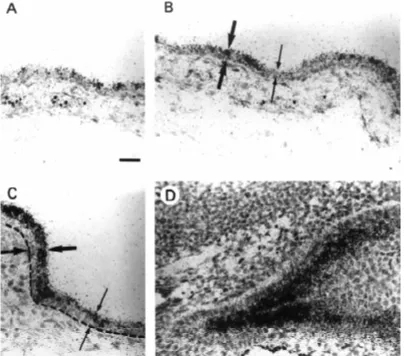
in the dorsal and ventral regions (not shown). At E14.5 (Fig. 1), hybridization signal of galectin-7 transcripts was still weak but more widespread, including the dorsal (Fig. 1A) and ventral (Fig. 1B) epidermis. At this stage we also saw stron-ger signal in regions such as the tail or lateral body epidermis (not shown) where stratification was advanced and the suprabasal cells were morphologically differentiated (i.e., flattened). A striking example of the correlation between the stratification stage and the level of galectin-7 expression was found in the ventral epidermis (Fig. 1C). In this region, galectin-7 expression was clearly correlated with the thickness of the suprabasal compartment which is indicated by arrows. Galectin-7 expression was strong in areas where stratification was more advanced (i.e., two or more suprabasal cell layers) while it was weak where stratification was less ad-vanced (i.e., only one suprabasal cell layer) (Fig. 1C). Figure 1D shows that the highest levels of mRNA were detected in the stratified epidermal region of the outer nostril. This do-main of strong signal did not extend into the adjacent monolayered olfactory epithelium (not shown).
Stratification does not occur simultaneously in all regions of epidermis (Byrne et al., 1994), it is an ongoing phenomenon which takes place over a period of several days. As a conse-quence, we found that the expression profile of galectin-7 mRNA was best recapitulated in the E16.5 embryo where some regions are still monolayered while some others are already stratified (Fig. 2). At this stage, transcripts could be detected in all areas of the epidermis that were included in a near-sagittal section of an E16.5 embryo (Fig. 2A,B), although the intensity of the signal varied considerably in different regions of the body. For instance, there was a marked difference between the very strong sig-nal on the ventral surface of the body, and the relatively weak signal in the dorsal area. In regions of the developing skin such as the E16.5 ventral body (Fig. 2C,D) and tail (Fig. 2E,F), galectin-7 expression was particularly strong. In these regions, the epidermis comprised three morphologically distinct compartments, an in-ner basal cell monolayer, a suprabasal com-partment several cells thick, and an outer single layer of flattened periderm cells which can be seen in Figure 2E,F. The most intense hybrid-ization signal in these areas was localized over the suprabasal cells. A much lower density of silver grains was present over the basal layer.
and the periderm), indicating that the process of epidermal stratifica-tion was at a relatively early stage. As previously described in the E14.5 epidermis (Fig. 1C), a high level of galectin-7 expression was observed in regions of the epidermis where the stratification process was most advanced (i.e., comprising two to three suprabasal cell No specific signal was detected over periderm cells.
In regions where the level of galectin-7 mRNA was relatively low, the epidermis was found to be less advanced in its development. In general, a weaker signal was found in areas where there were fewer cells in the suprabasal compartment (between the basal monolayer
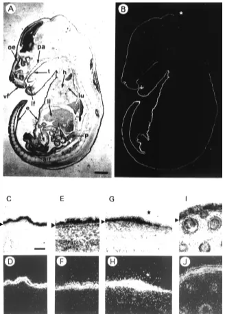
Fig. 2. Expression of galectin-7 mRNAs in the E16.5 mouse embryo. A near-sagittal section of E16.5 mouse embryo was hybridized in situ using an antisense galectin-7 riboprobe. (A) Bright field, (B), Dark field. Note that galectin-7 mRNA expression is mainly restricted to the epidermis. Also note that the intensity of the signal varies considerably in different regions of the body. The star indicates the abrupt transition between regions of high and low galectin-7 expression on the back of the head and the body. pa, palate; t, tongue; vf, vibrissal follicles; lf, lip furrow; h, heart; lu, lung; e, epidermis; li, liver; b, bladder; i, intestine; p, pancreas; a/r, ano-rectal region. Details of galectin-7 in situ hybridization in the E16.5 mouse embryo: bright field (C,E,G,I), dark field (D,F,H,J). (C,D) Ventral body skin; (E,F) tail skin; (G,H) dorsal skin over the back of the head and body; (I,J) mystacial whisker pad. In (G,H), note that the strongest hybridization signal is observed in the most stratified (anterior, i.e., left) area of epidermis whereas the weakest was observed in the less stratified (posterior, i.e., right) area. Arrowheads indicate the basal epidermal cell layer. Bar in A,B, 1.4 mm. Bar in B-J, 50 µm.
layers). Thus, at the back of the head of the E16.5 embryo, a marked reduction in hybridization signal coincided precisely with an abrupt change in the thickness of the epidermis (Fig. 2A,B,G,H, see star). Galectin-7 expression was elevated in the anterior most stratified region and was barely detectable in the adjacent dorsal region, where epidermal stratification was less advanced (i.e., comprising no or only one cell layer). A similar transition from strong to weak expression of galectin-7 could be seen in the facial region, in the epithelium covering the outer and inner parts of the lip (Fig. 2A,B). Galectin-7 was also expressed in both the surface epidermis and in the developing whisker follicles (Fig. 2I,J) apparently in the outer root sheath where galectin-7 was also found in adult skin (Magnaldo et al., 1998).
The overall increase in the level of galectin-7 mRNA in the embryonic epidermis with developmental age, and the correlation between local levels of galectin-7 mRNA and the thickness of the suprabasal compartment indicate that the expression of this gene is closely associated with the process of epidermal stratification.
Galectin-7 expression in non-epidermal epithelia
As shown in Figure 2, galectin-7 expression in the mouse embryo was not confined to the epidermis, but was also found in a limited number of other epithelia. Sections through the buccal cavity (Fig. 3A,B) showed that there were alternating stripes of
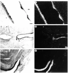
weak and strong galectin-7 expression in the ridged epithelium lining the upper palate. A strong signal was localized over the ridges of stratified epithelium, while the intervening stripes of simple epithelium were only very faintly labeled. Uneven hybridiza-tion signal was also seen on the surface of the tongue (Fig. 3A,B), where the signal was concentrated over the small blocks of stratified epithelium corresponding to developing filiform papillae (see bright field picture in panel 3A). We also observed the accumulation of transcripts in the region of the presumptive tooth bud (not shown). In the embryonic digestive tract, galectin-7 mRNA was detected in the stratified epithelium lining the upper part of the E16.5 oesophagus, but not in the monolayered epithelium further down (Fig. 3C,D). At the end of the digestive tract there was a similar marked transition from the non-expressing, monolayered mucous epithelium of the rectum to the heavily labeled, stratified epithelium of the anal area (Fig. 3E,F). No expression was de-tected in the mucous or glandular epithelia of the stomach or intestine.
In addition to the absence of galectin-7 expression in the mucous epithelia lining the hollow passages of the respiratory, gastro-intestinal and uro-genital tracts, we also noted that this gene was not expressed in any of the endoderm-derived "solid" or glandular organs such as the kidney, adrenal, liver, lung or pan-creas (Fig. 2 and data not shown). These results demonstrate that galectin-7 expression is restricted to all stratified epithelia.
Galectin-7 expression in the developing gonads
The earliest embryonic stage at which we detected galectin-7 transcripts was E11.5, when there was faint but specific expression in the sexually indifferent genital ridge (Fig. 4A,B). After one further day of development, when the gonads could be identified as either female or male, galectin-7 expression was found in both the ovary (Fig. 4C,D) and the testis (Fig. 4E,F). Later on, at E14.5, we observed a graded distribution of signal across the ovary, with maximal expression in the coelomic epithelium and cortex, and weaker expression in the medulla (not shown). In the E12.5 testis, hybridization signal was localized over the cords and no specific signal was detected over the interstitial cells. Expression in the testis cords persisted during late embryonic development (Fig. 4G,H) and into early postnatal life. Figure 4 (I,J) shows testicular expression on the day of birth (P0). After reaching a maximal level at approximately P4, expression declined to undetectable levels in the adult testis (not shown). The localization of signal over the centre of the testis cords suggested that galectin-7 mRNA was expressed in the developing Sertoli cells, but we could not exclude additional expression in germ cells.
The localization of galectin 7 transcripts in the embryonic gonads is consistent with expression in the presumptive granulosa and Sertoli cells. Galectin-7 expression in the sexually indifferent gonad at E11.5 could therefore indicate that this is one of the earliest known markers of the gonadal supporting cell lineage.
Discussion
Following the isolation of a galectin-7 cDNA from human epider-mal cells, and the demonstration of galectin-7 mRNA and protein in human, mouse and rat epidermis (Madsen et al., 1995; Magnaldo et al., 1995, 1998), we have carried out an in situ hybridization study on the mouse embryo. Our main objectives were to determine how widely this gene is expressed in different tissues, and to document its Fig. 3.Expression of galectin-7 mRNAs in non-epidermal epithelia.
expression in detail during embryonic develop-ment of the epidermis.
Tissue specificity of galectin-7
We have found that galectin-7 transcripts are expressed in the mouse embryo with a remark-able degree of tissue specificity. Expression of the galectin-7 gene is restricted to a subset of epithelia that are, or are destined to become, stratified. This was particularly evident in regions of transition or alternation from simple to stratified epithelium, as in the external nares, lip, upper palate, oesophagus, rectum and umbilicus, where we observed a sharp demarcation between cells expressing high (stratified) or very low (monolayered) level of galectin-7. It is interesting that this strict correlation between galectin-7 ex-pression and stratified organization holds true in keratinized epithelia such as the epidermis, the lining of the tongue and upper oesophagus, as well as in non-keratinizing epithelia such as the lining of the buccal cavity and the presumptive seminiferous epithelium. In contrast, we did not detect galectin-7 mRNA in simple, transitional or pseudostratified epithelia, or in organs such as the kidney, lung, liver or intestine.
In agreement with our previous work on mouse and human adult tissues (Magnaldo et al., 1998), these observations support the con-clusion that galectin-7 is a marker of epithelial stratification, regardless of other characteris-tics that may reflect the anatomical location or specialized function of a particular epithelium. This is also consistent with the observation that galectin-7 expression is only slightly decreased when adult keratinocytes switch from the ex-pression of epidermal to oesophageal markers upon treatment with retinoic acid (Magnaldo et al., 1995). Taken together these results distin-guish galectin-7 from other markers such as the suprabasal keratins, which are selectively ex-pressed according to the body location and function of a given epithelium (e.g., palmoplantar versus interfollicular epidermis) and from any other marker of the epithelial phenotype (Davies and Garrod, 1997).
The only apparent discrepancy in the overall tissue specificity of galectin-7 was the expression in the early embryonic gonad, where transcripts were detected in the mesenchymal stroma of the E11.5 undifferentiated genital ridge. However, the distribution of transcripts after morphological differentiation of the gonads suggests that the galectin-7 positive cells belong to the somatic
Fig. 4.Expression of galectin-7 mRNAs in developing gonads. In situ hybridization is shown using bright field illumination, (A,C,E,G,I); or dark field illumination, (B,D,F,H,J). (A,B) E11.5 sexually indifferent genital primordia; (C,D) E12.5 ovary; (E,F) E12.5 testis. (G,H) E17.5 testis; (I,J) P0 testis. In (G,H,I,J) note that galectin-7 expression is specifically detected in the testis cords. m, mesonephros; gr, genital ridge; ce, coelomic epithelium; tc, testis cords; Bar, 50 µm.
epithelial keratins K8, K18 and K19 in a dynamic pattern (Stosiek et al., 1990; Fridmacher et al., 1992, 1995).
Galectin-7 expression and epidermal development
The epidermis develops from a single layer of ectoderm, in a series of steps which vary in their precise character and timing in supporting cell lineage that gives rise to the granulosa cells of the
different regions of the embryo (for review see Byrne, 1997). Surface ectoderm starts to become a bilayered epithelium at approximately E9.5, the inner layer forming the basal compartment of the embryonic epidermis. The outer periderm layer will be shed from the skin surface later in development. From E13.5 onwards, a third layer of cells begins to appear between the basal and periderm layers. The cells in this suprabasal compartment arise by egression of cells from the basal layer by the process known as stratification. In contrast to adult epidermis, newly formed embry-onic suprabasal cells may continue to divide for a short period of time of before displaying the characteristic flattened morphology of suprabasal keratinocytes and piling up in multiple layers (Byrne et al., 1994).
Since we did not detect any galectin-7 expression in the ecto-derm between E9.5 and E13.5, we suggest that this gene is not involved in very early cell fate decisions such as the initial decision to develop either as a stratified, or as a simple epithelium. The main features of galectin-7 mRNA expression in developing epidermis were all represented in various regions of the body at E16.5 (Figs. 2 and 3). At this stage, there were areas of dorsal bilayered epidermis expressing very low levels of galectin-7 mRNA, areas of ongoing stratification where the signal was stronger, and regions of even more intense signal where the suprabasal compartment contained several layers of flattened cells. The dynamics of this pattern suggest that galectin-7 expression is regulated in a stepwise fashion as the embryonic epidermis develops. The onset of expres-sion occurs in basal cells at the late bilayered stage, coinciding with egression of the first cells from the basal to the suprabasal compartment. Expression is then up-regulated in the new suprabasal cells, and maintained or increased during their subsequent mor-phological differentiation. The stepwise increment of galectin-7 expression during epidermal stratification in the mouse embryo is reminiscent of the onset of expression of two peanut-binding glycoproteins during the same process in human embryonic epi-dermis (Fisher and Holbrook, 1987; Watt et al., 1989). One interesting possibility would be that galectin-7 interacts these glycoproteins in vivo (Watt et al., 1989).
From these observations we conclude that galectin-7 may be important during distinct stages of epithelial stratification; namely movement from the basal to the suprabasal compartment, differen-tiation, and the maintenance of a stratified organization. Clearly, the subcellular localization of galectin-7 protein holds vital clues to its function. It will be important to determine if galectin-7 protein is externalized, for instance at the basal pole of epithelial cells, where it could be involved in modulating carbohydrate-dependent attach-ment to the baseattach-ment membrane (Cooper et al., 1991; Sato and Hughes, 1992) or at other faces of the cell where it could affect intercellular adhesion. In this respect, it is intriguing that the pattern of galectin-7 mRNA expression in the embryo bears a close resemblance to that of the desmosomal cadherin DSG1 (King et al., 1996). The possibility that galectin-7, like galectin 4 (Chiu et al., 1992), might be associated with adherens junctions is currently being investigated. In addition, it will be important to compare galectin-7 distribution with that of galectin-3, which is also ex-pressed in developing epidermis (Fowlis et al., 1995).
The stepwise increments in galectin-7 expression that accom-pany embryonic keratinocyte development may reflect the shifting balance between proliferation and differentiation. As cells leave the basal layer, cease proliferating, and differentiate, there is a concomitant increase in the level of galectin-7 mRNA. Equally, this
may account for the fact that galectin-7 appears to be expressed at higher levels in the basal layer of adult (Magnaldo et al., 1995, 1998) than embryonic epidermis. An inverse relationship between galectin-7 expression and proliferation rate is further supported by two lines of evidence. First, galectin-7 expression is abrogated in cultured squamous cell carcinoma lines that have escaped pro-liferation control (Magnaldo et al., 1998). Second, basal and suprabasal cell layers of squamous and basal cell carcinomas exhibit very low levels of galectin-7 expression (Magnaldo et al., 1998). In addition, galectin-7 has recently been identified as one of 14 early response genes induced by the tumor suppressor gene, p53 (Polyak et al., 1997).
In conclusion, this work provides evidence that galectin-7 plays a role in the development and maintenance of all stratified epithe-lia, perhaps by modulating cell proliferation or cell interactions. Generation of galectin-7 mutations in mice should help elucidate its role. In addition, this gene is a new marker which can be used in the characterization of disorders and mutations affecting the skin and other epithelia. The galectin-7 gene should also provide an excel-lent model for the study of tissue-specific transcriptional regulation.
Materials and Methods
Probe for in situ hybridization
A full length rat galectin-7 cDNA (Magnaldo et al., 1998) inserted into Bluescript SK vector was used for synthesizing 35S cRNA probes following the procedure previously described in Fowlis et al. (1994).
Tissue preparation for in situ hybridization
Embryos were fixed in freshly prepared 3.7% paraformaldehyde (PFA) pH 7.4, at 4°C overnight. Samples were rinsed first in PBS and then in 0.85% NaCl. After successive dehydration steps, embryos were embedded in Paraplast X-TRA. Sections (6 µm) were cut and collected on superfrost-plus slides.
In situ hybridization procedure
In situ hybridization was carried out as described in Wilkinson (1992) with modifications according to Akhurst (1993). Briefly, the sections were deparaffinized, rehydrated and fixed for 20 min at 4°C in 3.7% PFA in PBS, pH 7.4. After washing in PBS, they were incubated for 7 min in proteinase K (40 µg/ml), rinsed in PBS, fixed for 5 min in the same PFA solution, and incubated for 10 min in 0.1 M triethanolamine-HCl, pH 8.0, containing 0.25% acetic anhydride, washed and dehydrated. Sections were then hybridized overnight at 55°C in the presence of the radiolabeled probe. The coverslips were removed by incubation in a solution of 5xSSC containing 10 mM DTT at 55°C. The sections were subsequently washed for 20 min at 65°C in 2xSSC-50% formamide-20 mM DTT, then twice in 0.5 M NaCl-10 mM Tris-5 mM EDTA (TEN) at 37°C followed by an incubation in 20 µg/ml of RNAse A in TEN for 30 min at 37°C, washed in TEN at 37°C and then again in 2xSSC-50% formamide-20 mM DTT at 65°C and finally in 2xSSC and 0.1xSSC both at 50°C for 15 min before dehydration. Slides were dipped in K5 Ilford emulsion, diluted 1:1 in 2% glycerol, dried and exposed for 3-7 days at 4°C. Development was carried out in Kodak D19 for 2 min, then in 1% acetic acid-1% glycerol for 1 min, and fixation in 30% sodium thiosulfate for 2 min. The sections were counterstained with 0.1% toluidine blue for 1-2 min, dehy-drated and mounted in DPX. The specificity of the galectin-7 antisense riboprobe was attested by the absence of hybridization signal when using the sense riboprobe and by the observation of expected hybridization signals when using unrelated antisense riboprobes (Colnot et al., 1997).
Akcnowledgments
Association pour la Recherche sur le Cancer (ARC 6935 from F.P. and ARC 1711 from T.M.). P.T. was a recipient of an E.E.C. post-doctoral fellowship.
References
AKHURST, R.J. (1993). Localization of growth factor mRNA in tissue sections by in situ hybridisation. In Growth factors: a practical approach. (Eds McKay, I. and Leigh, I.) Oxford: IRL Press.
ASSELINEAU, D. and DARMON, M. (1995). Retinoic acid provokes metaplasia of epithelium formed in vitro by adult human epidermal keratinocytes. Differentiation 58: 297-306.
BARONDES, S., COOPER, D., GITT, M. and LEFFLER, H. (1994). Galectins. Structure and function of a large family of animal lectins. J. Biol. Chem. 269: 20807-20810.
BYRNE, C. (1997). Regulation of gene expression in developing epidermal epithelia. BioEssays 19: 691-698.
BYRNE, C., TAINSKY, M. and FUCHS, E. (1994). Programming gene expression in developing epidermis. Development 120: 2369-2383.
BYSKOV, A.G. and HOYER, P.E. (1994). Embryology of mammalian gonads and ducts. In The physiology of reproduction Vol 1, (Eds. Knobil,E. and Neill, J.D.) Raven NY, pp. 487-540.
COLNOT, C., RIPOCHE, A., FOWLIS, D., CANNON, V., SCAEROU, F., COOPER, D. and POIRIER, F. (1997). The role of galectins in mouse development. Trends Glycosci. Glycotechnol. 9: 31-40.
COOPER, D., MASSA, S. and BARONDES, S. (1991). Endogenous muscle lectin inhibits myoblast adhesion to laminin. J. Cell. Biol. 115: 1437-1448.
CHIU, M., JONES, J. and O’KEEFE, E. (1992). Restricted tissue distribution of a 37-kD possible adherens junction protein. J. Cell. Biol. 119: 1689-1700.
DAVIES, J.A. and GARROD, D.R. (1997). Molecular aspects of the epithelial
phenotype. BioEssays 19: 699-704.
FISHER, C. and HOLBROOK, K.A. (1987). Cell surface and cytoskeletal changes associated with epidermal stratification and differentiation in organ culture of embryonic human human skin. Dev. Biol. 119: 231-241.
FOWLIS, D., COLNOT, C., RIPOCHE, M-A. and POIRIER, F. (1995). Galectin-3 is expressed in the notochord, developing bones and skin of the post-implantation embryo. Dev. Dynamics 203: 241-251.
FRIDMACHER, V., LE BERT, M., GUILLOU, F. and MAGRE, S. (1995). Switch in the expression of the K19/K18 keratin genes as a very early evidence of testicular differentiation in the rat. Mech. Dev. 52: 199-207.
FRIDMACHER, V., LOCQUET, O. and MAGRE, S. (1992). Differential expression of acidic cytokeratins 18 and 19 during sexual differentiation of the rat gonad. Development 115: 503-517.
FUCHS, E. (1990). Epidermal differentiation. Curr. Opin. Cell. Biol. 2: 1028-1035.
FUCHS, E. (1997). Of mice and men: genetic disorders of the cytoskeleton. Mol. Biol. Cell 8: 189-203.
HUGHES, R.C. (1992). Lectins as cell adhesion molecules. Curr. Opin. Struct. Biol. 2: 687-692.
KING, I., O’BRIEN, T. and BUXTON, R. (1996). Expression of the "skin-type" desmosomal cadherin DSC-1 is closely linked to the keratinization of epithelial tissues during mouse development. J. Invest. Dermatol. 107: 531-538.
LEFFLER, H. (1997). Introduction to galectins. Trends Glycosci. Glycotechnol. 9: 9-19.
MADSEN, P., RASMUSSEN, H., FLINT, T., GROMOV, P., KRUSE, A., HONORE, B., VORUM, H. and CELIS, J. (1995). Cloning, expression, and chromosome map-ping of human galectin-7. J. Biol. Chem. 270: 5823-5829.
MAGNALDO, T., BERNERD, F. and DARMON, M. (1995). Galectin-7, a human 14-kDa S-lectin, specifically expressed in keratinocytes and sensitive to retinoic acid. Dev. Biol. 168: 259-271.
MAGNALDO, T., FOWLIS, D. and DARMON, M. (1998). Galectin-7, a marker of all types of stratified epithelia. Differentiation 63: 159-168.
POLYAK, K., XIA, Y., ZWEIER, J., KINZLER, K. and VOGELSTEIN, B. (1997). A model for p53-induced apoptosis. Nature 389: 300-305.
SATO, S. and HUGHES, R. (1992). Binding specificity of the baby hamster kidney lectin for type I and II chains, polylactosamine glycans, and appropriately glycosylated forms of laminin and fibronectin. J. Biol. Chem. 267: 6983-6990.
STOSIEK, P., KASPER, M. and KARSTEN, U. (1990). Expression of cytokeratins 8 and 18 in human Sertoli cells of immature and atrophic seminiferous tubules. Differentiation 43: 66-70.
SUN, T., EICHNER, R., SCHERMER, A., COOPER, D., NELSON, W. and WEISS, R. (1984). Classification, expression and possible mechanism of evolution of mam-malian epithelial keratins: a unifying model. In Cancer cells 1. The transformed phenotype. (Eds. Levine, A.J., Van de Woude, G.F., Topp, W. and Watson, J.D.) Cold Spring Harbor Laboratory, New York, pp 169-176.
WATT, F. M., KEEBLE, S., FISHER, C., HUDSON, D., COOD, J. and SALISBURY, J. R. (1989). Onset of expression of peanut lectin-glycoproteins is correlated with stratification of keratinocytes during epidermal development in vivo and in vitro. J. Cell Sci. 94: 355-359.
WILKINSON, D. G. (1992). In situ hybridization to cellular RNA with radiolabelled RNA probes. In In situ hybridization. (Ed. Wilkinson D.G.) IRL Press, Oxford pp. 15-32.