Original Article
Regeneration of halved embryonic lower first mouse molars:
correlation with the distribution pattern of non dividing IDE cells,
the putative organizers of morphogenetic units, the cusps
ROMUALD COIN
1#, RÉGINE SCHMITT
2#, HERVÉ LESOT
1, JEAN-LUC VONESCH
3and
JEAN-VICTOR RUCH
1*
1INSERM U-424, Institut de Biologie Médicale, Faculté de Médecine, Strasbourg, 2INSERM U-424, U.F.R d’Odontologie de Strasbourg, and 3Institut de Génétique et de Biologie Moléculaire et Cellulaire, Illkirch, France
ABSTRACT Recently we demonstrated that non-cycling, cap-stage, mouse molar inner dental epithelial (IDE) cells corresponding to the primary enamel knot (EK) area underwent a coordinated temporo-spatial patterning leading to their patchy irregular segregation at the tips of the forming cusps. These non-cycling cells were suggested to perhaps represent the organizers of the morpho-genetic units (OMU), the cusps. The present study has analyzed the regenerative capacity of halved cap-stage first lower mouse molars through three dimensional (3D) reconstructions. Partial regeneration of the anterior half and possible complete regeneration of the posterior half were documented. Using BrdU (5-bromo-2'-deoxyuridine) labeling and 3D reconstructions of the IDE, we have correlated the patterns of cusp regeneration with the distribution of BrdU negative IDE cells. These data support a morphogenetic role for the non-cycling IDE cells.
KEY WORDS:
mouse molar, regeneration, enamel knot, non-cycling IDE cells
0214-6282/2000/$20.00
© UBC Press Printed in Spain
www.ehu.es/ijdb
*Address for reprints: Institut de Biologie Médicale, INSERM U-424, Faculté de Médecine, 11 rue Humann, 67085 Strasbourg cedex France. FAX: 03 88 25 78 17. e-mail: Ruch@odont3.u-strasbg.fr
#These authors contributed equally to this work.
Abbreviations used in this paper: BrdU, 5-bromo-2'-deoxyuridine; IDE, Inner Dental Epithelium; EK, Enamel Knot; EFA, Enamel Free Area; OMU, Organizer of Morphogenetic Unit; SHH, Sonic Hedgehog; BMP, Bone Morphogenetic Protein; FGF, Fibroblast Growth Factor; 3D, three dimensional.
Introduction
Tooth crown morphogenesis is controlled by the bud-cap staged dental mesenchyme (Kollar and Baird, 1969, 1970a,b; Schmitt et al., 1999). It has been hypothesized that the mesen-chyme induces the formation of the enamel knot (EK), a transitory epithelial structure, located at the tip of the cap, expressing signaling molecules including sonic hedgehog (SHH), bone mor-phogenetic proteins (BMP), fibroblast growth factor’ (FGF) (Keranen et al., 1998). According to Jernvall et al. (1994), this primary EK might represent a signaling center for tooth morpho-genesis causing unequal growth of the enamel epithelium and inducing the formation of secondary EKs located at the tips of the forming cusps.
Recently, we demonstrated using BrdU labeling (Coin et al., 1999) that Go cells of the inner dental epithelium (IDE) corre-sponding to the EK area of the first lower mouse molar underwent sequential segregation and that this led to the formation of distinct groups of cells, each one corresponding to a particular developing cusp. These observations suggested that within cells of the EK that act as the organizers of morphogenetic units (OMU), the activity could specifically lie in these Go cells of the IDE, giving rise
to the ameloblasts of the enamel free area (EFA) (Coin et al., 1999).
To experimentally test the possible correlation between the distribution of these OMU and cusp formation, we analyzed both the cusp pattern and the distribution of the OMU in regenerating, halved cap-stage first lower mouse molars using three dimen-sional (3D) reconstructions. As a matter of fact, Fisher (1971) analyzed the morphological development in vitro of halved lower mouse molar germs and she demonstrated convincingly partial regeneration of the anterior halves of E-13/E-14 first molars and complete regulation of 20% of corresponding posterior halves.
Results
The physiological cusp patterns of the first and second lower molars are shown in Figure 1. The cusps are named according to the terminology of Gaunt (1955, 1961). The M1 crown comprises seven cusps which have a marked biserial antero-posterior ar-rangement: two series of three buccal and three lingual cusps, essentially of a paired nature and one single posterior cusp. The crown surface is broken up by a longitudinal cleft, which separates the lingual and buccal ridges with each ridge showing a three-fold subdivision due to an anterior and median transverse valley. All cusps slope in an anterior direction (Fig. 1A).
The second lower molar (M2) does not show an anterior area of the crown equivalent to that of M1, but there is a narrow anterior cingulum present. Except for this difference, the median and the posterior part of the M2 shows a striking resemblance to the M1 (Fig. 1B).
Morphological observations throughout in vitro develop-ment
Fifty-eight intact left E-14 molars were cultured in vitro and fifty-five survived. Sixty-seven right E-14 molars were halved and eight anterior parts and three posterior parts demonstrated rapid invo-lution in vitro.
Crown morphogenesis of the intact molars was evident. The anterior parts of the halved molars demonstrated always limited crown morphogenesis. The posterior parts of the M1 developed systematically several cusps.
The posterior parts of the halved molar rudiments included the primordium of M2 in some cases, which also developed in vitro and this was observed in about 50% of the specimens.
Preliminary 3D reconstructions
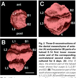
Sections from an intact left E-14 molar and the corresponding anterior and posterior parts of the halved right molar, cultured for 6 days were reconstructed. The posterior part developed an M2. Figure 2 shows the results for the halved molar and for the M2. The anterior part developed 4 cusps (Fig. 2A), two anterior (tentatively named L1,B1) and two posterior (L2 and B2). The corresponding posterior half developed 6 cusps (Fig. 2B). The tooth was subdi-vided by a deep transverse fissure into a larger anterior portion containing four cusps (L1,B1,B2,L2) and a smaller posterior domain containing two cusps (L3 and B3). The M2 demonstrated a normal cusp pattern: two anterior (L2,B2), two posterior (L3,B3) and a single postero-median cusp 4 (Fig. 2C).
3D reconstructions after BrdU labeling
To assess possible overlap between the dynamic pattern of distribution of BrdU negative IDE cells (Coin et al., 1999) and the pattern of cusp formation, we pulse labeled the intact and the halved teeth after various preincubation periods and performed 3D reconstructions.
Eight hour pulse labeling after 2 h of preincubation
The reconstructed intact left molar showed the presence of one distinct area of non-labeled IDE cells located at the center of the IDE with an antero-posterior orientation (Fig. 3A,D).
The corresponding anterior (Fig. 3B,E) and posterior (Fig. 3C,F) halves of the right molar showed respectively 1) a negative
Fig. 1. Scanning electron micrographs of E-23, post-natal, first (A) and second (B) right lower mouse molars. The cusps were named according to the terminology of Gaunt (1955, 1961). (A) Seven cusps exist: three lingual (L1,L2,L3), three buccal (B1,B2,B3), and a seventh named cusp 4. Cusps L1,B1,B2 and L2 express a «trefoil» like pattern. The crest connect-ing L1,B1,B2 and L2 has a lconnect-ingual discontinuity. (B) Five cusps are present: two lingual (L2,L3), two buccal (B2,B3) and a fifth named cusp 4. Cusps L2 and B2 are connected by a crest as cusps L3 and B3 are. A deep median transverse fissure subdivided the molar into an anterior part including cusps L2 and B2 and a posterior one with cusps L3,B3 and 4 are. Note that cusp 4 has also a median and posterior localization. ant, anterior; post, posterior. (x40).
Fig. 2. Three-D reconstructions of the dental mesenchyme of ante-rior (A) and posteante-rior (B) parts of a halved E-14 first lower mouse molar and of the corresponding contralateral second molar (C) cultured for 6 days. (A) After 6 days, the anterior part of the halved molar shows four cusps (L1,L2,B1 and B2). (B) The posterior part of the tooth shows regeneration: six cusps (L1,L2,L3,B1,B2 and B3) developed.
area still adjacent to the performed section and 2) a rather central negative domain with an antero-posterior orientation.
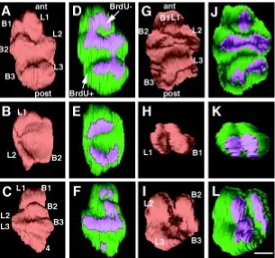
Eight hour pulse labeling after 96 h or 108 h of preincubation Two intact teeth and the contralateral halves were recon-structed (Fig. 4). After 96 h of preincubation the intact left M1 showed 6 cusps with corresponding BrdU negative areas of the IDE (Fig. 4A,D). The anterior half developed three cusps with three corresponding BrdU negative domains (Fig. 4B,E). The posterior half displayed a complete cusp pattern with seven cusps and seven, partially connected BrdU negative areas (Fig. 4C,F). The crown displayed crests rather than tubercles.
After 108 h of preincubation, the crown pattern of the intact left M1 showed six cusps: two anterior (L1,B1), which were probably fused, two median (L2,B2) and two posterior (L3,B3) cusps. A deep median transverse fissure subdivided the tooth into a large anterior portion and a smaller posterior one. Five partially fused BrdU negative domains of the IDE coincided with this cusp pattern (Fig. 4G,J). The corresponding anterior half showed the presence of only two cusps, probably L1 and B1 and two fused Brdu negative areas were observed (Fig. 4H,K). The crown of the corresponding posterior half comprised four cusps: two anterior (probably L2 and B2) and the two posterior (probably L3 and B3) and four corresponding BrdU negative domains were observed (Fig. 4I,L).
Twenty four hour continuous labeling after 84 h or 108 h of preincubation
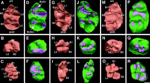
After 84 h of preincubation, one intact left M1 and the contralat-eral halves were reconstructed (Fig. 5A-F).The intact molar developed a normal cusp pattern of a left M1 cultured in vitro: six cusps (L1,L2,L3,B1,B2,B3) were observed. The crest connecting L1,B1,B2 and L2 had a lingual discontinuity. Corresponding BrdU negative areas of the IDE were observed (Fig. 5A,D). The anterior half displayed three cusps and three corresponding negative BrdU areas (Fig. 5B,E). The posterior half demonstrated a physi-ological cusp pattern of a left first lower molar. The crest connect-ing the four anterior cusps had a lconnect-ingual discontinuity orientated towards the right side and a supernumerary cusp was observed on the lingual side between L1 and L2. Corresponding BrdU negative areas of the IDE were superposed on the cusps (Fig. 5C,F).
After 108 h of preincubation, two intact left M1 and the contralat-eral halves were reconstructed (Fig. 5G-R). The two intact teeth developed six cusps: two anterior (L1,B1), two median (L2,B2) and two posterior (L3,B3) cusps. BrdU negative areas corre-sponded precisely to the localization of these six cusps (Fig. 5G,J,M,P). The two corresponding anterior halvesdemonstrated four cusps, tentatively: L1,B1,B2 and L2. Corresponding BrdU negative areas were observed (Fig. 5H,K,N,Q). The posterior halves contained five cusps: two lingual (L1,L2) and three buccal (B1,B2,B3) cusps. Five corresponding negative BrdU domains were present (Fig. 5I,L,O,R).
Table one summarizes the number of cusps which developed in the corresponding anterior and posterior parts of halved right lower molars.
Histological observations
After 2 h of preincubation and 8 h BrdU incorporation, the intact E-14 molars demonstrated obvious BrdU negative primary EKs
(Fig. 6A). In contrast, both the anterior and posterior parts of the halved teeth only showed disorganized EKs in which BrdU nega-tive IDE cells were present (Fig. 6E,I respecnega-tively).
After 24 h of preincubation and 8 h BrdU labeling, the intact molars revealed the presence of small, BrdU negative, primary EKs (Fig. 6B). The anterior parts of halved molars showed an apparent reorganization of the BrdU negative primary EKs (Fig. 6F). The corresponding posterior parts demonstrated the pres-ence of typical primary EKs (Fig. 6J).
After 84, 96 or 108 h of preincubation and 8 or 24 h BrdU incorporation, BrdU negative IDE cells were located in all the specimens at the tips of the forming cusps (Fig. 6C,G,K).
After 6 days in vitro, terminal differentiation of the odontoblasts was initiated in both intact and halved molars (Fig. 6D,H,L).
Discussion
Morphogenesis in general terms may be defined as a set of developmental processes leading to organ specific spatial distri-bution of functional cells. These processes include cell division, cell death, cell migration, dynamic cell adhesion and cell-matrix interactions. The organ specific control mechanisms share com-mon denominators: epigenetic, sequential and cyclical exchange of information between interacting cells as a result of paracrine factors and their receptors, substrate adhesion molecules and their ligands, cell adhesion molecules and their signaling. These inductive processes regulate the transcription of selected genes and morphoregulatory genes according to Edelman (1992) or of «genomorphens» according to Krasnow (1997). During the initial
Fig. 3. Three-D reconstructions of an intact E-14 first left lower molar (A,D) and of the anterior (B,E) and posterior (C,F) parts of the halved contralateral molar.
stages of organogenesis most organ rudiments are endowed with regenerative capacity.
As far as tooth morphogenesis is concerned, the bud-cap stage dental ecto-mesenchyme controls the tooth specific cusp pattern, eventually through BMP-4 regulated formation of the primary EK (Jernvall et al., 1998). The latter may control tooth crown morphogenesis through induction of secondary signaling EKs (Keranen et al., 1999). Recently, we demonstrated (Coin et al., 1999), using BrdU labeling, the progressive subdivision of the initial group of BrdU negative IDE cells of the primary EK area into as many distinct subgroups as cusps will develop. A cellular continuity exists between components of the primary and second-ary EKs and we suggested that these particular IDE cells might act as the organizer of morphogenetic units (OMU), each unit repre-senting a cusp. The signaling OMU’s could regulate the cuspidogenesis controlling local cell proliferation, cell death, cell adhesion and cell matrix interactions.
The possible regeneration of molars has been previously examined. Glasstone (1952) observed that rabbit molars when halved at initial stages of development and grown in vitro dis-played a capacity to regenerate and to form two miniature molar teeth. For the halved mouse molar (displaying limited growth), Fisher (1971) demonstrated a limited potential for regulation, since recovery and regeneration were never displayed by both parts of divided tooth germs.
At the histo-morphological level, the present data confirm Fisher’s observations. The anterior and posterior halves of E-14 first lower mouse molars demonstrated differential regulatory potential. The anterior parts never displayed complete regulation: 2,3 or 4 cusps developed. In contrast, the posterior halves developed 4 to 7 cusps. The delayed appearance of cusp 4 (after
6 days of culture) has been previously described (Schmitt et al., 1999). Normal terminal differentiation of the odontoblasts was observed. Terminal differentiation of ameloblasts would be initi-ated after longer periods of in vitro cultures (Schmitt et al., 1999). The regenerative capacity of the E-14 dental tissues appears to be irregular since the total number of developing cusps in corresponding halves fluctuated from 6 to 10 (Table 1). All the E-14 embryos are not exactly at the same developmental stage and the molars we used varied from the bud-cap transition to well formed cap. Apparently, during this period the potential for regu-lation decreased. Furthermore, this potential appears to switch off more rapidly in the anterior part. There exists a transitory antero-posterior increasing gradient of regenerative capacity. Butler (1982) stated that the molar cusps can be traced back to a specific arrangement of presumptive cusps in the cap-stage. Bringing together this possibility and our own observations, we suggest that at the initial cap-stage, the anterior (L1,B1) and median (L2,B2) cusps are determined while the presumptive posterior
Fig. 4. Three-D reconstructions of the dental mesenchyme and the corresponding IDE of whole E-14 first left lower molars (A,D,G,J) and anterior (B,E,H,K) and posterior parts (C,F,I,L) of the halved contralateral molars cultured in vitro for 96 h (A-F) or 108 h (G-L) and then labeled with BrdU for 8 h. For each specimen the cusp pattern can be compared with the distribution pattern of BrdU negative inner dental epithelial cells. (A,D), (B,E), (C,F), (G,J), (H,K), (I,L): the BrdU-cells cover the developing cusps. ant, anterior; post, posterior; BrdU+, 5-bromo-2'-deoxyuridine positive; BrdU-, 5-5-bromo-2'-deoxyuridine negative. Bar, 100 µm.
TABLE 1
NUMBER OF CUSPS IN THE ANTERIOR AND CORRESPONDING POSTERIOR PARTS OF RIGHT, HALVED M1 CULTURED IN VITRO
Number of teeth Number of cusps
Anterior halves Posterior halves
2 3 7
1 2 4
2 4 5
1 3 6
cusps (L3,B3 and 4) still manifest a regulative capacity. This hypothesis implies that depending on the level of the section, the 2 anterior (L1,B1) and 1 or 2 median (L2,B2) cusps developed according to a determined fate (without any regulation). Obvi-ously, our observations do not prove such a possibility.
The main goal of our observations was to assess whether the distribution of BrdU negative IDE cells, the hypothetical organizer of morphogenetic units (OMU), can be correlated with the pattern of regenerative cusps. Coin et al. (1999) have shown that after 8 h BrdU labeling the negative IDE cells include both slow cycling cells and Go cells. The Go cells were identified after continuous (72 h) BrdU labeling, which led, however, to partial inhibition of cusp formation. To avoid such a negative effect of BrdU, in the present work we restricted the incubation with BrdU to 8 or 24 h. The comparison of these two modalities (compare Fig. 4 and Fig. 5) shows that the areas of BrdU negative IDE cells are reduced after 24 h of labeling and include mainly (albeit not exclusively) Go cells. Different periods of pre-incubation were chosen to both follow the initial behavior of BrdU negative cells and their distribu-tion during cusp formadistribu-tion. According to Coin et al. (1999), even the longest preincubation time used in this work, followed by 24 h of BrdU labeling allowed to exclusion of the later presence of physiological post-mitotic ameloblasts arising from the IDE.
After 2 h of preincubation and 8 h BrdU labeling, a distinct BrdU negative EK was only present in the intact molars. In both parts of the halved molars, only BrdU negative IDE cells were observed. Apparently, the division led to disorganization of the EK, which was either partially or completely restored after 24 h of
preincuba-tion and 8 h BrdU incorporapreincuba-tion in the anterior and posterior halves respectively. This dissimilar EK restoration might be cor-related with unequal regenerative capacities. After the longer preincubations, allowing for the expression of the cusp pattern, and 8 or 24 h BrdU labeling a systematic coincidence between the cusp pattern and the patterned distribution of BrdU negative IDE cells (the putative OMU’s) was observed.
We postulated (Coin et al., 1999) that the primary EK controls the tooth specific temporo-spatial patterning of the OMU and consequently the tooth specific cusp pattern. According to this hypothesis, the limited reorganization of the primary EK in the anterior halves could explain the limited regulation. The Tabby tooth phenotype, i.e. reduced cusps number, has also been explained through reduced primary EK formation and hence limited signaling (Pispa et al., 1999).
The present observations demonstrate a correlation between the regenerative cusp pattern and the distribution of the putative OMU’s. Demonstration that this correspondence has causal implications awaits further investigation.
Materials and Methods
Tissues
Organ culture
The halved (posterior and anterior parts) and intact teeth were cultured on 2 ml of semi-solid control medium per Petri dish (Nunc, Roskilde, Denmark; 35x10 mm) for 2,84,96,108, and 144 h. The medium consisted of BGJ-B (Gibco, Fitton Jakson modified) supplemented with ascorbic acid (0,18 mg/ml, Merck), L-Glutamine (2 mM, Seromed), foetal calf serum (20%, Boehringer Bioproducts), kanamycin (0.1 mg/ml, Gibco) and Difco agar (0.5%) The teeth were incubated and grown at 37°C in a humidified atmosphere of 5% CO2 in air and the medium was changed every two days.
Bromodeoxyuridine labeling
Cell proliferation was analyzed by culturing the tissues in the presence of the thymidine analog 5-bromo-2'-deoxy-uridine (BrdU, cell proliferation kit; Amersham Life Science). The halved and intact teeth were cultured in the presence of 0.4 ml of BrdU at a concentration of 3 µg/ml for 8 or 24 h respectively after 2,84,96 or 108 h of culture in control medium.
Histology-immunohistochemistry
All the halved and intact teeth were fixed in Bouin-Hollande’s fluid, embedded in paraffin and serial 5 µm sections were performed. BrdU incorporated into DNA was identified on the 5 µm thick de-waxed sections with a specific mouse monoclonal antibody and immunoperoxidase labeling following the manufacturer’s instructions (Amersham Life Sci-ence). After immunostaining, sections were counterstained with eosin. The other sections were stained with Mallory’s stain.
Three-D reconstructions
Drawings of the contours of the mesenchyme and of the inner dental epithelium of the halved right teeth, of intact left teeth and in one case the second molar were made at 5 µm intervals from the serial histological sections, at a magnification of x250, using a Zeiss Jeneval microscope equipped with a drawing chamber. The inner dental epithelium was subdivided into different zones corresponding to the positive and negative cells. The digitalization of the serial drawings was achieved using a Hamamatsu C2400 camera connected to a digital imaging system. The digitalization of the serial drawings and correlation of successive images (Olivo et al., 1993) have been previously described (Lesot et al., 1996). Software packages allowing image acquisition and treatment were developed and adapted to this work. Three-dimensional images were generated using a volume rendering program (Sun Voxel, Sun Microsystems).
Acknowledgments
We wish to thank Dr. A.J. Smith for critical reading of this manuscript and Mr. A. Ackermann for technical help. This research was partially financed by the International Human frontier Science Program (grant TG-558/95 M), by the Faculty of Odontology-Strasbourg and by the Fondation Dentaire de France (UB/SS 500144-98002598).
References
BUTLER, P.M. (1982). Some problems of the ontogeny of tooth patterns. In Teeth: form, function, and evolution (Eds. Björn Kurtén). Columbia University Press, New York, pp. 44-51.
Fig. 6. Histological sections of 14 intact left molars (A-D) and E-14 anterior (E-H) and posterior (I-L) parts of halved right molars cultured for 2 h (A,E,I), 24h (B,F,J), 96 h (G) 108 h (C,K) and 144 h (D,H,L) in vitro. The intact and halved teeth were labeled with BrdU (A-C,E-G,I-K) or stained with Mallory (D,H,L). (A) The 8 h label-ing after 2 h of preincubation dem-onstrates the presence of negative cells in the EK of the intact cap-stage molar. (E) In the anterior and
COIN, R., LESOT, H., VONESCH, J.L., HAIKEL, Y. and RUCH, J.V. (1999). Aspects of cell proliferation kinetics of the inner dental epithelium during mouse molar morphogenesis: a reappraisal of the role of the enamel knot area. Int. J. Dev. Biol. 43: 261-267.
EDELMAN, G.M. (1992). Morphoregulation. Dev. Dyn. 193: 2-10.
FISHER, A. (1971). Morphological development in vitro of the whole and halved lower molar tooth germ of the mouse. Arch. Oral Biol. 16: 1481-1496.
GAUNT, W. (1955). The development of the molar pattern mouse. Acta. Anat. 24: 249-268.
GAUNT, W. (1961). The development of the molar pattern of the golden hamster (Mesocricetus Auratus W.), together with a re-assessment of the molar pattern of the mouse (Mus Musculus). Acta. Anat. 45: 219-251.
GLASSTONE, S. (1952). The development of halved tooth germs. A study in experimental embryology. J. Anat. (Lond.) 86: 12-15.
JERNVALL, J., ABERG, T., KETTUNEN, P., KERANEN, S. and THESLEFF, I. (1998). The life history of an embryonic signalling center: BMP-4 induces p21 and is associated with apoptosis in the mouse tooth enamel knot. Development 125: 161-169.
JERNVALL, J., KETTUNEN, P., KARAVANOVA, I., MARTIN, L.B. and THESLEFF, I. (1994). Evidence for the role of the enamel knot as a control center in mammalian tooth cusp formation: non-dividing cells express growth stimulating Fgf-4 gene. Int. J. Dev. Biol. 38: 463-469.
KERANEN, S.V., ABERG, T., KETTUNEN, P., THESLEFF, I. and JERNVALL, J. (1998). Association of developmental regulatory genes with the development of different molar tooth shapes in two species of rodents. Dev. Genes Evol. 208: 477-486.
KERANEN, S.V., KETTUNEN, P., ABERG, T., THESLEFF, I. and JERNVALL, J. (1999). Gene expression patterns associated with suppression of odontogenesis in mouse and vole diastema regions. Dev. Genes Evol. 209: 495-506.
KOLLAR, E.J. and BAIRD, G. (1969). The influence of the dental papilla on the development of tooth shape in embryonic mouse tooth germs. J. Embryol. Exp. Morphol. 21: 131-148.
KOLLAR, E.J. and BAIRD, G. (1970a). Tissue interactions in embryonic mouse tooth germs. I. Reorganization of the dental epithelium during tooth-germ reconstruc-tion. J. Embryol. Exp. Morphol. 24: 159-171.
KOLLAR, E.J. and BAIRD, G. (1970b). Tissue interactions in embryonic mouse tooth germs. II. The inductive role of the dental papilla. J. Embryol. Exp. Morphol. 24: 159-171.
KRASNOW, M.A. (1997). Genes that control organ form: lessons from bone and branching morphogenesis. Cold. Spring. Harb. Symp. Quant. Biol. 62: 235-240.
LESOT, H., VONESCH, J.L., PETERKA, M., TURECKOVA, J., PETERKOVA, R. and RUCH, J.V. (1996). Mouse molar morphogenesis revisited by 3D reconstruction. II. Spatial distribution of mitoses and apoptosis in cap to bell staged first and second upper molar teeth. Int. J. Dev. Biol. 40: 1017-1031.
OLIVO, J.C., IZPISUA-BELMONTE, J.C., TICKLE, C., BOULIN, C. and DUBOULE, D. (1993). Reconstruction from serial sections: a tool for developmental biology. Application to Hox genes expression in chicken wing buds. Bioimaging 1: 151-158.
PISPA, J., JUNG, H.S., JERNVALL, J., KETTUNEN, P., MUSTONEN, T., TABATA, M.J., KERE, J. and THESLEFF, I. (1999). Cusp patterning defect in Tabby mouse teeth and its partial rescue by FGF. Dev. Biol. 216: 521-534.
SCHMITT, R., LESOT, H., VONESCH, J.L. and RUCH, J.V. (1999). Mouse odontogenesis in vitro: the cap-stage mesenchyme controls individual molar crown morphogenesis. Int. J. Dev. Biol. 43: 255-260.