Genetic Analysis of Bleeding Disorders
Eunice Edison,1 Barbara A. Konkle,2 Anne C. Goodeve3,4
1Department of Haematology, Christian Medical College, Vellore, India; 2Bloodworks Northwest and University of Washington, Seattle, WA, USA; 3Sheffield Diagnostic Genetics Service, Sheffield Children's NHS Foundation Trust and 4 Department of Infection, Immunity and Cardiovascular Disease, University of
Sheffield, UK
Running head: Genetic Laboratory for Bleeding Disorders
Corresponding author: Prof Anne Goodeve, BSc, PhD Haemostasis Research Group
Department of Infection, Immunity and Cardiovascular Disease Faculty of Medicine, Dentistry and Health
Beech Hill Road
SHEFFIELD, S10 2RX, UK
Word count: 3188, including abstract- references References: 14
Tables: 3
Keywords: bleeding disorder, confirmation-sensitive gel electrophoresis, dosage analysis, genetic
analysis, next generation sequencing, pathogenicity prediction, F8 inversion.
Abstract
Molecular genetic analysis of inherited bleeding disorders has been practiced for over 30 years. Technological changes have enabled advances, from analyses using extragenic linked markers to next-generation DNA sequencing and microarray analysis. Two approaches for genetic analysis are described, each suiting their environment. The Christian Medical Centre in Vellore, India uses confirmation
sensitive gel electrophoresis mutation screening of multiplexed PCR products to identify candidate mutations, followed by Sanger sequencing confirmation of variants identified. Specific analyses for F8
intron 1 and 22 inversions are also undertaken.
The MyLifeOurFuture US project between the American Thrombosis and Hemostasis Network, the National Hemophilia Foundation, Bloodworks Northwest and Biogen uses molecular inversion probes (MIP) to capture target exons, splice sites plus 5’ and 3’ sequences and to detect F8 intron 1 and 22 inversions. This allows screening for all F8 and F9 variants in one sequencing run of multiple samples (196 or 392). Sequence variants identified are subsequently confirmed by a diagnostic laboratory.
predict likely pathogenicity and using a standard suite of tools can help standardise their documentation.
Introduction
Genetic analysis is a well-established process in the analysis of inherited bleeding disorders. New technology continually enables the process to become more automated in laboratories that can access these new techniques. A number of variations on next generation DNA sequencing (NGS) are being used for bleeding disorder genetic analysis. Simpler screening techniques also retain utility in many
laboratories and although their mutation detection rate may be slightly lower, these remain an important contribution to genetic analysis, facilitating identification of mutations in many laboratory situations. The approaches to analysis of diverse bleeding disorders in Vellore, India, and of haemophilia A and B simultaneously in Seattle, US are presented.
Once candidate pathogenic mutations have been identified in index cases, the possible pathogenicity of the variant(s) can be assessed and where relevant, reported back to the requesting clinician with an assessment of likely contribution to the patient’s disease. An approach to undertaking pathogenicity assessment and its documentation is described.
Proficient genetic laboratory for diagnosis of bleeding disorders at Christian Medical College, Vellore,
India-Eunice Edison
diagnosis. The Centre has therefore gradually developed advanced hematological and molecular genetic diagnostic facilities over the last 20 years.
Evaluating molecular genetics of hemophilia and other hemostatic disorders was initiated in 1995. Analysis was initially confined to intron 22 inversion genotyping and linkage-based analysis of mutations in hemophilia A and B [1]. In 2000, with support from Dr Carol Kasper, ex-Vice President of the World Federation of Hemophilia, enhancement of the technologies available was initiated and Dr Shaji who undertook training in haemophilia genetic analysis at the international haemophilia training centre (IHTC) in Sheffield, UK.
hemostasis have contributed to better understanding of the biology of these disorders, facilitating accurate detection of carriers and genetic counselling in many families (Table 1).
The establishment of the molecular genetic facilities has not only aided accurate diagnosis, but furthered understanding the complexity of genetic changes, genotypic-phenotypic heterogeneity and also devising therapeutic options in patients with bleeding disorders. This is well documented in scientific contributions to national (n=12) and international conferences (n=13).
The laboratory provides training in molecular diagnosis of bleeding disorders. We have instructed fourteen individuals (South Africa-1, South Korea-3, Malaysia-2, Sri Lanka-2, Bangladesh-2 and India-4) in molecular diagnosis of haemophilia; four of these were trained through IHTC fellowships.
With the increase in the number of laboratories offering molecular diagnosis, the need for a proficiency testing program was felt. An external quality assurance scheme for haemophilia (A and B) and genetic markers of thrombosis was initiated in 2006 when three laboratories were included. This recently increased to six laboratories; surveys are sent twice yearly. Thirty four samples (haemophilia A and B) have been dispatched in fifteen surveys. Response rate is between 70 and 80% while accuracy ranges between 70-80%.
The techniques have been combined to provide comprehensive genetic analysis of bleeding disorders and facilities and techniques are continually expanded to enable CMC Vellore to be the most proficient hemostasis genetic laboratory in the country.
The MyLifeOurFuture Haemophilia Genetic Analysis Project-Barbara Konkle
The MyLifeOurFuture (MLOF) initiative (www.mylifeourfuture.org) was begun in the US in 2012 through partnership between the American Thrombosis and Hemostasis Network (ATHN), the National
data repository and samples to facilitate research in haemophilia and its complications. At that time, surveys of consumers by NHF and of providers by ATHN found that approximately 20% of patients had
F8 or F9 DNA analysis performed, confirming the need for genetic analysis to support clinical care. In addition, the program was built on the commitment by the partners to expand research in haemophilia and its treatment.
Each of the four partners brings distinct expertise and together they govern the project. ATHN, working
with 135+ affiliated Haemophilia Treatment Centers (HTC), provides HTC provider education, secure data collection infrastructure known as ATHN Clinical Manager, and point of access for future research proposals. Participating HTC providers contracted through ATHN enroll patients, obtain samples for genotyping, and provide clinical results to patients. NHF, a US national patient advocacy organization with 52 chapters, educates the bleeding disorders community about the initiative through its
publications, annual meeting and local chapter events. This keeps the community informed about project status and supports recruitment. BWNW facilitates receipt of samples from sites, serves as the central genotyping laboratory and houses the research sample repository. Biogen, a biotechnology company with products for treatment of haemophilia, provides scientific collaboration and financial support.
Patients and for minors, their parents (patients/parents), are informed of the project through NHF and their area HTC, which establishes a contract with ATHN. Patients/parents are offered enrollment in the institutional review board approved research repository. Blood for DNA extraction is sent to BWNW for genetic analysis and, for those consenting to research, additional samples are stored in the repository. Haemophilia severity is determined by local laboratory testing. Initial F8 and F9 DNA analysis is
PCR methodology described by Rossetti et al [12]. This allows screening for all F8 and F9 variants in one sequencing run of multiple samples (196 or 392). Variants identified by NGS are confirmed in the BWNW CLIA-certified laboratory by another method specific to the variant. In males where no likely deleterious DNA variant is identified, and in females with moderate/severe disease and only one identified deleterious variant, MLPA is used. A clinical report is returned to the HTC, and results are transmitted electronically through ATHN to the HTC’s Clinical Manager database. Variant results are classified per American College of Medical Genetics and Genomics (ACMG) guidelines for pathogenicity [13].
As of February 2016, 4651 patients have enrolled in MLOF, and 3694 of those participants/parents consented to the sample and data research repository. Ninety four HTCs were engaged in the project, with 81 sites actively enrolling patients. Of the 4649 patients, 3636 have haemophilia A [1924 severe (S) Male (M):1913, Female (F):11], 659 moderate (Mo) (M:650, F:9), 1053 mild (Mi)(M:907, F:146)], 906 have haemophilia B [205 S (M:203, F:2), 336 Mo (M:335, F:1), 365 Mi (M:319, F:46)], and 107 are unclassified as part of a carrier pilot. Once optimized, our NGS approach has detected all variants, including large deletions, except for partial gene duplications of which it detected 6 of 9 found in 16 patients, all with haemophilia A. In the first 3000 patient samples analyzed, 228 previously unreported variants were found. A potentially causative variant was found in all patients except 27 with
haemophilia A (S:7, Mo:2, Mi:18) and 2 with haemophilia B (S:0, Mo:1, Mi:1). In sequencing both genes simultaneously we found a potentially causative variant in the other gene in 5 patients, 3 of whom had been previously reported as associated with haemophilia. Factor levels in those patients and prevalence in a population database (ExAC) raise questions regarding their pathogenicity.
of pathogenicity or likely pathogenicity (>90% certainty) require additional evidence, and given the past practice to genetically test only one affected family member, required segregation data can be lacking. Within MLOF, additional data from the HTCs will be obtained to maximize knowledge regarding the variants, which will inform deter pathogenicity assessment and deepen knowledge concerning hemophilia-causing variants.
In the future, with enrollment of 5000 subjects in the research repository, investigators worldwide will be able to apply to access the de-identified genetic data, repository samples, and phenotypic data through ATHN. Coded data and biologic samples will be linked to coded clinical data from the
ATHNdataset, gathered separately from this study. An independent, multidisciplinary research review committee managed by ATHN will evaluate proposals for scientific integrity and feasibility of research proposal and govern release of the data and sample repository. In concert, ~2200 samples are undergoing whole genome sequencing through the US National Heart, Lung and Blood Institute
Transomics in Precision Medicine (TOPMed) program and results will be deposited in the NIH Database of Genotypes and Phenotypes (dbGap) for use for scientific investigation.
In summary, MLOF is providing genetic information for patients and their families to help inform reproductive planning and clinical care. In addition, through the research repository and the ATHN clinical database, an invaluable resource for research in hemophilia and associated disorders is being built.
Pathogenic or Not? Classification of genetic variants in haemostasis-Anne Goodeve
the variant can contribute substantially to evidence of pathogenicity. However, making a judgement on possible pathogenicity is more challenging when the variant has not been previously reported.
Organizations including the UK Association for Clinical Genetic Science (ACGS) and the ACMG have produced guidelines on sequence variant classification [13, 14].
Standardized methods to collect and document information available on the sequence variant can be very useful in determining its likely pathogenicity. The ACGS recommend a classification on a scale of 1-5, where a classification of 1 indicates that the variant is common in the normal population and therefore not pathogenic, whereas 5 indicates that the variant has been seen previously in other patients with the disorder and that there is evidence of its pathogenicity through other analyses such as
in vitro expression [14]. Categories between these extremes are 2; unlikely pathogenic, 3; variant of unknown significance and 4; likely pathogenic (Table 2). Both the ACGS and ACMG guidelines [13, 14] recommend combining different evidence types to reach a conclusion regarding pathogenicity.
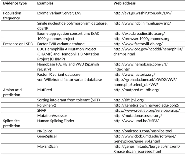
Two resources that can be most useful at the outset of these investigations are locus-specific mutation databases (LSDB) for the gene(s) of interest and sequence variant databases where many individuals have been analyzed for their sequence variation. Resources are listed in Table 3. Classification can be most challenging when a variant has not been previously reported. In silico predictions should be employed to help predict possible pathogenicity in these instances. Many different algorithms using different methodologies have been designed for both missense mutations and possible splice site mutations (PSSM) and these resources are listed in Table 3 and in the guidelines [13, 14].
Amino acid conservation examines the extent of conservation of the residue across several species, typically using sequences from several mammals, but also including chicken, frog and fish to encompass around 500 million years of evolution. Strongly conserved residues are much more likely to be
different algorithm types should be used, and generally at least five different tools are recommended to help reach a consensus. For these analyses to be most useful for future patients, a standardized
documentation format can be used to record the findings from each part of the data analysis that can be updated when further information is identified.
For a small proportion of mutations, functional analysis may be available through publications that can include in-vitro mutagenesis and missense mutation expression analysis. This can be helpful in
pathogenicity prediction, as can analysis of mRNA expression analysis, seeking any aberrant transcript(s) produced as a result of a mutation affecting splicing. In some instances, where predicted pathogenicity is inconclusive (category 3, variant of unknown significance), the report can suggest analysis of additional family member(s) to determine possible co-segregation of the variant with the disorder. Similar analysis of relatives can also be undertaken where the phase of inheritance is uncertain when two or more potentially pathogenic variants are present.
Utilization of the laboratory analytical techniques along with application of external quality assessment and systematic analysis of variant pathogenicity can lead to robust systems for bleeding disorder genetic analysis.
Acknowledgements
References
1. Jayandharan G, Shaji RV, George B, Chandy M, Srivastava A. Informativeness of linkage analysis for genetic diagnosis of haemophilia A in India. Haemophilia 2004; 10: 553-9.
2. Rossetti LC, Radic CP, Larripa IB, De Brasi CD. Genotyping the hemophilia inversion hotspot by use of inverse PCR. Clin Chem 2005; 51: 1154-8.
3. Bagnall RD, Waseem N, Green PM, Giannelli F. Recurrent inversion breaking intron 1 of the factor VIII gene is a frequent cause of severe hemophilia A. Blood 2002; 99: 168-74.
4. Williams IJ, Abuzenadah A, Winship PR, Preston FE, Dolan G, Wright J, et al. Precise carrier diagnosis in families with haemophilia A: use of conformation sensitive gel electrophoresis for mutation screening and polymorphism analysis. Thromb Haemost 1998; 79: 723-6.
5. Jayandharan G, Viswabandya A, Baidya S, Nair SC, Shaji RV, George B, et al. Six novel mutations including triple heterozygosity for Phe31Ser, 514delT and 516T-->G factor X gene mutations are
responsible for congenital factor X deficiency in patients of Nepali and Indian origin. J Thromb Haemost
2005; 3: 1482-7.
6. Jayandharan GR, Viswabandya A, Baidya S, Nair SC, George B, Mathews V, et al. Mutations in coagulation factor XIII A gene in eight unrelated Indians. Five novel mutations identified by a novel PCR-CSGE approach. Thromb Haemost 2006; 95: 551-6.
7. Jayandharan G, Spreafico M, Viswabandya A, Chandy M, Srivastava A, Peyvandi F. Mutations in the MCFD2 gene are predominant among patients with hereditary combined FV and FVIII deficiency (F5F8D) in India. Haemophilia 2007; 13: 413-9.
8. David S, Jayandharan GR, Abraham A, Jacob RR, Devi GS, Patkar N, et al. Molecular basis of Wiskott-Aldrich syndrome in patients from India. Eur J Haematol 2012; 89: 356-60.
9. Ghosh K, Shetty S. Epidemiology, diagnosis, and management of von Willebrand disease in India.
10. Turner EH, Lee C, Ng SB, Nickerson DA, Shendure J. Massively parallel exon capture and library-free resequencing across 16 genomes. Nature Meth 2009; 6: 315-6.
11. Boyle EA, O'Roak BJ, Martin BK, Kumar A, Shendure J. MIPgen: optimized modeling and design of molecular inversion probes for targeted resequencing. Bioinformatics 2014; 30: 2670-2.
12. Rossetti LC, Radic CP, Larripa IB, De Brasi CD. Developing a new generation of tests for
genotyping hemophilia-causative rearrangements involving int22h and int1h hotspots in the factor VIII gene. J Thromb Haemost 2008; 6: 830-6.
13. Richards S, Aziz N, Bale S, Bick D, Das S, Gastier-Foster J, et al. Standards and guidelines for the interpretation of sequence variants: a joint consensus recommendation of the American College of Medical Genetics and Genomics and the Association for Molecular Pathology. Genet Med 2015; 17: 405-24.
14. Wallis Y, Payne S, McAnulty C, Bodmer D, Sistermans E, Robertson K, et al. Practice Guidelines for the Evaluation of Pathogenicity and the Reporting of Sequence Variants in Clinical Molecular Genetics. Association for Clinical Genetic Science and the Dutch Society of Clinical Genetic Laboratory Specialists. 2013. Available from
Table 1. Summary of genetic diagnosis carried out in the CMC Vellore laboratory on patients with
disorders of hemostasis from 1996-2016.
Disorder Patients Analyzed
(n)
Prenatal Diagnosis
(n)
Haemophilia A 667 232
Haemophilia B 143 26
FVII Deficiency 5 4
FX Deficiency 2
-FXIII Deficiency 6 2
Glanzmann Thrombasthenia 18 6
Bernard-Soulier Syndrome 4
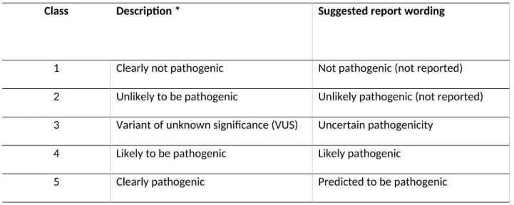
Table 2. Five ACGS variant pathogenicity classes
Class Description * Suggested report wording
1 Clearly not pathogenic Not pathogenic (not reported)
2 Unlikely to be pathogenic Unlikely pathogenic (not reported) 3 Variant of unknown significance (VUS) Uncertain pathogenicity
4 Likely to be pathogenic Likely pathogenic
5 Clearly pathogenic Predicted to be pathogenic
Table 3. Classes and examples of evidence used to help determine predicted pathogenicity.
Evidence type Examples Web address
Population frequency
Exome Variant Server; EVS http://evs.gs.washington.edu/EVS/ Single nucleotide polymorphism database;
dbSNP
http://www.ncbi.nlm.nih.gov/snp/ Exome aggregation consortium; ExAC http://exac.broadinstitute.org/
1000 genomes project http://browser.1000genomes.org
Presence on LSDB Factor FVIII variant database http://www.factorviii-db.org/ CDC Hemophilia A Mutation Project
(CHAMP) and Hemophilia B Mutation Project (CHBMP)
http://www.cdc.gov/ncbddd/hemophilia/ champs.html
Hemobase HA, HB and VWD (Spanish registry)
http://www.hemobase.com/EN/ index.htm
Factor IX variant database http://www.factorix.org/
von Willebrand factor variant database https://grenada.lumc.nl/LOVD2/VWF/ home.php?select_db=VWF
Amino acid prediction
MutPred http://mutpred.mutdb.org/
Sorting intolerant from tolerant (SIFT) http://sift.jcvi.org/
PolyPhen-2 http://genetics.bwh.harvard.edu/pph2/
SNAP https://www.rostlab.org/services/snap/
MutationAssessor http://mutationassessor.org/
Splice site prediction
Human Splicing Finder http://www.umd.be/HSF3/
NNSplice http://omictools.com/nnsplice-tool
GeneSplicer http://www.cbcb.umd.edu/software/
GeneSplicer/gene_spl.shtml
MaxEntScan http://genes.mit.edu/burgelab/maxent/