R E S E A R C H
Open Access
Differential expression of interferon-induced
microRNAs in patients with chronic hepatitis C virus
infection treated with pegylated interferon alpha
Carolina Scagnolari
1*, Pompea Zingariello
2, Jacopo Vecchiet
2, Carla Selvaggi
1, Delia Racciatti
2, Gloria Taliani
3,
Elisabetta Riva
4, Eligio Pizzigallo
2, Guido Antonelli
1Abstract
There have been reports of in-vitro interferon (IFN)-mediated antiviral activity against the hepatitis C virus (HCV) through microRNAs (miRNAs). The main aim of this study was to evaluate the expression of several miRNAs (miR-1, miR-30, miR-128, miR-196, miR-296) in peripheral blood mononuclear cells (PBMCs) from healthy individuals after in vitro IFN-treatment and in PBMCs from patients with chronic hepatitis C (CHC) before and 12 hours after the first injection of pegylated IFN alpha. We demonstrated that expression of these miRNAs could be recorded in PBMCs collected from healthy individuals before and after in-vitro IFN alpha treatment. Our analysis revealed that the levels of expression of all miRNAs investigated in patients with CHC were different to those in healthy individuals. When levels of the miRNAs were measured 12 hours after the first IFN injection, increases in expression levels of IFN-induced miRNAs were observed in 25-50% of patients, depending on the type of miRNA examined. No correla-tions were observed between HCV viral load, alanine aminotransferase status and expression of miRNA. Together these findings suggest that: (i) IFN alpha in-vitro treatment of PBMCs leads to a transcriptional induction of all miR-NAs investigated; (ii) miRmiR-NAs can be induced differentially by IFN treatment in patients with HCV. Given the impor-tance of miRNAs in defending the host against virus infections, it is possible that IFN-induced miRNAs may represent an important determinant of the clinical outcome of IFN therapy in HCV infection.
Introduction
MicroRNAs (miRNAs) are an important class of small non-coding RNA molecules that have recently come to prominence as critical regulators in a wide array of mechanisms of cell physiology. There is increasing evi-dence that miRNAs may also have an important func-tion in viral replicafunc-tion and may be used by host cells to control viral infection [1,2]. Indeed, it has been demon-strated that viral RNAs and the miRNA machinery may interact in various ways. First, mammalian viruses encode miRNAs that can act on both the control of viral genes and of cellular genes by repressing their expression. Second, cellular miRNAs may recognize viral RNAs and silence them, or control the expression of a cellular protein necessary for the virus life cycle.
It has also been suggested that miRNAs may be an effector in the classical vertebrate innate immune system [3], and recently an even more direct link between IFN and miRNAs has emerged [4]. Interferon (IFN) beta has been reported as modulating the expression of several cellular miRNAs that are capable of inhibiting hepatitis C virus (HCV) replication and infection, because they have sequence-predicted targets within the HCV geno-mic RNA. In addition, Pederson and co-authors reported that IFN beta downregulated the expression of miR-122, which has been implicated in the control of HCV RNA replication. This finding could lead to a bet-ter understanding of the factors involved in the failure of IFN therapy in patients with chronic hepatitis C (CHC). Due to different viral, environmental and host factors, a sustained virological response is achieved in about 50% of patients infected with HCV genotype 1 and in about 80% of patients infected with HCV geno-types 2 or 3; more importantly, despite extensive * Correspondence: [email protected]
1
Department of Molecular Medicine, Laboratory of Virology,“Sapienza” University of Rome; Rome, Italy
Full list of author information is available at the end of the article
examination of the biological and clinical effects of IFN in patients with CHC, the prediction of treatment responses in individual patients still remains difficult [5,6].
In the framework of a study aimed at further charac-terizing the state of responder, and at improving our knowledge and understanding of IFN therapy effects on patients with CHC, we undertook in-vitro and ex-vivo expression analyses of cellular miRNAs that had pre-viously been reported as being involved in IFN-mediated antiviral activity against HCV [4], using real-time quan-titative reverse transcription polymerase chain reaction
(RT-PCR) assay. The ex-vivoanalysis was undertaken
before and 12 hours after the first injection of pegylated IFN alpha in CHC patients. Gene expression analysis of MxA, a well-characterized IFN type I gene, was also undertaken as a control. The association between miRNA expression and alanine aminotransferase (ALT) status, HCV genotype, HCV-RNA and response to ther-apy was evaluated.
Methods
Patients and healthy blood donors
Peripheral blood samples were obtained from 12 patients with hepatitis C and ten healthy volunteers. The patients with HCV were treated by subcutaneous
injection with either 180 μg PegIFN alpha-2a
(PEGASYS; Hoffmann-LaRoche, Basel, Switzerland) (n = 9) or 1.5μg/kg PegIFN alpha-2b (PegIntron; Schering-Plough, Kenilworth, NJ, USA) (n = 3) plus ribavirin. Treatment duration was 24 or 48 weeks according to HCV genotype. Patients who were HCV-RNA negative after 24 weeks of post-treatment follow-up were consid-ered sustained viral responders. The demographic and clinical data of patients at the time of sample collection are summarized in Table 1. None of the patients had been treated previously with IFNs or other immunosup-pressive therapy (treatment-naïve patients). Written informed consent was obtained from each patient, and the study was approved by the Ethics Committees and/ or Institutional Review Boards of the participating insti-tutions. PBMCs from healthy donors were treated with 100 international unit (IU)/ml of IFN alpha [leukocyte, Alfaferone (AlfaWassermann, Bologna, Italy)] for 20 hours, the incubation time selected in previous studies aimed at the measurement of IFN-stimulated genes (ISGs) [7,8].
PBMCs from CHC patients were collected at baseline and 12 hours after the first injection of pegylated IFN alpha. The timing was determined by the following: first, only two sample collections (i.e., pre- and post-dose) were considered to be suitable by the Ethics Com-mittee; second, previous reports had shown significant changes in ISGs expression 12 hours after IFN type I
administration in patients with different chronic dis-eases, including CHC [9-14].
Blood sampling
Venous peripheral blood from each patient and healthy control was drawn into tubes containing ethylenediami-netetraacetic acid. Peripheral blood mononuclear cells (PBMCs) were separated using Ficoll-Hypaque gradient sedimentation; 5 × 10⁶ PBMCs were collected, pelleted and frozen at -80°C until examined. After centrifuging, plasma samples were stored at -80°C until required.
Taqman quantitative RT-PCR for MxA-mRNA
MxA gene transcripts in PBMCs from patients with CHC and healthy individuals were quantified by a real
time 5’ exonuclease RT-PCR Taqman assay using an
ABI 7000 sequence detector (Applied Biosystems, Monza, Italy). Briefly, the total cellular RNA was extracted from cells using the Trizol reagent, following the manufacturer’s instructions, and was retrotran-scribed as previously deretrotran-scribed [15]. Next the following primer pair and probe for MxA were added to the uni-versal PCR master mix (Applied Biosystems) at 300 and 100 nM, respectively, in a final volume of 50 mL.
(for-ward primer, 5’-CTGCCTGGCAGAAAACTTACC-3’;
reverse primer, 5’-CTCTGTTATTCTCTGGTGAGTCT
CCTT-3’; probe, 5’CATCACACATATCTGTAAATCTC TGCCCCTGTTAGA-3’). Co-amplification of the beta-glucuronidase gene (Assay-On-Demand, Hs99999908_mL, Applied Biosystems) was used to normalize the amount of total RNA present. The relative amount of each transcript, normalized to beta-glucuronidase mRNA, was calculated using the arithmetic formula (2 -ΔCt) or (2 -ΔΔCt) according to the supplier’s guidelines (Applied Biosystems).
Taqman quantitative RT-PCR for microRNAs
MicroRNAs (1, 30, 128, 196, miR-296) in PBMCs collected from patients with CHC and healthy individuals were quantified by a real time 5’ exo-nuclease RT-PCR Taqman assay.
All primer and probes of each miRNA investigated were present in the TaqMan microRNA assays pur-chased from Applied Biosystems.
MiRNAs were extracted from the cells using the mir-Vana miRNA Isolation Kit (Ambion, Austin, TX), according to the manufacturer’s protocol. Applied Bio-systems TaqMan MicroRNA Reverse Transcription Kit (Applied Biosystems, Monza, Italy) was used (following the manufacturer’s protocol) for reverse transcription (RT) of extracted total miRNAs. Each RT reaction con-tained 5 ng of extracted total miRNA, 3 μL of TaqMan
MicroRNA assays, 1.50 μL of RT10x buffer, 0.25 mM
transcriptase and 0.25 U/μL RNase inhibitor. The 15μL reactions were incubated in a Biometra T3 Thermocycler (MMedical, Italy) in a 96-well plate for 30 minutes at 16°C, 30 minutes at 42°C, 5 minutes at 85°C, and then held at 4°C. For the real-time PCR step, amplification was carried out using TaqMan MicroRNA assays (Applied Biosystems) on the Applied Biosystems 7000 Real-Time PCR system. The 20μL reaction included 1.33 μL RT product, 10μL of TaqMan Universal PCR Master
Mix with no UNG and 1 μL of TaqMan MicroRNA
assays. The reactions were incubated in a 96-well optical plate at 95°C for 10 minutes, following by 40 cycles of 95° C for 15 s and 60°C for 1 minute. Real-time PCRs for each miRNA were run in triplicate. The relative expres-sion levels of each miRNA were measured using the con-stitutively expressed RNU6B as endogenous control The expression of each miRNA relative to RNU6B was deter-mined using the arithmetic formula (2 - ΔCt) or (2
-ΔΔCt) according to the supplier’s guidelines (Applied Biosystems).
Statistical analysis
All results are expressed as the mean ± standard devia-tion (median). The coefficient of variadevia-tion (CV) was used to measure the interpatient variability in blood concentrations of miRNAs and MxA. Levels of miRNAs and MxA observed in PBMCs from three healthy indivi-duals before and after the stimulation in vitrowith IFN alpha were compared using a T-test as suggested by Bland and Altman [16]. Differences between patients with CHC and healthy individuals, and between patient groups, in terms of blood concentrations in miRNAs and MxA, were compared using the Wilcoxon test. The same test was used to assess differences between miR-NAs and MxA expression levels in patients with CHC.
A Spearmen rho coefficient was calculated to assess the correlation between pre-dose and HCV viral load, ALT status. Significance was fixed at the 5% level. Analysis was performed using spss version 13.0 for Windows.
Results
Baseline and in vitro IFN alpha induced expression of microRNAs in PBMCs collected from healthy individuals As there are no published reports about expression pro-files of miR-1, miR-30, miR-128, miR-196, and miR-296 in PBMCs from normal individuals, our first investiga-tion was of their expression in PBMCs from healthy volunteers using real-time quantitative RT-PCR. The expression of MxA in these individuals was also evalu-ated for control purposes.
PBMCs isolated from healthy donors were found to express all the miRNA considered with varying expression levels, depending on the examined miRNA type. Specifi-cally, the baseline miRNA values in PBMCs that were determined using the equation (2 -ΔCt), according to the supplier’s guidelines, ranged between 0.30 and 128.96. MxA-mRNA levels were also found in PBMCs from all healthy donors (Table 2).
We then examined whether leukocyte IFN alpha could stimulate in-vitro expression of the miRNAs listed above as previously reported for IFN beta. PBMCs, freshly iso-lated from three healthy individuals, were treated in
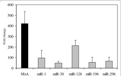
vitro with IFN alpha at 100 IU/ml (leukocyte, Alfafer-one), and levels of miRNA and MxA-mRNA were mea-sured 20 hours later by quantitative real-time RT-PCR. Again, levels of MxA transcripts were measured as posi-tive controls for IFN action. The results showed that IFN alpha in-vitro treatment of PBMCs leads to a tran-scriptional induction of all miRNAs investigated as well as MxA-mRNA (Figure 1). In particular, of the miRNAs
Table 1 Demographic and clinical characteristics of patients with chronic hepatitis C
Patient
8 M 47 2a/2c 3347068 445000 NR 937560 NR 1000 80 Alfa 2b 93 172 NR
9 M 49 2a 2859813 437000 NR 378000 NR 1000 84 Alfa 2b 110 115 NR
10 F 44 1a 2505703 17500 NR 327000 NR 1200 68 Alfa 2a 23 26 NR
11 F 66 1b 21967894 1200000 NR 2550 NR 800 51 Alfa 2a 44 80 NR
12 F 42 1a 1469947 161000 NR 16100 NR 1000 72 Alfa 2b 73 110 NR
detected 20 hours post treatment, miR-1 and miR-128 had increased the most relative to untreated PBMCs, whereas miRNA-30 had increased the least.
Expression of microRNAs in PBMCs collected from patients with CHC before and after the first injection of IFN alpha
Having established that a baseline expression of miR-1, miR-30, miR-128, miR-196 and miR-296 could be recorded in PBMCs collected from healthy donors before and after in-vitro IFN alpha treatment, we decided to analyse the expression of the same miRNAs, as well as MxA, in 12 patients with CHC, of whom 7 were classified as responders and five as non-responders to Peg-IFN alpha plus ribavirin therapy. Blood samples
were collected before and 12 hours after the first Peg-IFN alpha administration.
Patients with CHC expressed baseline levels of all examined miRNAs but the levels were highly variable (CV > 100%). Importantly, the levels of expression of miRNAs were different for patients with CHC compared with healthy controls. There were higher levels of almost all miRNAs in patients with CHC compared with healthy individuals with the exception of miR-196 (Table 2). The differences did not reach statistical signif-icance, probably because of the low number of patients and the wide variability in miRNA expression observed in them. As expected, the same trend was also observed for MxA.
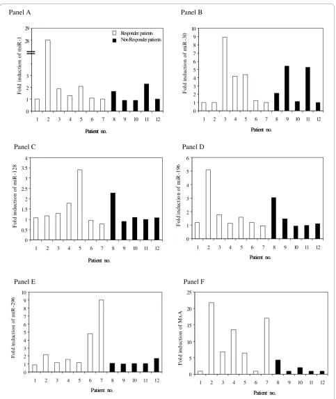
We then examined the expression of IFN-induced miRNAs and MxA in patients with CHC after the first injection of IFN alpha. The results are shown in Figure 2. It can be seen that 12 hours after IFN alpha adminis-tration, a greater than 1.5-fold increase in MxA was recorded in 58% of patients with CHC, whereas IFN induction of miRNAs varied between 25% and 50%, depending on the type of miRNA examined. The great-est increase in miRNA and MxA levels after IFN injec-tion were observed independently in patients no. 2 1, miR-196 and MxA), no. 3 30), no. 5 (miR-128) and no.7 (miR-296).
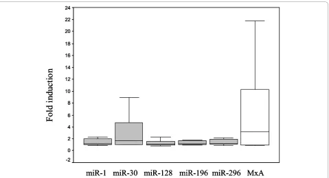
In addition, different increases after IFN treatment relative to baseline were observed for miR-1, miR-30, miR-296 and MxA (p < 0.05) (Figure 3).
The baseline levels of miRNAs were also analysed to determine whether the expression of these molecules could be associated with the clinical outcome of IFN therapy. The analyses showed that baseline levels of miRNAs were not significantly different between responders and non-responders (Table 3). However, a trend toward higher baseline expression of miR-296 was observed in non-responder compared with responder
Table 2 Baseline expression of microRNAs and MxA-mRNA in healthy controls and in patients with chronic hepatitis C (CHC)
Healthy controls* n = 10 Patients with CHC* n = 12 Mean ratio CHC/healthy controls
miR-1 0.30±1.43
(0.36)
2.82±5.09 (0.10)
9.4
miR-30 128.96±93.99
(128.61)
255.57±466.62 (27.41)
1.98
miR-128 1.39±3.31
(0.05)
19.54±36.15 (0.03)
14.05
miR-196 1.85±3.21
(0.73)
0.81±1.52 (0.04)
0.43
miR-296 3.39±7.12
(0.46)
* Data are expressed as mean ± standard deviation (median).
Figure 1Interferon (IFN) induced expression of microRNAs (miR-1, miR-30, miR-128, miR-196, miR-296) in peripheral blood mononuclear cells collected from three healthy individuals after in-vitro treatment with IFN alpha (100 international unit (IU)/ml). Expression of MxA-mRNA was also evaluated. Significant increases, relative to baseline, after in vitro IFN treatment were observed for miR-1,
patients. In contrast, miR-128 and miR-196 tended to be higher in responders than in non-responder patients.
Although we were unable to reach a definitive conclu-sion because there were too few patients, the overall expression of miRNA induced after IFN administration was observed to be no different in responders than in non-responder patients. In contrast, and as expected [17], responder patients were characterized by a higher induction of MxA after IFN administration compared with non-responders (p = 0.07).
We also analysed the baseline expression and the level of increase of miRNAs and MxA after IFN alpha treat-ment in relation to the HCV genotype, ALT levels and RNA viral load, but no significant association was found (data not shown).
Discussion
We have demonstrated for the first time that miRNAs, previously reported to be involved in IFN-mediated anti-viral activity against HCV, are expressed in PBMCs
Figure 3Fold induction of microRNAs (miR)-1, miR-30, miR-128, miR-196, miR-296 and MxA-mRNA in peripheral blood mononuclear cells collected from patients with chronic hepatitis C after the first injection of interferon. Significant increases, relative to baseline, after IFN treatment were observed for miR-1, miR-30, miR-296 and MxA-mRNA (p < 0.05 using Wilcoxon test).
Table 3 Baseline and IFN-induced expression of microRNAs and MxA-mRNA in patients with chronic hepatitis C according to the response to antiviral therapy (Peg-interferon (IFN) and ribavirin)
Baseline expression* Fold IFN induction*
Responders Non-responders Responders Non-responders
miR-1 2.61 ± 4.03
(0.52)
3.12 ± 6.84 (0.07)
5.26 ± 10.23 (1.29)
1.35 ± 0.60 (1.00)
miR-30 222.10 ± 446.32
(19.61)
302.42 ± 543.38 (31.52)
3.09 ± 2.97 (1.20)
2.99 ± 2.21 (2.12)
miR-128 31.25 ± 42.77
(0.04)
0.01 ± 0.01 (0.007)
1.48 ± 0.90 (1.15)
1.27 ± 0.57 (1.08)
miR-196 1.19 ± 1.78
(0.05)
0.05 ± 0.03 (0.04)
1.85 ± 1.46 (1.21)
1.51 ± 0.87 (1.12)
miR-296 0.77 ± 1.01
(0.31)
34.23 ± 68.12 (0.21)
2.96 ± 2.97 (1.56)
1.18 ± 0.29 (1.07)
MxA 9.96 ± 11.17
(7.16)
8.67 ± 11.09 (2.99)
9.68 ± 8.02 (6.86)
1.82 ± 1.49 (0.97)
collected from healthy individuals, and that their expres-sion in such cells may be induced by IFN-alpha to vary-ing degrees. Specifically, greater increases in miR-1 and miR-128 and a lower increase in miR-30 were recorded in IFN alpha-treated PBMCs. This is in agreement with Pedersen and co-authors, who observed a high-fold increase in miR-1 and a low-fold increase in miR-30 in experiments performed in vitrowith Huh7 cells treated with IFN beta [4]. However, the same authors also observed that, in primary hepatocytes, 1 and miR-30 expression increased at the same levels after IFN beta treatment. The differences in the levels of miRNAs induced after in-vitro IFN treatment and the Pedersen study may reflect the different sensitivities of each cell type to IFN action in terms of miRNA induction, as also reported by others [18,19].
Having established that PBMCs from healthy controls expressed the above miRNAs before and after IFN alpha treatment, the study then focused on evaluating whether PBMCs collected from patients with CHC expressed baseline levels of miRNAs and how IFN administration could modulate their expression. The results showed that PBMCs from patients with CHC had a trend towards greater expressions of these miRNAs compared with healthy controls, with the exception of miR-196. These findings are new but not surprising because, in agreement with earlier studies, they could indicated greater endogenous activation of IFN-induced pathways in patients with CHC than in healthy controls [20-23].
As far as the influence of baseline expression of these miRNAs on the clinical outcome of IFN therapy in patients with CHC is concerned, slight, although not significant, differences were observed between respon-ders and non-responrespon-ders. Several studies have shown that HCV-positive patients with elevated ISGs expres-sion tend to respond poorly to therapy compared with patients with low baseline expression [17,24-26]. The cause of these different responses to therapy is not understood. It can be speculated that patients with CHC who have elevated initial expression were refractory to further stimulation of ISGs by exogenous IFN. We observed in our previous study that there was an inverse correlation between the relative increase in IFN-induced biomarkers and their baseline levels in patients with CHC or multiple sclerosis [23]. However, in this study, no inverse correlation was found between baseline expression of miRNAs and their levels after IFN induc-tion (data not shown). Moreover, although IFN alpha was seen to induce changes in miRNA expression in patients with CHC, and that the highest increase in each miRNA was seen only in responder patients, no significant differences were found in the expression levels of IFN-induced miRNAs between responders and non-responders, and HCV-RNA levels appeared to have
no influence on the baseline expression of IFN-induced miRNAs.
It is reasonable, therefore, to speculate that IFN treat-ment as well as HCV infection could affect the expres-sion of these miRNAs in the liver more than in the PBMCs. In this regard, it has recently been shown that patients with CHC who had no virological response dur-ing later IFN therapy had markedly low baseline levels of miR-122, whereas only limited changes were seen for the other miRNAs investigated [27]. However, the authors did not compare the expression of IFN-induced miRNAs in the liver and in the PBMCs collected from the same patients with CHC undergoing IFN treatment. In addition, Meier and co-authors reported recently that while IFN-alpha treatment led to the induction of type I IFN regulated genes in PBMCs, such an induction appeared not to occur in the livers of patients with hepatitis C, which suggests that the mechanism by which IFN-alpha treatment causes viral clearance might be independent of hepatic activation of type I IFN regu-lated genes [28]. All this indicates more clearly the com-plexities of the analysed phenomena and the difficulties in interpreting our data. A better understanding of the regulation of HCV-specific miRNA induction both in the liver and in PBMCs is required to shed light on these important and critical issues. Unfortunately, we consider that no firm conclusions can be drawn with regard to the relationship between baseline or IFN-induced miRNA expression and the clinical outcome of IFN alpha therapy in patients with CHC.
additional results, as suggested by Sarasin-filipowicz and co-authors [30].
This study extends previous investigations into the activation of the IFN system in patients with CHC and, specifically, the ability of type I IFN to regulate miRNA expression [4,27]. In particular, we have demonstrated that IFN alpha in-vitro treatment of PBMCs leads to transcriptional induction of miRNAs. We have also demonstrated, for the first time to our knowledge, that miRNA expression could be measured in PBMCs col-lected from patients with CHC both before and after IFN alpha administration. Larger longitudinal studies are required to gain a better understanding of the activation of IFN-induced miRNAs in patients affected by CHC.
Acknowledgements
This work was supported by grants to GA from“Sapienza”University (Projects“Ateneo Federato”); PRIN 2008 (number 20085JWPK3) and Founds of Research ex 60% from G. d’Annunzio University, School of Medicine, Chieti, 2008-09.
Author details
1Department of Molecular Medicine, Laboratory of Virology,“Sapienza” University of Rome; Rome, Italy.2Department of Medicine and Science of Aging, Infectious Disease Clinic, G. d’Annunzio University, School of Medicine, Chieti, Italy.3Department of Infectious and Tropical Diseases,
“Sapienza”University of Rome, Rome, Italy.4Virology section, University Campus Bio-Medico, Rome, Italy.
Authors’contributions
CS was responsible for design of the study, execution of the Taqman experiments, performing data analysis and writing the manuscript; PZ was responsible for performing selection of patients with CHC and analysing of clinical data; JV was responsible for performing selection of patients with CHC and analysis of clinical data; CS was responsible for executing the TaqMan experiments and analysis the HCV-positive patient data, DR was responsible for performing selection of patients with CHC and analysing of clinical data; GT was responsible for analysis of the data and revising of the manuscript; ER was responsible for helping into the design of study and analysis of the miRNAs data; EP was responsible for selection of patients with CHC, analysis of clinical data, revising of the manuscript and grants owner; GA was responsible for helping into the design of the study, writing of the manuscript, and grants owner. All authors read and approved the final manuscript.
Competing interests
The authors declare that they have no competing interests.
Received: 22 June 2010 Accepted: 12 November 2010 Published: 12 November 2010
References
1. Corbeau P:Interfering RNA and HIV: reciprocal interferences.PLoS Pathog 2008,4:e1000162.
2. Grassmann R, Jeang KT:The roles of microRNAs in mammalian virus infection.Biochim Biophys Acta2008,1779:706-711.
3. Obbard DJ, Gordon KH, Buck AH, Jiggins FM:The evolution of RNAi as a defence against viruses and transposable elements.Philos Trans R Soc Lond B Biol Sci2009,364:99-115.
4. Pedersen IM, Cheng G, Wieland S, Volinia S, Croce CM, Chisari FV, David M:
Interferon modulation of cellular microRNAs as an antiviral mechanism. Nature2007,449:919-922.
5. Hadziyannis SJ, Koskinas JS:Differences in epidemiology, liver disease and treatment response among HCV genotypes.Hepatol Res2004,29:129-135.
6. Tai AW, Chung RT:Treatment failure in hepatitis C: mechanisms of non-response.J Hepatol2009,50:412-420.
7. Fernández M, Quiroga JA, Martín J, Herrero M, Pardo M, Horisberger MA, Carreño V:In vivo and in vitro induction of MxA protein in peripheral blood mononuclear cells from patients chronically infected with hepatitis C virus.J Infect Dis1999,180:262-267.
8. Erickson AK, Seiwert S, Gale M Jr:Antiviral potency analysis and functional comparison of consensus interferon, interferon-alpha2a and pegylated interferon-alpha2b against hepatitis C virus infection.Antivir Ther2008,
13:851-862.
9. Pawlotsky JM, Hovanessian AG, Roudot-Thoraval F, Robert N, Bouvier M, Babany G, Duval J, Dhumeaux D:Effect of alpha interferon (IFN-alpha) on 2’-5’oligoadenylate synthetase activity in peripheral blood mononuclear cells of patients with chronic hepatitis C: relationship to the antiviral effect of IFN-alpha.Antimicrob Agents Chemother1996,40:320-324. 10. Pawlotsky JM, Hovanessian A, Roudot-Thoraval F, Lebon P, Robert N,
Bouvier M, Babany G, Duval J, Dhumeaux D:Activity of the interferon-induced 2’,5’-oligoadenylate synthetase in patients with chronic hepatitis C.J Interferon Cytokine Res1995,15:857-862.
11. Chung RT, Gale M Jr, Polyak SJ, Lemon SM, Liang TJ, Hoofnagle JH:
Mechanisms of action of interferon and ribavirin in chronic hepatitis C: Summary of a workshop.Hepatology2008,47:306-320.
12. Hoffmann AL, Krumbholz M, Faber H, Kuempfel T, Starck M, Pöllmann W, Meinl E, Hohlfeld R:Multiple sclerosis: Relating MxA transcription to anti-interferon-β-neutralizing antibodies.Neurology2007,68:958-959. 13. Imam H, Janson ET, Gobl A, Alm G, Oberg K:Induction of MxA mRNA in
patients with neuroendocrine tumors after interferon treatment. Lack of correlation with antitumor response.Anticancer Res1995,15:2191-2195. 14. Feld JJ, Lutchman GA, Heller T, Hara K, Pfeiffer JK, Leff RD, Meek C,
Rivera M, Ko M, Koh C, Rotman Y, Ghany MG, Haynes-Williams V, Neumann AU, Liang TJ, Hoofnagle JH:Ribavirin improves early responses to peginterferon through improved interferon signalling.Gastroenterology 2010,139:154-162.
15. Scagnolari C, Selvaggi C, Chiavuzzo L, Carbone T, Zaffiri L, d’Ettorre G, Girardi E, Turrizziani O, Vullo V, Antonelli G:Expression levels of TLRs involved in viral recognition in PBMCs from HIV-1-infected patients failing antiretroviral therapy.Intervirology2009,52:107-114.
16. Bland JM, Altman DG:Analysis of continuous data from small samples. BMJ2009,338:a3166.
17. Antonelli G, Simeoni E, Turriziani O, Tesoro R, Redaelli A, Roffi L, Antonelli L, Pistello M, Dianzani F:Correlation of interferon-induced expression of MxA mRNA in peripheral blood mononuclear cells with the response of patients with chronic active hepatitis C to IFN-alpha therapy.J Interferon Cytokine Res1999,19:243-251.
18. Hubbell HR, Liu RS, Maxwell BL:Independent sensitivity of human tumor cell lines to interferon and double-stranded RNA.Cancer Res1984,
44:3252-3257.
19. Roos G, Leanderson T, Lundgren E:Interferon-induced cell cycle changes in human hematopoietic cell lines and fresh leukemic cells.Cancer Res 1984,44:2358-2362.
20. Patzwahl R, Meier V, Ramadori G, Mihm S:Enhanced expression of interferon-regulated genes in the liver of patients with chronic hepatitis C virus infection: detection by suppression-subtractive hybridization.J Virol2001,75:1332-1138.
21. Ji X, Cheung R, Cooper S, Li Q, Greenberg HB, He XS:Interferon alpha regulated gene expression in patients initiating interferon treatment for chronic Hepatitis C.Hepatology2003,37:610-621.
22. MacQuillan GC, Mamotte C, Reed WD, Jeffrey GP, Allan JE:Upregulation of endogenous intrahepatic interferon stimulated genes during chronic hepatitis C virus infection.J Med Virol2003,70:219-227.
23. Scagnolari C, Bellomi F, Trombetti S, Casato M, Carlesimo M, Bagnato F, Lavolpe V, Bruno R, Millefiorini E, Antonelli L, Girardi E, Turriziani O, Antonelli G:Expression of biomarkers of interferon type I in patients suffering from chronic diseases.Clin Exp Immunol2007,147:270-276. 24. MacQuillan GC, de Boer WB, Platten MA, McCaul KA, Reed WD, Jeffrey GP,
Allan JE:Intrahepatic MxA and PKR protein expression in chronic hepatitis C virus infection.J Med Virol2002,68:197-205.
25. Giannelli G, Guadagnino G, Dentico P, Antonelli G, Antonaci S:MxA and PKR expression in chronic hepatitis C.J Interferon Cytokine Res2004,
26. Gerotto M, Dal Pero F, Bortoletto G, Realdon S, Ferrari A, Boccato S, Alberti A:PKR gene expression and response to pegylated interferon plus ribavirin therapy in chronic hepatitis C.Antivir Ther2004,9:763-770. 27. Sarasin-Filipowicz M, Krol J, Markiewicz I, Heim MH, Filipowicz W:
Decreased levels of microRNA miR-122 in individuals with hepatitis C responding poorly to interferon therapy.Nat Med2009,15:31-33. 28. Meier V, Mihm S, Ramadori G:Interferon-alpha therapy does not
modulate hepatic expression of classical type I interferon inducible genes.J Med Virol2008,80:1912-1918.
29. Taylor MW, Tsukahara T, Brodsky L, Schaley J, Sanda C, Stephens MJ, McClintick JN, Edenberg HJ, Li L, Tavis JE, Howell C, Belle SH:Changes in gene expression during pegylated interferon and ribavirin therapy of chronic hepatitis C virus distinguish responders from nonresponders to antiviral therapy.J Virol2007,81:3391-3401.
30. Sarasin-Filipowicz M, Oakeley EJ, Duong FH, Christen V, Terracciano L, Filipowicz W, Heim MH:Interferon signaling and treatment outcome in chronic hepatitis C.Hepatology2008,48:1330-1333.
doi:10.1186/1743-422X-7-311
Cite this article as:Scagnolariet al.:Differential expression of interferon-induced microRNAs in patients with chronic hepatitis C virus infection treated with pegylated interferon alpha.Virology Journal20107:311.
Submit your next manuscript to BioMed Central and take full advantage of:
• Convenient online submission
• Thorough peer review
• No space constraints or color figure charges
• Immediate publication on acceptance
• Inclusion in PubMed, CAS, Scopus and Google Scholar • Research which is freely available for redistribution