R E S E A R C H
Open Access
New insights regarding HCV-NS5A structure/
function and indication of genotypic differences
Lilian HT Yamasaki
1,4*, Helen A Arcuri
2, Ana Carolina G Jardim
1, Cintia Bittar
1, Isabel Maria VG de Carvalho-Mello
3and Paula Rahal
1Abstract
Background:HCV is prevalent throughout the world. It is a major cause of chronic liver disease. There is no effective vaccine and the most common therapy, based on Peginterferon, has a success rate of ~50%. The mechanisms underlying viral resistance have not been elucidated but it has been suggested that both host and virus contribute to therapy outcome. Non-structural 5A (NS5A) protein, a critical virus component, is involved in cellular and viral processes.
Methods:The present study analyzed structural and functional features of 345 sequences of HCV-NS5A genotypes 1 or 3, usingin silicotools.
Results:There was residue type composition and secondary structure differences between the genotypes. In addition, second structural variance were statistical different for each response group in genotype 3. A motif search indicated conserved glycosylation, phosphorylation and myristoylation sites that could be important in structural stabilization and function. Furthermore, a highly conserved integrin ligation site was identified, and could be linked to nuclear forms of NS5A. ProtFun indicated NS5A to have diverse enzymatic and nonenzymatic activities,
participating in a great range of cell functions, with statistical difference between genotypes.
Conclusion:This study presents new insights into the HCV-NS5A. It is the first study that using bioinformatics tools, suggests differences between genotypes and response to therapy that can be related to NS5A protein features. Therefore, it emphasizes the importance of using bioinformatics tools in viral studies. Data acquired herein will aid in clarifying the structure/function of this protein and in the development of antiviral agents.
Keywords:Hepatitis C virus, Non-structural 5A protein, Bioinformatics, Genotype, Quasispecies, IFN response
Introduction
Hepatitis C is a major health problem; it is highly preva-lent worldwide and has a high probability of persistence [1,2]. Chronic persistence can result in cirrhosis and hepatocellular carcinoma [3,4]. Hepatitis C virus (HCV) is member of the Flaviviridae family within the Hepaci-virus genus, although many of its features are distinct from other family members including the structural organization of the protein and the 5’-cap independent translation [5].
On the basis of viral variability, HCV is classified into seven genotypes and more than 50 subtypes [6]. In
addition, an infected patient will harbour different but related viral genomes known as quasispecies. This high variability can be explained by a combination of three factors: (1) viral RNA polymerase acts without proof-reading [7]; (2) HCV has co-evolved with human popu-lations for millions of years [8]; (3) the viral life cycle is fast, resulting in the production of approximately 1.3 × 10¹² virions per patient per day [9].
The HCV RNA genome translates a polyprotein that is cleaved by viral and host proteases to generate ten structural and non-structural proteins [10-12]. Among the non-structural proteins, NS5A is a phosphoprotein critical for the HCV life cycle. It is composed of approximately 447 amino acids and may participate in viral RNA replication, modulation of cell signaling path-ways, interferon response, pathogenesis and apoptosis
* Correspondence: [email protected] 1
Department of Biology, Sao Paulo State University–UNESP, Sao Jose do Rio Preto, SP, Brazil
Full list of author information is available at the end of the article
regulation. Its enzymatic functions and its complete structure have yet to be elucidated. However, evidence suggests that it functions through interaction with other HCV and host cell proteins [13-16].
NS5A is divided into three domains [17,18]. Domain I contains a membrane binding domain [19,20] and a zinc finger domain that are essential for HCV replication [17]. Domain II and III are naturally unstructured, per-forming function by interacting with several proteins [15,21].
The importance of NS5A protein in disease caused by hepatitis C is unquestioned. However, difficulties with experimental methods used to determine the structures of highly flexible proteins have resulted in a poor under-standing of the overall structure and functions of NS5A. Such difficulties have led to the development of bioin-formatic tools that are helpful in obtaining reliable data for these types of proteins. Ab initio tools are also important for studying proteins with low or no homol-ogy, and can be used to compare them with experimen-tally determined structures.
In the present study, complete sequences from HCV NS5A genotypes 1 and 3 were analyzed. These sequences were obtained from Brazilian patients who showed different responses to Peginterferon (PegIFN) therapy. Using these sequences, the aims were to ana-lyze structural and functional features. The knowledge obtained should aid in the design of new drugs and vac-cines, and in developing other resources to improve HCV therapy.
Results
Amino acid composition and secondary structure of NS5A
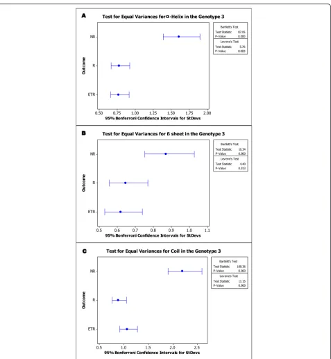
Therapy response did not differ according to the amino acids composition or the secondary structure type com-position. However, considering the genotypes, the aver-age percentaver-ages of alanine, glutamic acid, glutamine and tyrosine present in the NS5A protein were different between the genotypes 1a, 1b and 3. The average percen-tages of cysteine, valine and threonine differed between genotype 1 and 3. All sequences obtained from genotype 1b presented with 2% tyrosine (data not shown). Second-ary structure analysis demonstrated that distribution of each secondary structure type followed a normal distri-bution. Statistical analysis (t-test) suggests that the three secondary arrangements are high significant factors (p -value < .001) to differ the genotypes. Composition of helix, sheet or coil in percentage did not result in signifi-cant difference when we compared these three arrange-ments the therapy outcome responses (Figure 1A-C). In contrast, when we compared the variation of these com-positions among the response groups, there is a great (and statistical) difference, especially in genotype 3. Test for equal variances (Bartlett’s test and Levene’s test)
resulted in different variation behaviour comparing the three outcome responses sequences in genotype 3 (Figure 2A-C). The same test applied for analysis intra-response group indicates that even when sequences are from dif-ferent patients, if they are from the same therapy out-come group, they are not significantly different (p> .05) (Table 1).
Transmembrane regions and pattern/motif search
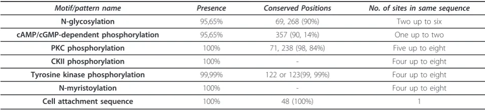
MEMSAT3 analysis demonstrated that all NS5A and reference sequences contained a possible transmem-brane region between residues 32 and 51, and Prosite recognized seven functional patterns in each NS5A sequence. No relationship was observed between therapy response or genotype and pattern number or motif loca-tion. Table 2 presents a summary of each conserved pat-tern/motif encountered in the NS5A analysis. Between two and seven N-glycosylation sites were present within the same sequence. In two positions (69 and 268), this pattern was conserved in 90% of sequences; this motif was absent at these positions in 4% of cases. cAMP- and cGMP-dependent protein kinase phosphorylation sites were identified; 90% of sequences had a minimum of one such motif at position 357, but 4% of sequences from one patient lacked this motif. Protein kinase C (PKC) phosphorylation sites were present in all sequences at positions 71 and 238. The number of these sites in the same sequence ranged from five to eight. Casein kinase II phosphorylation sites were recognized in non-conserved positions, being present between 4 and 8 times in the same sequence. Tyrosine kinase phosphorylation sites were identified in a conserved position (residue 122 or 123). N-myristoylation sites were present in various numbers and several positions, with NS5A sequences possessing between four and eight of these motifs in the same sequence. A cell attachment sequence (RGD sequence) was present in all sequences, conserved in number and position; NS5A sequences possessed this motif at position 48. Figure 3 summarizes the results of the MEMSAT3 and Prosite analyses in the reference sequence AF009606.
Prediction of functional features
The results from NS5A domain I suggested that this region is related to the functional category of energy metabolism, with enzymatic activity from unknown class. For domain I, prediction of gene ontology category
varied among transcription regulation, growth factor, immune response, transcription and none of these cate-gories. To calculate the chi-square, categories below 5 sequences were excluded. Significant difference was
observed between genotypes but not between therapy response groups, with statistical power of 0.999.
Prediction for domain II sequences varied in func-tional category, enzymatic function and gene ontology category. Resulted functional category includes energy metabolism and translation, with statistical difference
between therapy outcome groups (except between non-responders–NR and end of therapy responders- ETR groups) and genotype. Enzymatic function prediction and gene ontology (GO) category prediction had signifi-cant difference between genotypes, with statistical power of 0.801 and 1, respectively.
(75 5 15
2X
WF
R
P
H
%RQIHUURQL&RQILGHQFH,QWHUYDOVIRU6W'HYV
7HVW6WDWLVWLF
39DOXH
7HVW6WDWLVWLF
39DOXH
%DUWOHWWV7HVW
/HYHQHV7HVW 7HVWIRU(TXDO9DULDQFHVIRUD+HOL[LQWKH*HQRW\SH
(75 5 15
2X
WF
R
P
H
%RQIHUURQL&RQILGHQFH,QWHUYDOVIRU6W'HYV
7HVW6WDWLVWLF
39DOXH
7HVW6WDWLVWLF
39DOXH
%DUWOHWWV7HVW
/HYHQHV7HVW 7HVWIRU(TXDO9DULDQFHVIRUVKHHWLQWKH*HQRW\SH
(75 5 15
2X
WF
R
P
H
%RQIHUURQL&RQILGHQFH,QWHUYDOVIRU6W'HYV
7HVW6WDWLVWLF
39DOXH
7HVW6WDWLVWLF
39DOXH
%DUWOHWWV7HVW
/HYHQHV7HVW 7HVWIRU(TXDO9DULDQFHVIRU&RLOLQWKH*HQRW\SH
Į
A
B
C
For domain III analysis, all the sequences were pre-dicted to be similar to proteins related to transport and binding, with nonenzyme function. The gene ontology category varied among growth factor, immune response, stress response, hormone, voltage-gated ion channel and unknown. These differences were significant different between end of therapy responders (ETR) sequences and the other two outcome groups (non-responders– NR, responders–R) and between genotypes. Statistical power values were 0.6 and 1, respectively.
Discussion
Infection with genotype 1 results in lower therapy suc-cess rates than other genotypes [22]. No previous study was found connecting NS5A amino acids composition or secondary structure type (Figure 1, 2 and Table 1) to this difference in response rate. The present study sug-gests that these two characteristics present genotypic differences. These differences could affect NS5A func-tions, by modifying its interactions with other viral and host proteins, or its stability. Consequently, these differ-ences could affect viral resistance, replication and other properties linked to NS5A that differ among genotypes.
Indeed, some observations of the viral genotype speci-fic features are reported between genotype 1 and 2. Viral dynamics was the first property detected, in a study that collected viral load data in patients receiving
IFN therapy. Viral kinetics was greatly different. In gen-otype 1 infected patients, IFN effectiveness, free virion clearance rate and cell death rate were lower than geno-type 2 HCV hosts. In contrast, percentage of individuals that reached an undetectable level during 14 days of therapy was higher in genotype 2 infected individuals [23]. Our group also foundin vivoindication of diver-gences in NS5A quasispecies composition and muta-tional profiles between genotypes 1 and 3 in baseline specimens [24,25]. Using the sequences derived from these studies, we showed that there is indication of structural and functional differences between NS5A-1a/ b and NS5A-3 (Table 3). In addition, at least for geno-type 3, there were differences in variance of structure between the different responses groups (Figure 2). This divergence is not observed when we compare the sequences extracted from patients with the same therapy response. Possibly resulting from the structural differ-ences, the functional prediction profile was also different between genotypes.
In vivoand derivedin silicoresults point to a relation-ship between NS5A and genotypic IFN response rate. However,in vitroresearches results are still controversial. In 2008, it was reported that cells infected by 2a-NS5A-containing replicons presented lower degree of IFN antag-onism than the counterpart containing 1b-NS5A. The same study also found suggestions that the V3 domain and the C-terminal region of the NS5A are related to IFN differential reaction [26]. Other study in 2010 did not reached similar conclusions. In this case, cells expressing 1b and 2a NS5A protein presented analogous capability of IFN responses inhibition and IL-8 expression induction. The author suggests that other viral factors and/or host factors may be involved in the genotypic difference of HCV [27]. Posterior, it was also described that HCV with recombinant NS5A from different genotypes presents dif-ferent sensibility to NS5A inhibitors, but not for IFN [28]. These hypothesis does support our group results, since it seems that NS5A has still numerous unknown direct and indirect interactions with host and viral proteins, conse-quence from the high structural flexibility observed in domains II and III of the protein [15,21].
Table 1 Statistical results (t-test) for comparison between genotype 1 and 3 secondary structure composition
Genotype No. of sequences (n) Mean StDev SE Mean p-value a-helix
1 109 18.06 0.52 0.05 0.00
3 143 17.51 0.41 0.03
b-sheet
1 109 19.58 0.27 0.03 0.00
3 143 18.61 0.33 0.03
Coil
1 109 62.35 0.52 0.05 0.00
3 143 63.88 0.38 0.03
StDev: Standard Deviation,SE: Standard Error
Table 2 Conservation of patterns/motifs predicted on NS5A, described in percentage of sequences
Motif/pattern name Presence Conserved Positions No. of sites in same sequence
N-glycosylation 95,65% 69, 268 (90%) Two up to six
cAMP/cGMP-dependent phosphorylation 95,65% 357 (90, 14%) One up to two
PKC phosphorylation 100% 71, 238 (98, 84%) Five up to eight
CKII phosphorylation 100% - Four up to eight
Tyrosine kinase phosphorylation 99,99% 122 or 123(99, 99%) Four up to eight
N-myristoylation 100% - Four up to eight
High structural flexibility is the key of the multifunc-tionality in promiscuous proteins [29]. The NS5A pro-tein, as other promiscuous proteins, presents an intrinsic disorder. Recent studies showed that the domain III can be unfold or partially fold in helix [21], another feature of promiscuous proteins, which can pre-sent different conformations depending on the interac-tions made [29]. Our secondary structure prediction
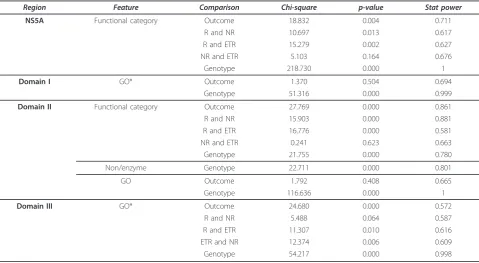
shows a high percentage of coils in NS5A, which results in the high structural flexibility of the NS5A. In addition with ProtFun predictions, we observed that NS5A may have genotypic differences in performed functions, vary-ing also between the domains, with a great statistical difference (p < .001) and power (> .95) (Table 3). Despite of the essential functions for HCV infection, NS5A from different genotypes may have different
Table 3 Statistical results for comparison between therapy outcome and genotype in ProtFun analysis
Region Feature Comparison Chi-square p-value Stat power
NS5A Functional category Outcome 18.832 0.004 0.711
R and NR 10.697 0.013 0.617
R and ETR 15.279 0.002 0.627
NR and ETR 5.103 0.164 0.676
Genotype 218.730 0.000 1
Domain I GO* Outcome 1.370 0.504 0.694
Genotype 51.316 0.000 0.999
Domain II Functional category Outcome 27.769 0.000 0.861
R and NR 15.903 0.000 0.881
R and ETR 16.776 0.000 0.581
NR and ETR 0.241 0.623 0.663
Genotype 21.755 0.000 0.780
Non/enzyme Genotype 22.711 0.000 0.801
GO Outcome 1.792 0.408 0.665
Genotype 116.636 0.000 1
Domain III GO* Outcome 24.680 0.000 0.572
R and NR 5.488 0.064 0.587
R and ETR 11.307 0.010 0.616
ETR and NR 12.374 0.006 0.609
Genotype 54.217 0.000 0.998
*values < 05 were excluded for the statistical calculation
secondary functions. These functions may lead directly or indirectly to the different SVR rate in genotypes. Also, it would explain why NS5A from different geno-types have different behaviour when the mutational and quasispecies profile are analysed [24,25].
Prosite analysis demonstrated the presence of several potential co-and post-translational modification sites that are well conserved in the sequence, including N-glycosylation sites (Figure 3 and Table 2). Carbohydrate binding can confer a different function on a protein. For example, it can lead to the addition of epitopes that facilitate the recognition of other proteins [30,31]. There are no studies that describe glycosylation of NS5A, but in some viruses, glycosylated proteins can be essential for viral assembly [32]. Helenius (1994) demonstrated that glycosylation promoted an increase in solubility, and possibly in interactions with chaperones on the endoplasmatic reticulum, thereby affecting folding and stabilization of the protein. Proteins without this modifi-cation could assemble in a non-reversable form or exit the endoplasmatic reticulum [33]. N- and O-linked gly-cosylation are also described in non-reticulum compart-ments, such as nucleus and cytosol [34]. Since this discover, several cytosolic proteins involved in the pro-cess of adding carbohydrate to proteins were character-ized [35,36]. Proteins which undergo these unusual glycosylation processes are linked with several functions, including nuclear membrane structure and transcription factors [37].
Following this hypothesis, NS5A glycosylation may be essential for maintaining its functional structure, as these modifications sequence appear to be conserved. In addition, this modification may play an important role in nuclear localization of NS5A mutants.
Potential phosphorylation sites were identified in NS5A. This modification has been experimentally described and is important for interaction with core proteins and for viral assembly [38]. Phosphorylation is a reversible modification process, and may be key to the multifunctionality of NS5A. Several proteins were identi-fied as playing a role in NS5A phosphorylation including AKT, p70s6K, MEK, MKK1, CKI, CKIIe and Syk [39-41] but we found no study has described phosphorylation by PKC or cAMP-/cGMP-dependent protein kinase. These proteins are still candidates for this process, as details concerning NS5A phosphorylation have yet to be fully elucidated.
Possible myristoylation sites with qualitative conserva-tion were recognized in NS5A. Covalent myristate bind-ing is not reversible and alters the protein’s hydrophobicity. In viral proteins such as Arenavirus and Arterivirus, this modification is related to functions such as protein cell localization and protein-protein interac-tions [42,43]. There are no studies describing
myristoylation in the NS5A protein. However, we sug-gest that this process is important in structural/func-tional stabilization of NS5A. If experimental data demonstrated that these modifications are present in NS5A, these sites could be possible targets for new anti-viral agents.
Interestingly, a cell attachment site (RGD) was present in all sequences between residues 48 and 52. This region is inside the trasmembrane region predicted by MEM-SAT3. RGD is a sequence for interaction with integrins, proteins located on the cell surface that act on cell-cell and cell-extracellular matrix interactions [44]. Although the intracellular functions of RGD require further inves-tigation, studies concerning proprotein convertase 1 showed that this sequence is essential for correct folding in the endoplasmatic reticulum and transport to secre-tory glands [45].
Micelles with cyclic RGD peptides transfected into HeLa cells tend to congregate in the perinuclear region [46]. Therefore, the RGD sequence in NS5A genotypes 1 and 3 could have a role in (1) folding and intracellular transport and/or (2) nuclear and perinuclear localiza-tion. The NS5A protein has a functional nuclear loca-tion signal (NLS) at its carboxy terminal [47]. The complete protein form is predominantly located in the cytoplasm and/or in the perinuclear region [47,48]. However, forms in which the NS5A amino terminal region (residues 1-31) has been deleted predominate in the nucleus [49]. These deleted forms occur naturally during infection, resulting in cell caspase activity [50,51]. The function of these nuclear forms requires further study but they have been shown to be transcriptional regulators [52-54]. Furthermore, these forms can trans-port other proteins complexed with NS5A. The c-Raf protein interacts with the NS5A carboxy terminal and is detected in the cell nucleus with these deleted forms of the NS5A protein [54].
Regarding these studies, we suggest that the factors participating in NS5A nuclear localization are (1) dele-tion of the amino terminal region, which inhibits the NLS region; (2) presence of the NLS without mutations; (3) possible interactions between RGD and proteins related to transport through the nuclear envelope (4) possible glycosylation of NS5A similar to other nuclear functional proteins.
It is important to highlight that if other proteins are transported to the nucleus with c-Raf, nuclear NS5A could be important to the regulation and modulation of cell processes.
Conclusions
study of proteins that are difficult to investigate by other experimental procedures. There was no relationship between the response to therapy and primary structure, but for genotype 3 secondary structure variances were different between the three outcome groups. In addition, there is evidence that the primary/secondary structure differs among genotypes and that this could be impor-tant during the infection process. Functional prediction also indicated that NS5A may have functional difference between genotypes. Altogether, structural and functional properties show that the two genotypes behaviour dur-ing infection have differences. The acquired data can be compared with future experimental data regarding the NS5A protein and may help in developing new antiviral strategies, considering the genotypic differences present in Hepatitis C virus.
Materials and methods
Sequence bank
The sequence bank included 345 NS5A complete sequences that were obtained from previous studies by our group [24,25] and nine reference sequences from Genbank. These sequences were extracted from 23 Bra-zilian patients infected with HCV genotypes 1a, 1b or 3.
The accession numbers in Genbank are
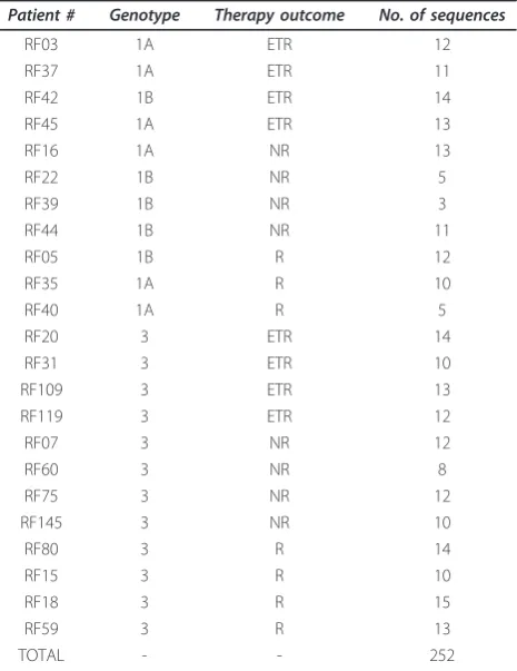
EU309511aEU309673 for genotype 1; EU826174 to EU826233 and from EU826249 to EU826352 for geno-type 3. These samples comprised patients who had a sustained virological response (SVR), non response (NR) or end of treatment response (ETR) after conventional therapy based on Interferon (genotype 3) or Peginter-feron (genotype 1) plus Ribavirin. Details of the study population are presented in Table 4. Redundant amino acid sequences were excluded, using the software LOCQSPEC 1.0 [55], resulting in 252 different sequences of complete NS5A.
Amino acids and secondary structure analysis
The percentage of each amino acid type was calculated and secondary structures investigated in all complete NS5A sequences from the sequence bank. These calcu-lations were performed using the PROF program [56], using the Predict Protein Server http://www.predictpro-tein.org[57].
Transmembrane region prediction
Prediction of transmembrane regions was developed by the MEMSAT3 program [58]http://bioinf.cs.ucl.ac.uk/ psipred/. All NS5A sequences from the sequence bank were analyzed using this program.
Prediction of sites
All sequences were analyzed using the PROSITE pro-gram [59], from the Predict Protein Server [57]. Prosite
is a pattern data bank, based on scientific publications or research describing the function of determined pro-tein groups [59].
Prediction of functional features
Sequences were submitted to ProtFun 2.2 Server. The method is based on sequence derived protein features such as predicted post translational modifications, pro-tein sorting signals and physical/chemical properties calculated from amino acid composition. This allows prediction of functionality for proteins which no homology can be found [60,61]. Acquired data were organized in tables (not shown) to posterior statistical analysis.
Statistical analysis
In order to establish if there were differences between the prediction results between the response groups or genotypes, test of homogeneity (chi-square test), t-test and equal variance test was calculated using the soft-ware MiniTab®15 (Minitab Inc., USA). Values under 5 (five) were excluded from statistical calculation, since these results could be deviation from the sample. Statis-tical power calculation was performed using online soft-ware Russ Lenth’s power and sample size page [62]. Table 4 Characteristics of the study population and number of NS5A different sequences
Patient # Genotype Therapy outcome No. of sequences
RF03 1A ETR 12
RF37 1A ETR 11
RF42 1B ETR 14
RF45 1A ETR 13
RF16 1A NR 13
RF22 1B NR 5
RF39 1B NR 3
RF44 1B NR 11
RF05 1B R 12
RF35 1A R 10
RF40 1A R 5
RF20 3 ETR 14
RF31 3 ETR 10
RF109 3 ETR 13
RF119 3 ETR 12
RF07 3 NR 12
RF60 3 NR 8
RF75 3 NR 12
RF145 3 NR 10
RF80 3 R 14
RF15 3 R 10
RF18 3 R 15
RF59 3 R 13
Acknowledgements
This work was supported by grants from FAPESP, CNPq and CAPES.
Author details
1Department of Biology, Sao Paulo State University–UNESP, Sao Jose do Rio
Preto, SP, Brazil.2Department of Clinical Medicine, Sao Paulo University–USP, Medicine College, Sao Paulo, SP, Brazil.3Viral Immunology Laboratory, Butantan Institute, Sao Paulo, SP, Brazil.4Rua Cristovao Colombo, 2265 Laboratorio de Estudos Genomicos, Jardim Nazareth, CEP: 15054-000, Sao Jose do Rio Preto, SP, Brazil.
Authors’contributions
LHTY carried out all experiments, acquisition of data, analysis and interpretation of data, and drafting the manuscript; HAA helped with the acquisition of data and interpretation of results; ACGJ, CB and IMVGCM participated in the study and the writing of the manuscript, PR conceived the study, participated in its analysis and coordination, and supplied suggestions for this manuscript. All authors read and approved the final manuscript.
Competing interests
The authors declare that they have no competing interests.
Received: 5 July 2011 Accepted: 12 January 2012 Published: 12 January 2012
References
1. Gale M Jr, Foy EM:Evasion of intracellular host defence by hepatitis C virus.Nature2005,436:939-945.
2. Appel N, Zayas M, Miller S, Krijnse-Locker J, Schaller T, Friebe P, Kallis S, Engel U, Bartenschlager R:Essential role of domain III of nonstructural protein 5A for hepatitis C virus infectious particle assembly.PLoS Pathog
2008,4:e1000035.
3. Alter HJ, Purcell RH, Shih JW, Melpolder JC, Houghton M, Choo QL, Kuo G: Detection of antibody to hepatitis C virus in prospectively followed transfusion recipients with acute and chronic non-A, non-B hepatitis.N Engl J Med1989,321:1494-1500.
4. Saito I, Miyamura T, Ohbayashi A, Harada H, Katayama T, Kikuchi S, Watanabe Y, Koi S, Onji M, Ohta Y,et al:Hepatitis C virus infection is associated with the development of hepatocellular carcinoma.Proc Natl Acad Sci USA1990,87:6547-6549.
5. Simmonds P:Genetic diversity and evolution of hepatitis C virus-15 years on.J Gen Virol2004,85:3173-3188.
6. Kuiken C, Simmonds P:Nomenclature and numbering of the hepatitis C virus.Methods Mol Biol2009,510:33-53.
7. Moradpour D, Cerny A, Heim MH, Blum HE:Hepatitis C: an update.Swiss Med Wkly2001,131:291-298.
8. Pybus OG, Charleston MA, Gupta S, Rambaut A, Holmes EC, Harvey PH:The epidemic behavior of the hepatitis C virus.Science2001,292:2323-2325. 9. Neumann AU, Lam NP, Dahari H, Gretch DR, Wiley TE, Layden TJ,
Perelson AS:Hepatitis C viral dynamics in vivo and the antiviral efficacy of interferon-alpha therapy.Science1998,282:103-107.
10. Moradpour D, Blum HE:A primer on the molecular virology of hepatitis C.Liver Int2004,24:519-525.
11. Lyra AC, Fan X, Di Bisceglie AM:Molecular biology and clinical implication of hepatitis C virus.Braz J Med Biol Res2004,37:691-695.
12. Kamar N, Bendall RP, Peron JM, Cintas P, Prudhomme L, Mansuy JM, Rostaing L, Keane F, Ijaz S, Izopet J, Dalton HR:Hepatitis E virus and neurologic disorders.Emerg Infect Dis2011,17:173-179.
13. Reed KE, Xu J, Rice CM:Phosphorylation of the hepatitis C virus NS5A protein in vitro and in vivo: properties of the NS5A-associated kinase.J Virol1997,71:7187-7197.
14. Macdonald A, Harris M:Hepatitis C virus NS5A: tales of a promiscuous protein.J Gen Virol2004,85:2485-2502.
15. Liang Y, Ye H, Kang CB, Yoon HS:Domain 2 of nonstructural protein 5A (NS5A) of hepatitis C virus is natively unfolded.Biochemistry2007, 46:11550-11558.
16. Love RA, Brodsky O, Hickey MJ, Wells PA, Cronin CN:Crystal structure of a novel dimeric form of NS5A domain I protein from hepatitis C virus.J Virol2009,83:4395-4403.
17. Tellinghuisen TL, Marcotrigiano J, Gorbalenya AE, Rice CM:The NS5A protein of hepatitis C virus is a zinc metalloprotein.J Biol Chem2004, 279:48576-48587.
18. Moradpour D, Brass V, Penin F:Function follows form: the structure of the N-terminal domain of HCV NS5A.Hepatology2005,42:732-735.
19. Brass V, Bieck E, Montserret R, Wolk B, Hellings JA, Blum HE, Penin F, Moradpour D:An amino-terminal amphipathic alpha-helix mediates membrane association of the hepatitis C virus nonstructural protein 5A.
J Biol Chem2002,277:8130-8139.
20. Liu S, Ansari IH, Das SC, Pattnaik AK:Insertion and deletion analyses identify regions of non-structural protein 5A of Hepatitis C virus that are dispensable for viral genome replication.J Gen Virol2006,87:323-327. 21. Verdegem D, Badillo A, Wieruszeski JM, Landrieu I, Leroy A,
Bartenschlager R, Penin F, Lippens G, Hanoulle X:Domain 3 of NS5A protein from the hepatitis C virus has intrinsic {alpha}-helical propensity and is a substrate of Cyclophilin A.J Biol Chem2011.
22. Wohnsland A, Hofmann WP, Sarrazin C:Viral determinants of resistance to treatment in patients with hepatitis C.Clin Microbiol Rev2007,20:23-38. 23. Neumann AU, Lam NP, Dahari H, Davidian M, Wiley TE, Mika BP,
Perelson AS, Layden TJ:Differences in viral dynamics between genotypes 1 and 2 of hepatitis C virus.J Infect Dis2000,182:28-35.
24. Jardim AC, Yamasaki LH, de Queiroz AT, Bittar C, Pinho JR, Carareto CM, Rahal P, Mello IM:Quasispecies of hepatitis C virus genotype 1 and treatment outcome with peginterferon and ribavirin.Infect Genet Evol
2009,9:89-698.
25. Bittar C, Jardim AC, Yamasaki LH, de Queiroz AT, Carareto CM, Pinho JR, de Carvalho-Mello IM, Rahal P:Genetic diversity of NS5A protein from hepatitis C virus genotype 3a and its relationship to therapy response.
BMC Infect Dis2010,10:36.
26. Tsai YH, Kuang WF, Lu TY, Kao JH, Lai MY, Liu CJ, Chen PJ, Hwang LH:The non-structural 5A protein of hepatitis C virus exhibits genotypic differences in interferon antagonism.J Hepatol2008,49:899-907. 27. Kang SM, Won SJ, Lee GH, Lim YS, Hwang SB:Modulation of interferon
signaling by hepatitis C virus non-structural 5A protein: implication of genotypic difference in interferon treatment.FEBS Lett2010, 584:4069-4076.
28. Scheel TK, Gottwein JM, Mikkelsen LS, Jensen TB, Bukh J:Recombinant HCV variants with NS5A from genotypes 1-7 have different sensitivities to an NS5A inhibitor but not interferon-alpha.Gastroenterology2011, 140:1032-1042.
29. Schreiber G, Keating AE:Protein binding specificity versus promiscuity.
Curr Opin Struct Biol2011,21:50-61.
30. Taylor ME, Drickamer K:Structural requirements for high affinity binding of complex ligands by the macrophage mannose receptor.J Biol Chem
1993,268:399-404.
31. Petrescu AJ, Milac AL, Petrescu SM, Dwek RA, Wormald MR:Statistical analysis of the protein environment of N-glycosylation sites: implications for occupancy, structure, and folding.Glycobiology2004,14:103-114. 32. Graff J, Zhou YH, Torian U, Nguyen H, St Claire M, Yu C, Purcell RH,
Emerson SU:Mutations within potential glycosylation sites in the capsid protein of hepatitis E virus prevent the formation of infectious virus particles.J Virol2008,82:1185-1194.
33. Helenius A:How N-linked oligosaccharides affect glycoprotein folding in the endoplasmic reticulum.Mol Biol Cell1994,5:253-265.
34. Hart GW, Haltiwanger RS, Holt GD, Kelly WG:Glycosylation in the nucleus and cytoplasm.Annu Rev Biochem1989,58:841-874.
35. Pedemonte CH, Sachs G, Kaplan JH:An intrinsic membrane glycoprotein with cytosolically oriented n-linked sugars.Proc Natl Acad Sci USA1990, 87:9789-9793.
36. Kreppel LK, Blomberg MA, Hart GW:Dynamic glycosylation of nuclear and cytosolic proteins. Cloning and characterization of a unique O-GlcNAc transferase with multiple tetratricopeptide repeats.J Biol Chem1997, 272:9308-9315.
37. Hart GW:Dynamic O-linked glycosylation of nuclear and cytoskeletal proteins.Annu Rev Biochem1997,66:315-335.
39. Huang Y, Staschke K, De Francesco R, Tan SL:Phosphorylation of hepatitis C virus NS5A nonstructural protein: a new paradigm for
phosphorylation-dependent viral RNA replication?Virology2007,364:1-9. 40. Tellinghuisen TL, Foss KL, Treadaway JC, Rice CM:Identification of residues
required for RNA replication in domains II and III of the hepatitis C virus NS5A protein.J Virol2008,82:1073-1083.
41. Neddermann P:NS5A phosphorylation and hyperphosphorylation.
Methods Mol Biol2009,510:95-110.
42. Gordon JI, Duronio RJ, Rudnick DA, Adams SP, Gokel GW:Protein N-myristoylation.J Biol Chem1991,266:8647-8650.
43. Farazi TA, Waksman G, Gordon JI:The biology and enzymology of protein N-myristoylation.J Biol Chem2001,276:39501-39504.
44. Ruoslahti E, Pierschbacher MD:Arg-Gly-Asp: a versatile cell recognition signal.Cell1986,44:517-518.
45. Rovere C, Luis J, Lissitzky JC, Basak A, Marvaldi J, Chretien M, Seidah NG: The RGD motif and the C-terminal segment of proprotein convertase 1 are critical for its cellular trafficking but not for its intracellular binding to integrin alpha5beta1.J Biol Chem1999,274:12461-12467.
46. Oba M, Aoyagi K, Miyata K, Matsumoto Y, Itaka K, Nishiyama N, Yamasaki Y, Koyama H, Kataoka K:Polyplex micelles with cyclic RGD peptide ligands and disulfide cross-links directing to the enhanced transfection via controlled intracellular trafficking.Mol Pharm2008,5:1080-1092. 47. Ide Y, Zhang L, Chen M, Inchauspe G, Bahl C, Sasaguri Y, Padmanabhan R:
Characterization of the nuclear localization signal and subcellular distribution of hepatitis C virus nonstructural protein NS5A.Gene1996, 182:203-211.
48. Kim JE, Song WK, Chung KM, Back SH, Jang SK:Subcellular localization of hepatitis C viral proteins in mammalian cells.Arch Virol1999,144:329-343. 49. Song J, Nagano-Fujii M, Wang F, Florese R, Fujita T, Ishido S, Hotta H:
Nuclear localization and intramolecular cleavage of N-terminally deleted NS5A protein of hepatitis C virus.Virus Res2000,69:109-117.
50. Hidajat R, Nagano-Fujii M, Deng L, Hotta H:Cleavage of the hepatitis C virus NS5A protein by caspase-3 in the interferon sensitivity-determining region in a sequence-dependent manner.Kobe J Med Sci2004, 50:153-166.
51. Kalamvoki M, Georgopoulou U, Mavromara P:The NS5A protein of the hepatitis C virus genotype 1a is cleaved by caspases to produce C-terminal-truncated forms of the protein that reside mainly in the cytosol.J Biol Chem2006,281:13449-13462.
52. Tanimoto A, Ide Y, Arima N, Sasaguri Y, Padmanabhan R:The amino terminal deletion mutants of hepatitis C virus nonstructural protein NS5A function as transcriptional activators in yeast.Biochem Biophys Res Commun1997,236:360-364.
53. Fukuma T, Enomoto N, Marumo F, Sato C:Mutations in the interferon-sensitivity determining region of hepatitis C virus and transcriptional activity of the nonstructural region 5A protein.Hepatology1998, 28:1147-1153.
54. Sauter D, Himmelsbach K, Kriegs M, Carvajal Yepes M, Hildt E:Localization determines function: N-terminally truncated NS5A fragments accumulate in the nucleus and impair HCV replication.J Hepatol2009,50:861-871. 55. Marucci EA, Zafalon GF, Jardim AC, Yamasaki LH, Bittar C, Rahal P,
Machado JM:Routine libraries for pattern recognition in quasispecies.
Genet Mol Res2008,7:970-981.
56. Rost B, Sander C:Improved prediction of protein secondary structure by use of sequence profiles and neural networks.Proc Natl Acad Sci USA
1993,90:7558-7562.
57. Rost B, Yachdav G, Liu J:The predictprotein server.Nucleic Acids Res2004, 32:W321-W326.
58. Jones DT:Improving the accuracy of transmembrane protein topology prediction using evolutionary information.Bioinformatics2007,23:538-544. 59. Bairoch A, Bucher P, Hofmann K:The PROSITE database, its status in 1997.
Nucleic Acids Res1997,25:217-221.
60. Jensen LJ, Gupta R, Blom N, Devos D, Tamames J, Kesmir C, Nielsen H, Staerfeldt HH, Rapacki K, Workman C,et al:Prediction of human protein function from post-translational modifications and localization features.J Mol Biol2002,319:1257-1265.
61. Jensen LJ, Ussery DW, Brunak S:Functionality of system components: conservation of protein function in protein feature space.Genome Res
2003,13:2444-2449.
62. Lenth RV:Java Applets for Power and Sample Size [Computer software].
Java Applets for Power and Sample Size2006-9[http://www.stat.uiowa.edu/ ~rlenth/Power/].
doi:10.1186/1743-422X-9-14
Cite this article as:Yamasakiet al.:New insights regarding HCV-NS5A structure/function and indication of genotypic differences.Virology Journal20129:14.
Submit your next manuscript to BioMed Central and take full advantage of:
• Convenient online submission
• Thorough peer review
• No space constraints or color figure charges
• Immediate publication on acceptance
• Inclusion in PubMed, CAS, Scopus and Google Scholar
• Research which is freely available for redistribution