Open Access
Case report
Primary small-cell neuroendocrine carcinoma of the duodenum – a
case report and review of literature
Naohiro Sata*
1, Munetoshi Tsukahara
1, Masaru Koizumi
1, Koji Yoshizawa
1,
Katsumi Kurihara
1, Hideo Nagai
1, Tsutomu Someya
2and Ken Saito
2Address: 1Department of Surgery, Jichi Medical School, 3311-1 Yakushiji Minami-kawachi Tochigi, Japan and 2Department of Pathology, Jichi Medical School, 3311-1 Yakushiji Minami-kawachi Tochigi, Japan
Email: Naohiro Sata* - sata@jichi.ac.jp; Munetoshi Tsukahara - m-tsuka@jichi.ac.jp; Masaru Koizumi - mkoizumi@jichi.ac.jp; Koji Yoshizawa - k-yoshi@jichi.ac.jp; Katsumi Kurihara - kurihara@jichi.ac.jp; Hideo Nagai - nagaihd@jichi.ac.jp;
Tsutomu Someya - sata@jichi.ac.jp; Ken Saito - sata@jichi.ac.jp * Corresponding author
Abstract
Background: Small-cell neuroendocrine carcinoma in the duodenum is an extremely rare neoplasm with poor prognosis.
Case presentation: A 57-year-old man presented with sudden onset gastrointestinal bleeding and fainting attacks. Duodenoscopy and hypotonic duodenography revealed a 3 × 3 cm protruding tumor with ulcerations situated opposite the ampulla of Vater in the second part of the duodenum. Local excision of the tumor was performed, followed by adjuvant chemotherapy with 5-fluoro uracil and leucovorin. Examination of the tumor by immunohistochemistry and electron microscopy indicated it to be neuroendocrine in nature, expressing synaptophysin and AE1/AE3, and containing dense core granules. The patient showed no sign of recurrence and has been disease-free for more than 48 months after surgery.
Conclusions: Most cases of small-cell neuroendocrine carcinoma in the duodenum show rapid progression of the disease, and even radical surgery with or without chemotherapy do not prevent death. We report a rare subtype of small-cell neuroendocrine carcinoma. This subtype appears to have a much better prognosis, and may be amenable to local excision, if the lesion is away from the ampulla of Vater.
Background
Duodenal Neuroendocrine tumors constitute 5% of all gastrointestinal neuroendocrine tumors [1,2]. Most of these show well-differentiated features and are classified as carcinoids or somatostatinomas [3-6]. Occurrence of carcinoma is rare, and carcinomas with anaplastic charac-ter, which are classified as small-cell carcinomas, are even less frequent [7-12]. The most common small-cell neu-roendocrine carcinoma (NEC) is the small-cell
undiffer-entiated carcinoma of the lung [13,14]. Although the features of these pulmonary tumors are well defined, the characteristics of their extrapulmonary counterparts are still unknown. We report a case of small-cell NEC in the duodenum that had unique morphological features and exceptionally good clinical outcome.
Published: 15 August 2004
World Journal of Surgical Oncology 2004, 2:28 doi:10.1186/1477-7819-2-28
Received: 06 May 2004 Accepted: 15 August 2004
This article is available from: http://www.wjso.com/content/2/1/28
© 2004 Sata et al; licensee BioMed Central Ltd.
World Journal of Surgical Oncology 2004, 2:28 http://www.wjso.com/content/2/1/28
Case presentation
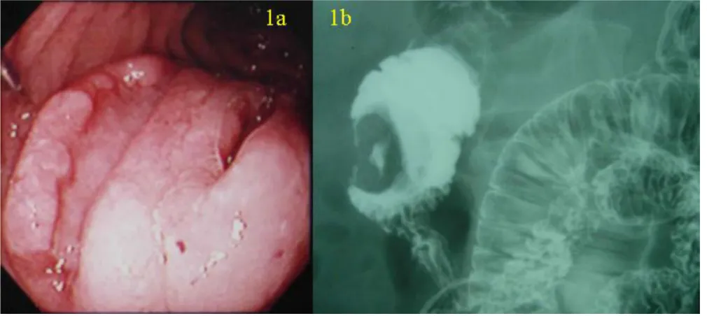
A 57-year-old man presented with sudden gastrointestinal tract bleeding and episode of fainting. Duodenoscopy (Figure 1a) and hypotonic duodenography (Figure 1b) revealed a 3 × 3 cm protruding tumor with two ulcerations located opposite the ampulla of Vater in the second part of the duodenum. Laboratory data showed no abnormal-ities in blood chemistry, tumor markers (CEA, CA19-9, NSE, proGRP) and endocrine markers (somatostatin, gas-trin, glucagons, serotonin, VIP) except a moderate anemia (9.5 g/dl hemoglobin). No abnormal findings were observed in the chest X-ray and computed tomography (CT).
A laparotomy was performed. As there was no serosal invasion or regional lymphadenopathy wide local exci-sion of the tumor was performed. On gross examination, the tumor showed two ulcerations and two different mor-phological components (Figure 2a and 2b). One compo-nent (compocompo-nent A) was round in shape with a round ulceration on the top, and the other component (compo-nent B), which enclosed the round compo(compo-nent, was cres-cent in shape with a spindle-shaped ulceration on the top. The two components showed different histopathological and immunohistochemical features (Table 1). The round component contained fibrous tissue, small nuclei, and clear nucleoli. Histopathologically, the crescent
compo-nent had more anaplastic features typical of small-cell car-cinoma, such as sheets of tightly packed anaplastic cells with round nuclei and scanty cytoplasm (Figure 2c, 2d). Neuroendocrine differentiation was investigated using immunohistochemical and ultrastructural techniques. Both components showed neuroendocrine features, with immunochemistry identifying synaptophysin and AE1/ AE3 (Figure 3a and 3b), and electron microscopy identify-ing dense core granules (Figure 4). Immunochemistry also showed that the crescent component expressed less cytok-eratin, vimentin and CD56, and more MIB-1 than the round component.
The patient was discharged three weeks after operation with uneventful postoperative period. Four cycles of monthly adjuvant chemotherapy with 5-fluoro uracil (5-FU) (325 mg/m2) and leucovorin (20 mg/m2) were
administered. The patient showed no sign of recurrence and is disease-free 48 months after surgery.
Discussion
Neuroendocrine carcinomas (NEC) in the duodenum are extremely rare, and are classified as either 'small-cell' or 'non small-cell' types. The small-cell NEC occurring in the duodenum and elsewhere in gastrointestinal tract are sim-ilar to the small-cell carcinoma of the lung [7,8]. Only eight cases of small-cell NEC in the duodenum have been
(a) Duodenoscopy showing a 3 × 3 cm protruding tumor with two ulcerations located opposite the ampulla of Vater in the second portion of the duodenum
Figure 1
reported, with the present case being the ninth (Table 2) [7-12]. Most cases occurred in middle-aged or geriatric males with lesions in the ampulla of Vater. Extra-ampul-lary small-cell NECs in the duodenum are extremely rare, with only two cases reported previously [7,8].
The natural course of small-cell NECs in the duodenum is still not clear. Most cases reported in the literature show rapid progress of the disease, with radical surgery and/or chemotherapy not altering the clinical course, and thus a poor prognosis. In six of the previous eight cases, patients underwent pancreaticoduodenectomy for removal of the tumor, while the remaining two did not undergo surgery
Macroscopic and microscopic findings of the tumor
Figure 2
World Journal of Surgical Oncology 2004, 2:28 http://www.wjso.com/content/2/1/28
because of multiple liver metastasis or poor general con-dition. In spite of radical resection with or without adjuvant chemotherapy, most cases showed rapid recur-rence and metastasis. Of the eight reported cases, one was unusual as it occurred in a middle-aged female with rapid progress of the disease but effective response to adjuvant chemotherapy using 5FU, tumor necrosis factor, and interferon this patient survived for more than eighteen months [10]. The present case was treated by a local exci-sion of the tumor followed by adjuvant chemotherapy using 5-FU and leucovorin, and showed a distinctively
unique clinical course, with the patient surviving for more than 48 months without any sign of recurrence. This case was presented with gastrointestinal bleeding, which con-tributed to early diagnosis, whereas the other previous cases in literature presented either with abdominal pain or jaundice. Hence, the good prognosis in present case could also be due to its earlier presentation. The lesion in the present case showed a different immunohistological char-acter from that in other cases, such as no immunoreactiv-ity to neuron-specific enolase (NSE) or chromogranin A.
Table 1: Immunochemical characteristics of the two components of the tumor.
Synaptophysi n
AE1/AE3 Vimentin CD56 chromograni n A
LCA, L26, UCHL1, CD3, ASMA, M-actin, desmin, CD34, NF, GFAP, and S100 were negative in both components.
Table 2: Profiles of the cases of primary small-cell neuroendocrine carcinoma in the duodenum reported in the literature.
Author
Morphology Location Metastasis Surgery Chemotherapy Prognosis (month)
1 Swanson 1986 [7]
76 M Abdominal pain, anorexia, weight loss
15 ulceration adjacent to the ampulla*
LN, liver biospy 5-FU, doxorubicin, mitomycin
Dead (1.5)
2 Zamboni 1990 [8]
62 M jaundice, weight loss
25 polypoid the papilla of Vater
20 ulceration the papilla of Vater
LN PD - Dead (6)
4 Zamboni 1990 [8]
51 M jaundice, weight loss, abdominal pain
30 soft fungating mass
? polypoid Peri ampullary
? - - (>5)
6 Sarker 1992 [10]
53 F jaundice, weight loss, back pain
74 M jaundice 35 polypoid the papilla of Vater
? PPPD - ?
8 Shim 2000 [12]
54 M jaundice 30 ulceration the papilla of Vater
5-FU leucovorin disease free (>48)
These differences too might partly explain the different character of this case.
Conclusions
This report identifies a new subtype of small-cell NEC in the duodenum. This subtype appears to have a much bet-ter prognosis, and may be amenable to local excision, if the lesion is away from the ampulla of Vater.
Competing interests
None declared.
Authors' contributions
NS, MT, MK, KY, KK, HN took part in the operation, per-formed the literature search and drafted the manuscript for submission. HN supervised the preparation of the manuscript and edited the final version for publication. TS, KS performed pathological investigations and contrib-uted to the pathological content of the manuscript.
All authors read and approved the manuscript.
Immunostaining for AE1/AE3 showing (a) diffuse cytoplasmic positivity in the component A, and (b) no reactivity in the compo-nent B
Figure 3
World Journal of Surgical Oncology 2004, 2:28 http://www.wjso.com/content/2/1/28
Ultrastructural study showed cytoplasmic dense-core granules in the component A
Figure 4
Publish with BioMed Central and every scientist can read your work free of charge "BioMed Central will be the most significant development for disseminating the results of biomedical researc h in our lifetime."
Sir Paul Nurse, Cancer Research UK
Your research papers will be:
available free of charge to the entire biomedical community
peer reviewed and published immediately upon acceptance
cited in PubMed and archived on PubMed Central
yours — you keep the copyright
Submit your manuscript here:
http://www.biomedcentral.com/info/publishing_adv.asp
BioMedcentral
Acknowledgement
The patient's consent was obtained for publication of his case records, duo-denoscopy, barium, and histopathological pictures.
References
1. Marcial MA, Pinkus PS, Scaring A: Ampullary somatostatinoma. Am J Clin Pathol 1983, 80:755-761.
2. Sanchez-Sosa S, Angeles AA, Orozco H, Larriva-Sahd J: Neuroendo-crine carcinoma of the ampulla of Vater. A case of absence of somatostatin in a vasoactive intestinal polypeptide-, bombesin-, and cholecystokinin-producing tumor.Am J Clin Pathol 1991, 95:51-54.
3. Capella C, Riva C, Rindi G, Usellini L, Chiaravalli A, Solcia E: Endo-crine tumors of the duodenum and upper jejunum. A study of 33 cases with clinico-pathological characteristics and hor-mone content.Hepatogastroenterology 1990, 37:247-252. 4. Feurle GE, Anlauf M, Hamscher G, Arnold R, Kloppel G, Weihe E:
Xenin-immunoreactive cells and extractable xenin in neu-roendocrine tumors of duodenal origin.Gastroenterology 2002,
123:1616-1626.
5. Sawady J, Katzin WE, Mendelsohn G, Aron DC: Somatostatin-pro-ducing neuroendocrine tumor of the ampulla (Ampullary somatostatinoma) evidence of prosomatostatin production. Am J Clin Pathol 1992, 97:411-415.
6. Rios A, Fernandez JA, Rodriguez JM, Lujan JA, Martinez E, Parrilla P:
Massive upper gastrointestinal bleeding as a manifestation of somatostatinoma of the ampulla of Vater.Dig Dis Sci 2001,
46:2162-2165.
7. Swanson PE, Dykoski D, Wick MR, Snover DC: Primary duodenal small-cell neuroendocrine carcinoma with production of vasoactive intestinal polypeptide. Arch Pathol Lab Med 1986,
110:317-320.
8. Zamboni G, Franzin G, Bonetti F, Scarpa A, Chilosi M, Colombari R, Menestrina F, Pea M, Iacono C, Serio G, Fiore-Donati L: Small-cell neuroendocrine carcinoma of the ampullary region. A clin-icopathologic, immunohistochemical, and ultrastructural study of three cases.Am J Surg Pathol 1990, 14:703-13.
9. Lee CS, Machet D, Rode J: Small cell carcinoma of the ampulla of Vater.Cancer 1992, 70:1502-1504.
10. Sarker AB, Hoshida Y, Akagi S, Hayashi K, Murakami I, Jeon HJ, Taka-hashi K, Akagi T: An immunohistochemical and ultrastructural study of case of small-cell neuroendocrine carcinoma in the ampullary region of the duodenum. Acta Pathol Jpn 1992,
42:529-535.
11. Sato T, Yamamoto K, Ouchi A, Imaoka Y, Tokumura H, Matsushiro T: Undifferentiated carcinoma of the duodenal ampulla. J Gastroenterol 1995, 30:517-519.
12. Shim CS, Moon JH, Cho YD, Lee MS, Jeon HB, Jin SY, Lee HK: Neu-roendocrine carcinoma of the ampulla of Vater.Gastrointest Endosc 2000, 51:593.
13. Bensch KG, Corrin B, Pariente R, Spencer H: Oat cell carcinoma of the lung: its origin and relationship to bronchial carcinoid. Cancer 1968, 22:1163-1172.