Vol. 4, Special Issue 6, May 2015
Unsupervised Active Learning Classification
for Detecting Retinal Hemorrhage in Digital
Fundus Images
P.Latha
1, Prof.R.Vijayalakshmi
2, P.Dheevambiga
3P.G. Student, Dept of Computer Engineering, Muthayammal Engineering College, Rasipuram, Namakkal, India1,3 Professor, Dept of Computer Engineering, Muthayammal Engineering College, Rasipuram, Namakkal, India2
ABSTRACT The development of automated screening systems for practice needs reliable detection of retinal hemorrhages. Automated detection of diabetic retinopathy (DR) is important for allowing timely treatment increasing accessibility to and productivity of eye care providers. Supervised splat feature classification method involves processing the retinal color images are partitioned into non overlapping segments covering entire image. Address existing problems using higher level entity splat a collection of pixels with similar color and spatial location each segment, i.e., splat, contains pixels with similar color and spatial location set of features is extracted from each splat. The supervised classification requires high cost in acquiring labeled data for training and it does not include active learning training set. The proposed work presents unsupervised active learning classification for detecting retinal hemorrhage in digital fundus images. The classifier automatically identifies the continuous hemorrhage connected with retinal vessel. Annotations help in comparing the variability of experts in interpreting same set of images in terms of clinical relevance and better definition of reference standard. Extend splat feature classification to learn from both labeled and unlabelled training data. The proposed active learning classification models choose the hemorrhage from retinal vessels portions. The performance evaluation of unsupervised active learning classifier for hemorrhage detection in retinal vessels is done with following parameters: fundus image size, active learning splat classes, hemorrhage splat size and false positive.
KEYWORDS: Unsupervised Active Learning, Active Splat Feature, Learning Classifier
1. INTRODUCTION
Vol. 4, Special Issue 6, May 2015
II.UNSUPERVISED ACTIVE LEARNING CLASSIFICATION (UALC) FOR DETECTION RETINAL HEMORRHAGE IN DIGITAL FUNDUS IMAGES
Retinal hemorrhage is the irregular bleeding of the blood vessels in the retina, the covering in the rear of the eye. In retinal image, make routine detection of hemorrhage is a main braving factor[9]. For automated detection of hemorrhage, a simplified structure is requiring to teach classifiers with optimal features studied into obtainable dataset. The fundus image screening automated system, reliable detection of retinal hemorrhage is required. Retinal hemorrhages are formed in diabetic retinopathy and hypertension. A splat feature classification method can be utilized to detect high level of abnormal retinal hemorrhages with large accuracy.
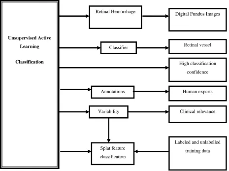
Fig 2.1. Architecture
Diagram of Unsupervised Active Learning Classification
Fig 2.1. described as retinal hemorrhage is the irregular bleeding of blood vessels in the retina, the covering in the rear of eye. Blood process to the retina is preserved in the retinal vein and route and a dense network of little blood vessels called capillaries. These blood vessels can become injured by damages and leans to bleed. This results in impermanent or permanent failure of vision.Splat features are removed into the splat segments by believing depicters such as color and force values. Generally RGB color space is believed for splat. From the color variation based on strength the aspiration splat are extracted. Regular strength variation across neighboring splat can be decided into the histogram. Splat features are chosen in two types of feature selection methods. Preface features are chosen by the filter criterion which is followed by covering method that selects the majority relevant features. The unsupervised active learning process categorized into three phase are:
Unsupervised Active
Learning
Classification
Retinal Hemorrhage
Digital Fundus Images
Classifier Retinal vessel
High classification confidence
Annotations Human experts
Variability Clinical relevance
Splat feature classification
Vol. 4, Special Issue 6, May 2015
a) Retinal Hemorrhage in Digital Fundus Images b) Active Splat Feature
c) Unsupervised Learning Classifier
a)Retinal Hemorrhage in Digital Fundus Images
Retinal images are obtained from the fundus photography. Fundus imaging is the technique of obtaining 2D representation of 3D retinal semi transparent tissues of eye. Image intensities represent amount of reflected quantity of light from the eye. Retinal hemorrhage is the abnormal bleeding of blood vessels in the retina membrane in the back of eye. Blood flow to the retina is maintained by retinal vein and artery and dense network of small blood vessels (capillaries). Blood vessels are damaged by injuries and tend to bleed results in temporary or permanent loss of vision. Retinal Hemorrhage is evaluated with acquired digital fundus images. Perform the operation of illuminating retina with white light and examined in full color. Imaging light is filtered to remove red color improves the contrast of the blood vessels. The gradient operators are used to attain high noise suppression in turn improve image smoothing derivatives. It removes the mirror circular reflection of fundus images caused by edge effects. Artifacts are removed by illumination of light with bright and contrast operations.
b)Active Splat Feature
Splat features are extracted from splat segments with color and intensity values. RGB color space is used for splat. Color variation on different intensity used in splat extraction Gradual intensity variation across neighboring splat is determined from histogram. Gaussian kernels used to extract regions with sharp and soft edges. Active splat features are extracted from filter[7] banks. Gaussian filter bank have no overshoot to step function minimizing rise and fall time. Texture analysis is made on the retinal hemorrhage regions in extracting active splat features. Texture analysis evaluate retinal image region characterized by texture content of intensity and gray level values. Active splat features are finally obtained from gray level Co occurrence matrix [6]. A splat feature classification[3] scheme can be utilized to detect high level of irregular retinal hemorrhages with larger accuracy. The retinal images are divided into non overlapping segments called splat. Splat encloses pixels with similar color and spatial location. Each splat into large range of features are extracted based on interface of each splat with its neighbor.
c) Unsupervised Learning Classifier
Unsupervised learning classifier is made on extracted active splat features in concurrence with annotation about the retinal hemorrhage from medical experts. Annotations from human used for generating precise classes of hemorrhage patterns. Annotations help in comparing the variability of experts in interpreting same set of images in terms of clinical relevance and better definition of reference standard. Learning classifier is trained to automatically identify continuous hemorrhage connected with retinal vessel. Learning classifiers provide accurate class labels for uncertain hemorrhages with decision surface boundary achieve high classification confidence. Separate normal components of retina from the abnormal hemorrhages. Active splat feature classes are derived from both labeled and unlabelled training data. Active learning classification depicts the continuation regions of hemorrhage in retinal vessels portions. Class-label is listed based on the medical expert annotations training samples.
III. PERFORMANCE METRICS
In this section evaluate the performance of unsupervised active learning classification is done for detecting retinal hemorrhage in digital fundus images through matlab environment. Performance can be compared based on lesions[2]. One of the major contributions of this work is improved classification techniques in digital fundus images. The performance metrics of the parameters is fundus image size, active learning splat classes, hemorrhage splat size and false positive.
Fundus image size
Active Learning splat classes
Vol. 4, Special Issue 6, May 2015
3.1. Active learning splat classes
Classification models are checked by employing it to data with identified objective value and combine the predicted values with well-known values. If the model executes well and meet the needs then be employed to the current data to expect the future. Active learning splat of classifier refers to the percentage of accurate calculations completed by the model when compared with the actual classifications in the test[5].
Table: 3.1. Fundus Image Size Vs Active Learning Splat Classes
Fundus Image Size
Active Learning splat classes (%)
SFA (Existing) UALC (Proposed)
30 60 79
60 58 77
90 55 76
120 53 73
150 50 71
Fig: 3.1. Fundus Image Size Vs Active Learning Splat Classes
Figure: 3.1. Demonstrate the active learning splat classes. X axis represents the fundus image size whereas Y axis denotes the active learning splat classes using both the Splat Feature Classification (SFA) Technique and proposed Unsupervised Active Learning Classification (UALC) Technique. When the fundus image size increased, active learning splat classes gets increases accordingly. The active learning splat classes is illustrated using the existing SFA and proposed UALC Technique. Figure 4.1.shows better performance of Proposed UALC method in terms of image size than existing SFA and proposed UALC. The Unsupervised Active Learning Classification (UALC) Technique achieves 15 to 25% high performance of active learning splat class’s variation when compared with existing system.
3.2. False Positive Rate
The rates of false positive measures were chosen based on the related work, where one or more publications handle these measures. The intensity of the fundus image was modified in the nonlinear curvature with power values of the hue saturation value (HSV) space[10]. In regulate to highlight brown regions; gamma correction was analyzed on red, green, and blue-bit image. For an obvious appreciating of these determines, it is essential to conceptualize a few
0 20 40 60 80
30 60
90 120
150
Active Learning
Splat
Classes
Fundus Image Size
SFA (Existing)
Vol. 4, Special Issue 6, May 2015
classifications utilized in experimental tests for detection of diseases. When an analysis effect is positive cannot convey it, which is known as "False Positive" (FP).
Table: 3.2. Fundus Image Size Vs False Positive (%)
Fundus Image Size
False Positive (%)
SFA (Existing) UALC (Proposed)
30 48 36
60 56 45
90 67 54
120 79 68
150 84 71
Fig: 3.2. Fundus Image Size Vs False Positive Rate
Figure: 3.2. Demonstrate the False Positive Rate. X axis represents the fundus image size whereas Y axis denotes the False Positive Rate using both the Splat Feature Classification (SFA) Technique and proposed Unsupervised Active Learning Classification (UALC) Technique. When the fundus image size increased, False Positive Rate gets decreases accordingly. The active learning splat classes is illustrated using the existing SFA and proposed UALC Technique. Figure 4.2.shows better performance of Proposed UALC method in terms of image size than existing SFA and proposed UALC. The Unsupervised Active Learning Classification (UALC) Technique achieves 10 to 15% high performance of false positive rate variation when compared with existing system.
IV.CONCLUSION
Finally conclude the problem is unsupervised active learning classification for detecting retinal hemorrhage in digital fundus images. Large hemorrhages are irregular in form with broad level of characteristics. A lot of hemorrhage splat overlies with the blood vessels and effects in misclassification. This process is done for classifier automatically identify the continuous hemorrhage connected with retinal vessel. Splat based image depiction builds it simpler for clinicians to explain the boundaries of goal objects which may decrease the rate of obtaining reference standard data for training. It provides the highest classification even in uncertain hemorrhages with decision surface boundary. As future
0 10 20 30 40 50 60 70 80 90
30 60 90 120 150
F
alse P
ositiveR
at
e
(%
)
Fundus Image Size
SFA (Existing)
Vol. 4, Special Issue 6, May 2015
work, it will be developed methods to detect other lesions related to diabetic retinopathy and other diseases that affect the retina.
REFERENCES
[1] Simon Tong and Daphne Koller, “Support Vector Machine Active Learning with Applications to Text Classification”, Computer Science Department, Stanford University, Stanford CA 94305-9010, USA
[2] Sérgio Bortolin Júnior and Daniel Welfer, “Automatic Detection of Micro aneurysms and Hemorrhages in Color Eye Fundus Images”, International Journal of Computer Science & Information Technology (IJCSIT) Vol 5, No 5, October 2013
[3] Athira R V and Ferlin Deva Shahila D, “Detection of Retinal Hemorrhage Using Splat Feature Classification Technique”, Journal of Engineering Research and Applications, ISSN: 2248-9622, Vol. 4, Issue 1( Version 3), January 2014, pp.327-330.
[4] Inbarathi.R and Karthikeyan.R, “Classification of Splat and GLCM Features in Fundus Images for Retinal Hemorrhage Detection”, Volume 4, Issue 2, February 2014.
[5] Vijayalakshmi R and Selvarajan S, (2014) “Comparative Studies on Canny and Sobel Edge Detection Techniques for Diabetic Retinopathy Eye Image” International Journal of Soft Computing, Vol. 9, Issue. 4, pp. 213-218.
6] Ando Shigeru, “Consistent Gradient Operators”, IEEE Transactions on Pattern Analysis and Machine Intelligence, Vol.22, No.3, March 2000.
[7] Monica Rose.V, “A Study on the Performance of Splat Based Feature Classification on Fundus Images for the Detection of Hemorrhage and Malarial Diseases”, International Journal of Emerging Technology and Advanced Engineering, Volume 4, Special Issue 2, April 2014).
[8] Nidhal Khdhair El Abbadi and Enas Hamood Al Saadi, “Improvement of Automatic Hemorrhages Detection Methods Using Shapes Recognition”, Iraq
[9] Karunya Karo Shanthi.Y, Karpagam.V, “Automatic Detection of Retinal Hemorrhage Based On Gabor Wavelet and Hybrid KNNSVM Algorithm for Fundus Images”, International Journal of Advanced Research in Computer Science Engineering and Information Technology, Volume: 2 Issue: 1 08-Feb-2014, ISSN_NO: 2321-3337.