Scholarship@Western
Scholarship@Western
Electronic Thesis and Dissertation Repository
8-13-2015 12:00 AM
Fibulin-3 Promotes Triple Negative Breast Cancer Cell Invasion
Fibulin-3 Promotes Triple Negative Breast Cancer Cell Invasion
Michelle M. Noonan
The University of Western Ontario Supervisor
Dr. Moshmi Bhattacharya
The University of Western Ontario
Graduate Program in Physiology and Pharmacology
A thesis submitted in partial fulfillment of the requirements for the degree in Master of Science © Michelle M. Noonan 2015
Follow this and additional works at: https://ir.lib.uwo.ca/etd
Part of the Cellular and Molecular Physiology Commons
Recommended Citation Recommended Citation
Noonan, Michelle M., "Fibulin-3 Promotes Triple Negative Breast Cancer Cell Invasion" (2015). Electronic Thesis and Dissertation Repository. 3031.
https://ir.lib.uwo.ca/etd/3031
This Dissertation/Thesis is brought to you for free and open access by Scholarship@Western. It has been accepted for inclusion in Electronic Thesis and Dissertation Repository by an authorized administrator of
(Thesis format: Monograph)
By
Michelle Marie Noonan
Graduate Program in Physiology and Pharmacology
A thesis submitted in partial fulfillment
of the requirements for the degree of
Master of Science
The School of Graduate and Postdoctoral Studies
The University of Western Ontario
London, Ontario, Canada
ii
Triple negative breast cancer (TNBC) is an aggressive subtype of breast cancer, and
metastasis is a leading cause of mortality in these patients. Fibulin-3, a secreted extracellular
matrix protein, has a pro-invasive role in other cancers. However, a role for fibulin-3 in
TNBC invasion is unknown. We have previously shown that KISS1R signaling promotes
TNBC cell invasion through EGFR and MMP-9 activity, via β-arrestin2. Thus, we
hypothesized that KISS1R signaling promotes TNBC cell invasion via fibulin-3. Our clinical
data suggests that plasma fibulin-3 levels are elevated in metastatic TNBC patients.
Additionally, we found that invasive breast cancer cells have increased expression of
fibulin-3 and treatment with kisspeptin, the KISS1R ligand, further increased fibulin-fibulin-3 expression
and secretion. Also, depletion of β-arrestins in TNBC cells decreased fibulin-3 expression.
Furthermore, fibulin-3 depletion in TNBC cells inhibited cell invasiveness through decreased
MMP-9 activity. These results identify fibulin-3 as a new signaling partner of KISS1R and
as a potential anti-metastasis target in TNBC.
Keywords: Metastasis, triple negative breast cancer, fibulin-3, EFEMP1, invasion, KISS1R,
iii
Firstly, I would like to express my sincere gratitude to my supervisor, Dr. Moshmi Bhattacharya, for her expertise, enthusiasm and patient guidance that has been invaluable to me throughout my graduate studies. Thank you for challenging me to always think critically, and encouraging me to strive for excellence. Without your supervision and constant help this thesis would not have been possible.
I would also like to thank the members of my advisory committee, Dr. Nica Borradaile, Dr. Lina Dagnino and Dr. Rommel Tirona for providing useful insight and suggestions along the way. Your guidance has been tremendously appreciated.
In addition, I would like to thank all past and present members of the Bhattacharya lab, especially Cameron Goertzen, Alexandra Blake and Magda Dragan. Cameron, thank you for sharing your technical expertise and infectious laughter with me, you made joining the lab an easy transition. Alexandra, you have become a cherished friend over the past two years. Thank you for the stimulating discussions, both science and otherwise, and for all the fun we have had in and out of the lab. Magda, your technical assistance, calm words and encouragement have been unbelievably helpful over the last two years. I have truly appreciated you every step of the way, as both a mentor and friend.
I would also like to extend my gratitude to our neighbours, the DiGuglielmo lab. Special thanks go to Eddie Chan for his wise advice and friendship.
iv
Abstract ... ii
Acknowledgments ... iii
Table of Contents ... iv
List of Tables... vii
List of Figures ... viii
List of Appendices ... x
List of Abbreviations ... xi
Chapter 1 - Introduction ...1
1.1 Breast Cancer ...2
1.1.1 Breast Cancer Staging ...2
1.1.2 Breast Cancer Classification: Hormone Receptor Status ...3
1.2 Breast Cancer Metastasis ...6
1.2.1 Metastatic Cascade ...6
1.3 Tumor Microenvironment ... 11
1.3.1 Fibulins ... 12
1.3.2 Fibulin-3 ... 12
1.3.3 Fibulin-3 in Cancer... 15
1.4 Matrix Metalloproteinases ... 19
1.4.1 Matrix Metalloproteinase-9 in Cancer ... 19
1.5 Metastasis Suppressor Genes ... 23
1.5.1 KISS1R Signaling ... 23
1.5.2 KISS1R Signaling in Cancer ... 27
v
1.6 Rationale, Hypothesis and Objectives ... 33
1.6.1 Rationale ... 33
1.6.2 Hypothesis ... 33
1.6.3 Objectives ... 33
1.7 References ... 34
Chapter 2 - Methods ... 44
2.1 cBioPortal Data Mining ... 45
2.2 Blood Collection and Fibulin-3 ELISA ... 45
2.3 Cell Culture ... 45
2.4 Stable Transfections and Gene Knockdown ... 46
2.5 Immunoblot Assays ... 49
2.6 Immunofluorescence Microscopy ... 50
2.7 Immunoblot Assays for Fibulin-3 Secretion ... 50
2.8 Scratch Assays for Motility ... 51
2.9 Cell Migration and Invasion Assays ... 51
2.10 Three-Dimensional (3D) Invasion Assays ... 52
2.11 Zymography ... 53
2.12 EGFR Immunoprecipitation ... 54
2.13 MTT Cell Viability Assays ... 54
2.14 Cell Growth Assays ... 55
2.15 Statistical Analysis ... 55
vi
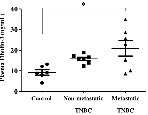
3.1 Plasma fibulin-3 levels increase metastatic TNBC patients ... 59
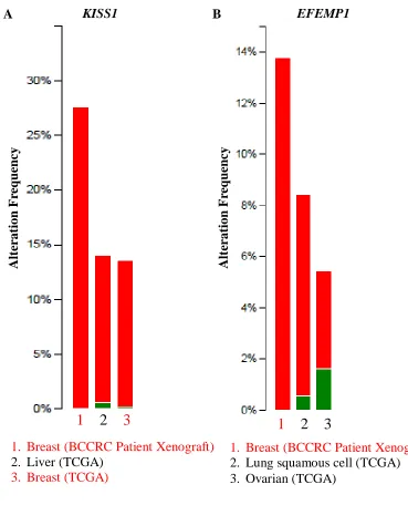
3.2 Altered EFEMP1 and KISS1 expression in invasive breast carcinoma ... 61
3.3 Fibulin-3 and KISS1R expression in breast cell lines ... 65
3.4 Kisspeptin-10 signaling stimulates fibulin-3 expression and secretion in ERα-negative breast cancer cells ... 68
3.5 β-arrestins regulate the expression of fibulin-3 protein in TNBC cells ... 71
3.6 Depletion of fibulin-3 diminishes TNBC cell migration ... 73
3.7 Depletion of fibulin-3 inhibits TNBC cell invasion ... 79
3.8 Depletion of fibulin-3 decreases MAPK signaling and MMP-9 expression and activity ... 83
3.9 Depletion of fibulin-3 inhibits kisspeptin-10-induced MMP-9 secretion and activity ... 86
3.10 Inhibition of ERK signaling further diminishes MMP-9 secretion and activity ... 88
3.11 Depletion of fibulin-3 decreases EGFR phosphorylation in TNBC cells ... 91
3.12 References ... 93
Chapter 4 – Discussion and Conclusions ... 95
4.1 Summary of Novel Findings and Conclusions ... 96
4.2 Contributions of Research to Current State of Knowledge ... 97
4.3 Limitations and Future Directions ... 102
4.4 Conclusions ... 104
4.5 References ... 106
Appendices ... 109
vii
Table 1.1: Role of fibulin-3 in various cancers ... 17
viii
Figure 1.1: Breast cancer staging ... 5
Figure 1.2: The metastatic cascade ... 10
Figure 1.3: Structure of the fibulins ... 14
Figure 1.4: Fibulin-3 activates EGFR in pancreatic cancer ... 18
Figure 1.5: Signaling cascades induced by pro-MMP-9 interaction with cell surface proteins ... 22
Figure 1.6: Kisspeptins (KP) and KISS1R signaling cascades ... 26
Figure 1.7: KISS1R signaling in ERα-negative breast cancer ... 31
Figure 3.1: Plasma fibulin-3 levels in TNBC patients... 60
Figure 3.2: Cross-cancer gene alteration summaries for KISS1 and EFEMP1 ... 63
Figure 3.3: Kaplan-Meier overall survival curves comparing cases of gene alteration to cases without gene alteration for KISS1 and EFEMP1 ... 64
Figure 3.4: Expression of fibulin-3 and KISS1R in breast cell lines ... 66
Figure 3.5: Endogenous fibulin-3 expression in breast cells ... 67
Figure 3.6: KP-10 stimulates the expression of fibulin-3 in ERα-negative breast cancer cells ... 69
Figure 3.7: KP-10 stimulates the secretion of fibulin-3 in ERα-negative breast cancer cells 70 Figure 3.8: β-arrestins regulate the expression of fibulin-3 protein in TNBC cells ... 72
Figure 3.9: Fibulin-3 knockdown in TNBC cells using shRNA ... 74
ix
Figure 3.12: Depletion of fibulin-3 inhibits MDA-MB-231 cell migration in transwell
chamber migration assay ... 77
Figure 3.13: Depletion of endogenous fibulin-3 does not affect cell growth ... 78
Figure 3.14: Fibulin-3 knockdown inhibits MDA-MB-231 TNBC cell invasion in
three-dimensional Matrigel assay ... 80
Figure 3.15: Fibulin-3 knockdown inhibits Hs578T TNBC cell invasion in three-dimensional
Matrigel assay ... 81
Figure 3.16: Depletion of fibulin-3 inhibits TNBC cell invasion in transwell chamber
Matrigel invasion assay ... 82
Figure 3.17: Depletion of fibulin-3 in TNBC cells reduces the phosphorylation of
downstream signaling molecules... 84
Figure 3.18: Depletion of fibulin-3 in TNBC cells decreases the expression and activity of
MMP-9 ... 85
Figure 3.19: Depletion of fibulin-3 inhibits KP-10-induced MMP-9 secretion and activity . 87
Figure 3.20: Inhibition of ERK signaling blocks basal and KP-10-induced MMP-9 secretion
and activity ... 89
Figure 3.21: EGFR inhibition decreases MMP-9 secretion and activity in scrambled control
but not in fibulin-3 depleted cells ... 90
Figure 3.22: Depletion of fibulin-3 decreases EGFR phosphorylation in TNBC cells ... 92
x
Health Science Research Ethics Approval ...110
xi
3D Three-dimensional
ANOVA Analysis of Variance
CAF Cancer-Associated Fibroblast
DAG Diacylglycerol
DAN Differential screening-selected gene Aberrative in
Neuroblastoma
DCIS Ductal Carcinoma in situ
DMSO Dimethyl Sulfoxide
DNA Deoxyribonucleic Acid
ECM Extracellular Matrix
EDTA Ethylenediaminetetraacetic acid
EFEMP1 EGF-containing Fibulin-like Extracellular Matrix Protein 1
EGF Epidermal Growth Factor
EGFR Epidermal Growth Factor Receptor
ELISA Enzyme-Linked Immunosorbent Assay
EMT Epithelial-to-Mesenchymal Transition
ER Estrogen Receptor
ERK1/2 Extracellular Signal-Regulated Kinase 1/2
FAK Focal Adhesion Kinase
FBS Fetal Bovine Serum
FRET Fluorescence Resonance Energy Transfer
FSH Follicle-Stimulating Hormone
GAPDH Glyceraldehyde 3-phosphate Dehydrogenase
xii
GRK2 G-Protein Coupled Receptor Kinase 2
HBSS Hank's Balanced Salt Solution
HER Human Epidermal Growth Factor Receptor
HIF-1 Hypoxia Inducible Factor-1
HRP Horseradish Peroxidase
IDC Invasive Ductal Carcinoma
ILC Invasive Lobular Carcinoma
IP3 Inositol Triphosphate
IQGAP1 IQ-motif-containing GTPase Activating Protein 1
KISS1R
KP
Kisspeptin 1 Receptor
Kisspeptin
KRAS Kristen Rat Sarcoma Viral Oncogene Homolog
LCIS Lobular Carcinoma in situ
LH Luteinizing Hormone
MAPK Mitogen-Activated Protein Kinase
MMP Matrix Metalloproteinase
MMTV Mouse Mammary Tumor Virus
mRNA Messenger Ribonucleic Acid
MTT 3-(4,5-dimethylthiazol-2-yl)-2,5-diphenyltetrazolium bromide
MVD Microvessel Density
NFκB Nuclear Factor Kappa-Light-Chain-Enhancer of activated B
Cells
PBS Phosphate Buffered Saline
xiii
PLC Phospholipase C
PR Progesterone Receptor
PyMT Polyoma Virus Middle T Antigen
RPMI Roswell Park Memorial Institute
RTK Receptor Tyrosine Kinase
SDS-PAGE Sodium Dodecyl Sulphate-Polyacrylamide Gel Electrophoresis
SEM Standard Error of the Mean
shRNA Small Hairpin Ribonucleic Acid
Src Proto-oncogene Tyrosine-Protein Kinase Src
TCGA The Cancer Genome Atlas
TGF-β Transforming Growth Factor-β
TIMP-3 Tissue Inhibitor of Metalloproteinases-3
TNBC Triple Negative Breast Cancer
TNM Tumor-Node-Metastasis
uPA Urokinase-type plasminogen activators
1.1 Breast Cancer
Breast cancer is the most common cancer diagnosed in Canadian women over the age
of 20 and it is estimated that approximately 1 in 9 women will develop breast cancer in her
lifetime†. In 2015, it is estimated that 25 000 Canadian women will be diagnosed with breast
cancer†. Additionally, breast cancer is the second leading cause of cancer deaths in Canadian
women with approximately 5 000 deaths estimated for 2015, representing 14% of all cancer
deaths†. It is evident that breast cancer is a very prevalent disease with devastating
consequences, and therefore it is imperative that the molecular processes of breast cancer
development and progression be better understood so that early diagnostics and clinical
therapies can be further advanced.
1.1.1 Breast Cancer Staging
Breast cancer staging is a means of determining the extent to which the cancer has
developed and spread to other tissues in the body [1]. Clinically, breast cancer staging is
utilized by physicians to estimate patient prognosis, since patient 10 year survival decreases
with increasing cancer stage [2]. The tumor-node-metastasis (TNM) staging system is the
clinical standard that takes into account the size of the primary tumor (T), the extent of
lymph node involvement (N), and the presence of metastases in distant tissues (M) [1].
Breast cancer commonly develops from the epithelium that lines the ducts or lobules of the
mammary glands and is defined as ductal carcinoma in situ (DCIS) or lobular carcinoma in
situ (LCIS). At this point, the cancer would be classified as stage 0, since the tumor has not
invaded into the surrounding tissue and remains confined by the basement membrane to the
ducts or lobules of the breast. However, as the cancer progresses these abnormal epithelial
surrounding tissue [3]. Thus, the aforementioned cancers become known as invasive ductal
carcinoma (IDC) and invasive lobular carcinoma (ILC). These cancers would be within the
range of stage I – III, which are classified based on the size of the primary tumor and the
extent that breast cancer cells have spread to lymph nodes in close proximity to the breast
(Figure 1.1). The final stage, stage IV, involves breast cancer that has metastasized to
distant tissues, such as the bone, lung, liver and brain [4].
1.1.2 Breast Cancer Classification: Hormone Receptor Status
Although the TNM staging system is an important tool in predicting patient
prognosis, a limitation of the system is that it only takes into account tumor burden and
excludes tumor biology [5]. It has been suggested that biomarkers and hormone receptor
status should be included in the staging system of breast cancer to assist clinicians in
optimizing treatments and to better predict survival [5]. Specifically, triple negative breast
cancer (TNBC), which represents 15-20% of all breast cancer cases, consists of tumor cells
that lack the estrogen receptor (ER), progesterone receptor (PR), and human epidermal
growth factor receptor (HER)2 [6]. Consequently, this breast cancer subtype does not
effectively respond to established hormone or HER2-targeted therapies. TNBC patients have
poorer prognosis, a high recurrence rate, and decreased survival compared to other breast
cancer subtypes [7,8,9]. TNBC patients are often younger than 50 years of age, African
American, and are more likely to have BRCA1 mutations [10,11,12,13]. Moreover,
approximately 60% of TNBC patients display overexpression of the epidermal growth factor
receptor (EGFR) [14].
EGFR (HER1), is a member of the EGF receptor family of receptor tyrosine kinases,
important cellular functions such as cell proliferation, survival and cell motility [15].
Activation of these receptors involves ligand binding, which induces homo- or
heterodimerization of EGFR with another receptor of the family, most frequently HER2 [15].
This dimerization promotes the autophosphorylation of the intracellular tyrosine residues,
thereby allowing for interactions with intracellular signaling effectors and the initiation of
signaling cascades [16]. Additionally, dysregulated expression and activity of EGFR has
been demonstrated to promote tumorigenesis and metastasis in several cancers, including
breast cancer [15].
Heitz and colleagues reported that women with TNBC displayed a higher rate and
earlier occurrence of cerebral metastases compared to ER-positive patients [17]. Currently,
chemotherapy is the only available pharmacological treatment used to treat TNBC; however,
due to the aggressive nature and genomic instability of the tumors, TNBC patients develop
chemoresistance [18]. Therefore, it is important that targeted therapies be further
investigated and developed for use in TNBC patients. Recently, the purportedly normal cells
of the tumor microenvironment have emerged as potential targets due to their ability to
1.2 Breast Cancer Metastasis
Metastasis is the process by which cancer cells detach from the primary tumor and
travel mainly through the blood stream or lymphatic system to distant parts of the body [21].
When breast cancer remains confined to the breast and does not undergo metastasis, 5-year
survival rates exceed 90% [22]. However, breast cancer patient prognosis is adversely
affected by the extent and site of metastases [23].
1.2.1 Metastatic Cascade
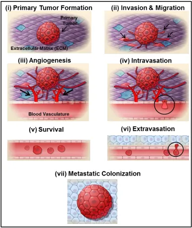
The metastatic cascade comprises a series of complex steps (Figure 1.2), the first step
in breast carcinoma development begins with cellular transformation of non-malignant
epithelial cells to a malignant phenotype, which has been suggested to be a result of acquired
genetic instability [24]. Cellular transformation involves uncontrolled growth regulation and
increased cell survival, due to evasion of apoptotic signaling [25].
Initial separation and spreading of cancer cells from the primary tumor involves a
process known as epithelial-to-mesenchymal transition (EMT), where cells lose expression
of epithelial markers and gain expression of mesenchymal markers, introducing a more
migratory and invasive phenotype [26]. Normally, epithelial cells are tightly organized in
continuous sheets of polarized cells, forming a barrier that is necessary to maintain
homeostasis and protect the body. Epithelial cells display an apical-basolateral polarity, with
distinct expression profiles at each plasma membrane [27]. Cell-cell adhesion and
communication is accomplished through the use of gap and tight junctions, desmosomes, and
adherens junctions [27]. However, during EMT cells lose polarity and cell-cell attachment
due to the loss of important adhesion molecules, such as E-cadherin [28]. At the same time,
markers, including N-cadherin, vimentin and matrix metalloproteases (MMPs) [29]. Cells
which have undergone EMT are capable of migrating away from the primary site through
cell-matrix interactions and cytoskeletal reorganization [30].
In addition to migration, cancer cells must invade through the basement membrane
that borders the tumor and into the surrounding extracellular matrix (ECM)(Figure 1.2) [31].
Thus, ECM remodeling must occur to allow cancer cell migration and invasion through the
tissue. This is accomplished by several proteolytic enzymes that are produced and secreted
by cancer cells [28]. The most common proteases involved in cancer metastasis are
urokinase-type plasminogen activators (uPA), cathepsins and MMPs [32,33,34]. Secretion
and activation of these proteolytic enzymes results in uncontrolled degradation of the
basement membrane and surrounding ECM, presenting the cancer cells with an avenue for
migration and metastasis. Additionally, the degradation of the ECM releases growth factors,
such as epidermal growth factor (EGF) and vascular endothelial growth factor (VEGF),
which can subsequently act on cancer cells to further promote angiogenesis and metastatic
progression [35].
The next step in the metastatic cascade is angiogenesis, the process by which new
blood vessels are formed from pre-existing vessels (Figure 1.2) [25]. Angiogenesis occurs
in normal physiological conditions, such as wound healing, the menstrual cycle and
placentation during pregnancy [36]. The process of angiogenesis is carefully controlled by a
balance of pro- and anti-angiogenic factors. However, in cancer, this balance is disrupted
and angiogenesis-inducing factors are abundantly released from tumor cells and the
surrounding stromal cells [36]. As the primary tumor continues to grow in size its need for
conditions are introduced. Hypoxia increases factors such as hypoxia inducible factor -1
(HIF-1), which has been shown to be increased in invasive breast carcinoma compared to in
situ tumors [37]. HIF-1 stimulates the release of pro-angiogenic factors, such as VEGF, from
tumor cells and from the degradation of the ECM [38]. VEGF subsequently binds to VEGF
receptors on endothelial cells of pre-existing vessels in the surrounding tissue, stimulating the
proliferation of endothelial cells and the development of a new vessel network within the
tumor itself and the surrounding tissue [37]. Furthermore, VEGF expression has been
inversely correlated to breast cancer patient survival [39]. Due to incomplete formation,
these new blood vessels have abnormal architecture and consequently there is increased
fenestration, enhancing the permeability and likelihood that cancer cells will enter the
circulation [36].
Once the tumor cells have invaded through the epithelial basement membrane and
surrounding ECM they will reach the newly formed tumor-associated vasculature. The
tumor cells must then transverse the endothelial cell basement membrane to reach the
vascular endothelial cells. It has been reported that blood vessel-associated macrophages
produce chemoattractants, such as EGF, to attract breast cancer cells to the vessel [40]. The
next step in the metastatic cascade is intravasation, the process by which these cancer cells
will enter the vessels through leaky endothelial cell junctions (Figure 1.2) [41]. Initially,
cancer cells adhere to endothelial cells, and then through endothelial cell retraction and
cancer cell migration, the cancer cell enters the vasculature by transendothelial migration
[41]. Additionally, cancer cells may also enter the circulation via the lymphatic system. In
contrast to the cardiovascular system, cells of the lymphatic system lack tight intracellular
junctions, and therefore lymph node metastases are more common earlier in tumor
circulation arrive in capillary beds in distant tissues, adhere to endothelial cells, and enter the
surrounding tissue by extravasation (Figure 1.2) [42]. Many of the metastasized cells
undergo apoptosis, however a subset of metastasized cells may survive and enter dormancy
or proliferation [43]. Like the primary tumor, the secondary tumor also requires
angiogenesis to occur to supply the growing tumor with oxygen and nutrients.
Paget’s century-old “seed and soil” theory proposes that the interaction between the
cancer cells or “seeds” and the organ microenvironment or “soil” explains the preference for
metastatic cells for certain organs [44]. This theory suggests that metastasis is multifactorial
and that the processes are influenced by factors independent from the tumor cell itself [44].
Additionally, it has been suggested that the organ microenvironment at the site of metastasis
can distinctly modify the gene-expression profile of cancer cells and consequently alter their
behaviour and growth [43]. Therefore, it is imperative that the tumor microenvironment and
its potential effects on the development of aggressive tumors be further investigated.
Specifically, new therapeutic strategies targeting the molecular changes that occur in regards
to expression of cell-cell and cell-ECM adhesion molecules and cell signaling cascades are
1.3 Tumor Microenvironment
The tumor microenvironment consists of the ECM, soluble factors and altered cell
types, including fibroblasts, endothelial cells and leukocytes [46]. These cells can interact
directly or through paracrine signaling with surrounding cells [47]. Like any organ, the
development and function of a tumor is the result of unique communication between
epithelial cells and surrounding stromal cells [47]. Evidence suggests that disruptions to the
ECM composition and structure may pave the way for tumor development or lead to
cancer-causing mutations [48]. In addition to degrading the normal ECM, stromal cells of the
microenvironment, primarily cancer-associated fibroblasts (CAFs), will deposit a new
tumor-associated ECM that consists of molecules such as fibronectin, tenascins and stiffer collagen
that will facilitate tumor cell motility [49]. Furthermore, studies have demonstrated that the
gene expression profile of the tumor-associated ECM is dramatically altered, including
upregulation of MMPs in invasive stroma compared to in situ stroma [50]. Additionally, due
to their altered signaling pathways, cancer cells may fail to interpret cues from the
microenvironment, and this can result in inappropriate cellular organization and growth [48].
For example, in three-dimensional (3D) culture, mammary epithelial cells will form
polarized acini-like structures, but malignant cells in 3D culture form disorganized colonies
[51]. These malignant cells will revert to organized polarized structures when cell-ECM β1
integrin signaling is inhibited [51]. Therefore, the influence of the tumor microenvironment,
especially cell-cell and cell-ECM interactions, has become an emerging target for therapeutic
1.3.1 Fibulins
The fibulin family consists of seven secreted glycoproteins associated with basement
membranes, elastic fibers and the ECM [52]. The fibulin family name is derived from the
Latin word fibula meaning clasp or buckle. It is fitting then, that fibulins function as
intramolecular bridges within ECM components, such as elastic fibres and basement
membranes [53]. Interestingly, the fibulins are highly conserved in species as evolutionarily
diverse as worms and humans [53]. Structural features of the fibulins include repeated
EGF-like domains and a unique C-terminal fibulin-type module (Figure 1.3), and these proteins
have been implicated in cell signaling and protein-protein interactions involved in cell
growth, adhesion and motility [54]. The physiological and pathological roles of many of the
fibulins have been extensively studied, however only within the last decade have roles for
fibulin-3 been discovered [54].
1.3.2 Fibulin-3
Fibulin-3, also known as EGF-containing fibulin-like extracellular matrix protein 1
(EFEMP1), interacts with tropoelastin and plays an important role in elastic fibre assembly
during development [52]. Fibulin-3 is strongly expressed in human heart and placenta and
weakly expressed in skeletal muscle, lung, brain and pancreas [55]. Fibulin-3 was also
shown to be expressed in the bone and cartilage of murine embryos, suggesting that fibulin-3
may play a role in skeletal development [56]. Additionally, fibulin-3 is highly expressed in
epithelial and endothelial cells [55]. The fibulin-3 promoter contains Sp1 transcription factor
binding sites, in addition to an estrogen response element, and estrogen has been shown to
ERα-positive MCF7 breast cancer cells repressed the expression of the gene for fibulin-3, referred
to as EFEMP1 [58].
The physiological roles of fibulin-3 in development and disease have been elucidated
by studying animal knockout models and heritable human disorders [54]. The phenotype of
the fibulin-3 knockout mouse involves reduced reproductivity, early aging, herniation and
lack of support for pelvic organs [59,60]. These observations are likely a result of reduced
elastic fibers in the fascia, a thin layer of connective tissue that separates and protects the
body’s organs [59]. Furthermore, a missense mutation in an EGF domain of fibulin-3 has
been discovered as the cause of early-onset macular degenerative diseases including Malattia
Leventinese and Doyne’s honeycomb retinal dystrophy [61]. These inherited macular
degenerative diseases resemble the most common cause of incurable blindness, age-related
macular degeneration, and are characterized by yellow-white deposits of ECM, called drusen,
that accumulate between Bruch’s membrane and the retinal pigment epithelium [52].
Interestingly, mutations in tissue inhibitor of metalloproteinases-3 (TIMP-3), an inhibitor of
MMPs, are also associated with macular degenerative diseases, and TIMP-3 has been shown
to bind fibulin-3 [62]. Lastly, fibulin-3 has been described as an indirect regulator of cell
growth through indirect protein interactions with the tumor suppressor differential
screening-selected gene aberrative in neuroblastoma (DAN) [63]. Fibulin-3 has also been shown to
promote cell proliferation by stimulating deoxyribonucleic acid (DNA) synthesis in vitro
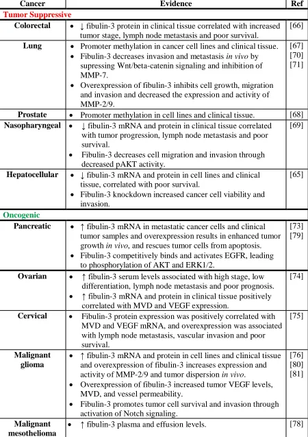
1.3.3 Fibulin-3 in Cancer
In the last five years, several studies have shown that fibulin-3 plays a role in
tumorigenesis. Fibulin-3 is differentially expressed in human cancers, and its effects have
been described as both tumor suppressive and oncogenic (Table 1.1) [65]. Fibulin-3
expression is decreased in colorectal [66], lung [67], prostate [68], nasopharyngeal [69] and
hepatocellular carcinomas [65]. In lung cancer, fibulin-3 downregulation promotes invasion
and metastasis through activation of Wnt/β -catenin signaling and MMP-7 [70]. Similarly,
Xu and colleagues showed that fibulin-3 negatively regulated lung cancer cell invasiveness
via p38-mitogen-activated protein kinase (MAPK) and MMP-2 and -9 [71]. In
nasopharyngeal carcinoma, fibulin-3 was shown to inhibit cell migration and invasion
through suppression of AKT phosphorylation [69]. In a fibulin-3 overexpression model in
pancreatic cancer cells, cancer stem cell markers were decreased, cells were resensitized to
cytotoxic agents and EMT was inhibited [72]. Thus, numerous studies have demonstrated
that fibulin-3 can have tumor suppressive roles in various human cancers.
Conversely, fibulin-3 has been reported to be upregulated in pancreatic [73],
ovarian[74] and cervical [75] carcinomas, as well as malignant gliomas [76] and malignant
mesothelioma [77]. Plasma and effusion fibulin-3 levels were reported to be increased in
patients with pleural mesothelioma compared to patients without pleural mesothelioma, and
therefore fibulin-3 has been proposed as a clinical biomarker for the disease [78].
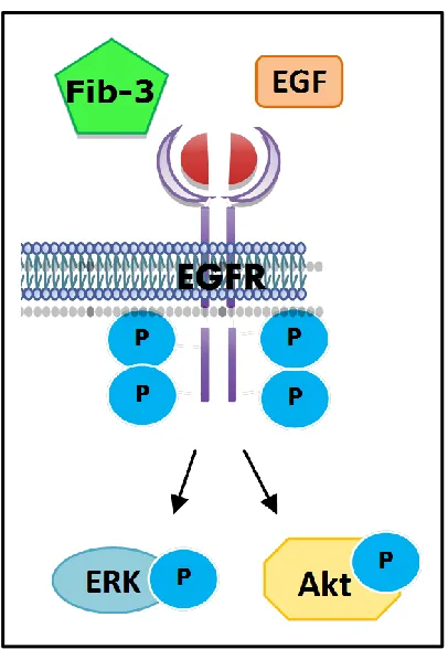
Interestingly, fibulin-3 has also been shown to bind cell surface receptors, specifically EGFR
in pancreatic carcinoma cells (Figure 1.4) [79]. Camaj and colleagues demonstrated that
fibulin-3 competes with EGF for binding to EGFR, and activates downstream extracellular
growth [79]. Fibulin-3 has pro-angiogenic roles in pancreatic [73] and ovarian [74]
carcinomas and malignant gliomas [80]. Fibulin-3 was upregulated in human ovarian
carcinoma tissues and positively correlated with microvessel density (MVD) and VEGF
expression [74]. Serum fibulin-3 levels were also elevated in ovarian carcinoma patients
compared to healthy controls and benign ovarian carcinoma patients, and were associated
with low differentiation, high stage and lymph node metastasis [74]. Pancreatic cancer cells
transfected with fibulin-3 displayed increased MVD, tumor growth and VEGF production in
vivo [73]. Additionally, tumor cells that were transfected with fibulin-3 displayed a decrease
in apoptosis and a shift from G0-G1 phase to S phase and mitosis, implicating fibulin-3 in
tumor cell survival and cell cycle progression [73]. Similar results were observed in an in
vivo model of malignant glioma in regards to VEGF secretion and MVD, and fibulin-3 was
found to be localized around tumor blood vessels [80]. Also, fibulin-3 stimulated endothelial
cell migration via a Notch-dependent signaling mechanism, a proliferative pathway
dysregulated in many cancers [80]. Malignant glioma cells and tumors overexpressing
fibulin-3 displayed increased expression and activity of MMP-2 and 9 [76]. Fibulin-3 has
also been shown to promote glioma cell survival and invasion through activation of Notch
signaling [81]. In contrast to Tong and colleagues, who suggested decreased fibulin-3 is
associated with poor prognosis in colorectal cancer, a recent study reports that fibulin-3
promotes colorectal cancer cell migration and invasion and tumor growth via a p38 MAPK
mechanism [82].
Although a role for fibulin-3 in breast cancer invasion is unknown, fibulin-3 has been
reported to be overexpressed in breast cancer effusions [83]. However, whether fibulin-3
signaling regulates TNBC cell migration and invasion, important processes required for
Table 1.1: Role of fibulin-3 in various cancers.
Cancer Evidence Ref
Tumor Suppressive
Colorectal ↓ fibulin-3 protein in clinical tissue correlated with increased
tumor stage, lymph node metastasis and poor survival.
[66]
Lung Promoter methylation in cancer cell lines and clinical tissue. Fibulin-3 decreases invasion and metastasis in vivo by
supressing Wnt/beta-catenin signaling and inhibition of MMP-7.
Overexpression of fibulin-3 inhibits cell growth, migration and invasion and decreased the expression and activity of MMP-2/9.
[67] [70] [71]
Prostate Promoter methylation in cell lines and clinical tissue. [68]
Nasopharyngeal ↓ fibulin-3 mRNA and protein in clinical tissue correlated
with tumor progression, lymph node metastasis and poor survival.
Fibulin-3 decreases cell migration and invasion through decreased pAKT activity.
[69]
Hepatocellular ↓ fibulin-3 mRNA and protein in cell lines and clinical tissue, correlated with poor survival.
Fibulin-3 knockdown increased cancer cell viability and invasion.
[65]
Oncogenic
Pancreatic ↑ fibulin-3 mRNA in metastatic cancer cells and clinical
tumor samples and overexpression results in enhanced tumor growth in vivo, and rescues tumor cells from apoptosis. Fibulin-3 competitively binds and activates EGFR, leading
to phosphorylation of AKT and ERK1/2.
[73] [79]
Ovarian ↑ fibulin-3 serum levels associated with high stage, low
differentiation, lymph node metastasis and poor prognosis. ↑ fibulin-3 mRNA and protein in clinical tissue positively
correlated with MVD and VEGF expression.
[74]
Cervical Fibulin-3 protein expression was positively correlated with MVD and VEGF mRNA, and overexpression was associated with lymph node metastasis, vascular invasion and poor survival.
[75]
Malignant glioma
↑ fibulin-3 mRNA and protein in cell lines and clinical tissue and overexpression of fibulin-3 increases expression and activity of MMP-2/9 and tumor dispersion in vivo.
Overexpression of fibulin-3 increased tumor VEGF levels, MVD, and vessel permeability.
Fibulin-3 promotes tumor cell survival and invasion through activation of Notch signaling.
[76] [80] [81]
Malignant mesothelioma
1.4 Matrix Metalloproteinases
MMPs are a family of zinc-dependent endopeptidases that are capable of degrading
other proteinases, proteinase inhibitors, cell surface receptors, cell-cell adhesion molecules
and essentially all ECM proteins [84]. Thus, since tumor cells must cross multiple ECM
barriers to metastasize to distant sites, and upregulation of MMPs is associated with invasive
ability in many cancers, MMPs can promote metastasis [85]. Additionally, a role for MMPs
has been demonstrated in EMT, angiogenesis and intravasation [86,87,88].
1.4.1 Matrix Metalloproteinase-9 in Cancer
MMP-9 is secreted as an inactive pro-MMP that can be activated by plasmin,
trypsin-2, MMP-trypsin-2, MMP-13 and MMP-3 [89]. There are numerous substrates for MMP-9, a few of
which include collagens, elastin, plasminogen and kisspeptins [90]. MMP-9 is primarily
produced and secreted by stromal cells, however cancer cells may stimulate stromal cell
production through paracrine signaling [34]. Increased MMP-9 expression and activity has
been demonstrated in many cancers, including brain, urogenital, lung, skin, colorectal and
breast [91]. Data from transgenic mice expressing an MMP-9 promoter-driven
β-galactosidase transgene demonstrate that the MMP-9 promoter activity is induced during
invasive cancer, but not in situ tumors [92]. MMP-9 has also been proposed as a prognostic
biomarker in breast cancer due to its significant association with aggressive subtypes of
breast cancer, such as TNBC, and its correlation with metastasis and relapse [93]. It has been
demonstrated that MMP-9 regulates cell invasion downstream of ERK signaling in glioma
[94] and breast cancer (ERα-positive MCF7 and TNBC MDA-MB-231) cells [95].
Moreover, it has been reported that EGF stimulates MMP-9 expression and activity in an
Despite its role in invasion, MMP-9 does more than just degrade ECM components,
MMP-9 can also regulate signaling pathways such as proliferation, survival and angiogenesis
[90]. The hemopexin (PEX) domain of both, inactive and active MMP-9 allows it to dock to
the hyaluronan receptor CD44 and initiate intracellular signaling cascades (Figure 1.5) [97].
Thus, in normal keratinocytes and malignant cells, proteolytically active MMP-9 binds to
CD44 on the cell surface and cleaves latent transforming growth factor-β (TGF-β) into an
active form, allowing it to activate its receptor and lead to enhanced tumor growth [97].
Catalytically inactive MMP-9 has been shown to increase leukemia cell survival after
docking to integrin and CD44 and activating intracellular anti-apoptosis pathways [98]. Both
active and pro-MMP-9 have been implicated in breast cancer cell migration through
interaction with CD44 [99,100]. Bourguignon and colleagues showed that CD44 is closely
associated with active MMP-9 in invadopodia, actin-rich protrusions of the plasma
membrane that are associated with degradation of the ECM [99]. Similarly, Dufour and
colleagues demonstrated that pro-MMP-9 interacts with CD44 on tumor epithelial cells,
leading to EGFR activation and phosphorylation of downstream signaling molecules, such as
ERK, AKT, and focal adhesion kinase (FAK) to increase cell migration [100]. MMP-9 has
also been shown to induce angiogenesis in pancreatic islet cells by stimulating VEGF release,
and MMP inhibitors and MMP-9 knockout decreased the number of angiogenic islets and
tumor burden [101]. MMP-9 has also been implicated in the process of intravasation, since
MMP inhibition significantly reduced intravasation and only cancer cells expressing MMP-9
intravasated [88].
Thus, due to the ability of MMPs to facilitate invasion and metastasis, inhibitors have
been developed to target their proteolytic activity in the hopes of preventing metastasis.
a result of their lack of selectivity towards MMPs [90]. However, our knowledge of MMPs
has grown considerably, particularly in regards to their non-proteolytic abilities, interactions
with cell surface proteins and resulting downstream signaling cascades. New
pharmacological inhibitors are focusing on blocking the PEX domain from binding to cell
surface proteins and therefore preventing subsequent effects on cell survival, migration and
angiogenesis [102]. This strategy has already been used to target MMP-9 [89,100].
Synthetic peptides that bind specific sites within the MMP-9 PEX domain were shown to
inhibit cell motility in both human fibrosarcoma and breast cancer cell lines [100]. Similarly,
a small inhibitor that prevents PEX from binding to cell surface integrins decreases cell
migration and tumor growth [103]. These studies suggest that pharmacologically targeting
the PEX domain of MMP-9 may be clinically beneficial alone or in combination with other
therapeutics to effectively prevent tumor growth and metastasis.
MMP-9 serum levels have been shown to be high in breast cancer compared to
benign breast disease and healthy controls [104]. Additionally, high MMP-9 serum levels
were associated with lymph node metastasis, increased tumor stage, and lower overall
survival [104]. Interestingly, fibulin-3 has been shown to regulate the expression and activity
of MMP-9 in malignant glioma, however the role of fibulin-3 in regulating MMP-9 in breast
1.5 Metastasis Suppressor Genes
Recently, new groups of genes have been classified as metastasis activator or
metastasis suppressor genes. These genes differ from oncogenes and tumor suppressor genes
in that they do not influence primary tumor growth, but rather are involved in metastasis of
cancer cells to distant tissues [105]. Metastasis requires cancer cells to undergo a series of
processes to successfully colonize secondary sites, therefore a gene that inhibits even one of
these processes would effectively block metastasis [106]. There have been over 150 genes
discovered to be involved in breast cancer tumor development and progression, however only
6 metastasis suppressor genes have been shown to function in vivo, in metastasis assays [22].
These include NME1, KAI1, BRMS1, MKK4, E-cadherin and KISS1 [22]. Evidently,
metastasis suppressor genes provide a promising target for pharmacological interventions to
prevent metastasis in cancer patients.
1.5.1 KISS1R Signaling
The metastasis suppressor gene, KISS1, encodes a 145-residue kisspeptin protein that
is rapidly cleaved into shorter secreted peptides termed kisspeptin-10 (KP-10), kisspeptin-13,
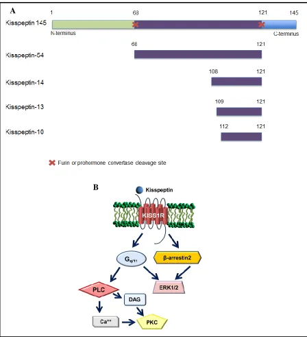
kisspeptin-14 and kisspeptin-54 (Figure 1.6A) [107]. The shorter kisspeptins are
N-terminally truncated peptides of kisspeptin-145 cleaved by furin, MMP-2, -9, and -14 or
prohormone convertases [108]. KP-10 is the shortest biologically active kisspeptin, and is
highly conserved among vertebrates [109]. Kisspeptins are the endogenous ligand for the
kisspeptin receptor (KISS1R) [107,110,111]. KISS1R has a single high affinity binding site
for kisspeptins, and all kisspeptins bind with similar affinities and potencies
including the placenta, pituitary, pancreas, brain, small intestine, liver, lung, kidney, testis,
ovary and breast [107,110,111].
KISS1R is a G-protein coupled receptor (GPCR) that signals via a Gq/11-coupled
mechanism leading to activation of phospholipase C (PLC) (Figure 1.6B). PLC activation
leads to increased production of inositol 1,4,5-triphosphate (IP3) and diacylglycerol (DAG)
which results in increased intracellular calcium and subsequent activation of protein kinase C
(PKC) and ERK1/2 [107,110,111]. In addition, KISS1R has been shown to activate ERK1/2
through a G-protein independent and β-arrestin2-dependent pathway [114]. KISS1R
contains residues in the second intracellular loop and cytoplasmic tail that allow it to
constitutively interact with β-arrestin1 and β-arrestin2 [115]. Also, desensitization and
internalization of KISS1R is mediated by G-protein-coupled receptor serine/threonine kinase
2 (GRK2) and β-arrestins [116]. Bianco and colleagues observed persistent expression of
KISS1R at the plasma membrane, even after KP-10 stimulation, and there was no
co-localization of KISS1R with lysosomal markers, indicating that ligand-induced KISS1R
internalization does not result in lysosomal degradation [117]. Instead, KISS1R is recycled
to the cell surface, prolonging KISS1R signaling, and suggesting that KISS1R may have
constitutive activity [117].
KP/KISS1R signaling plays an important physiological role in the regulation of the
reproductive axis and the initiation of puberty [118]. Studies have shown that
loss-of-function mutations in KISS1R result in hypogonadotropic hypogonadism in humans [116].
Hypogonadotropic hypogonadism is characterized by diminished functional activity of the
gonads and infertility due to the insufficiency of the pituitary to secrete gonadotropins:
signaling regulates the reproductive axis by stimulating the release of gonadotropin-releasing
hormone (GnRH) [118]. Pulsatile GnRH secretion then stimulates the pituitary to synthesize
and secrete FSH and LH [119]. KP/KISS1R signaling has also been implicated in pregnancy
and placentation. The circulating serum levels of KP increase approximately 1000 fold in the
first trimester and an amazing 10,000 fold in the third trimester, signifying an important role
for KP throughout pregnancy [120]. Moreover, KP has been shown to inhibit trophoblast
migration and invasion through decreased MMP-2 [121]. Consequently, KP has been
proposed to play a critical role in regulating placental invasion of the uterine wall during
implantation [121]. Due to the emergence of KISS1R as a clinically relevant GPCR, it is
vital that the molecular pathways regulating KISS1R signaling be further investigated in
Figure 1.6. Kisspeptins (KP) and KISS1R signaling cascades. (A) Cleavage of kisspeptin-145 results in the production of smaller peptides, termed KP-54, KP-14, KP-13 and KP-10. (B) The kisspeptin receptor (KISS1R) is a Gq/11-coupled receptor that upon binding of its
ligand, KP, will activate phospholipase C (PLC) and extracellular signal-regulated kinase (ERK1/2). Activation of PLC will lead to production of diacylglycerol (DAG) and increased intracellular calcium which will result in activation of protein kinase C (PKC). Additionally, KISS1R can also signal independently of G-proteins to activate ERK1/2 in a β-arrestin2 dependent mechanism.
A
1.5.2 KISS1R Signaling in Cancer
The KP/KISS1R system functions as a metastasis suppressor in melanoma [105],
pancreatic [122], bladder [123], ovarian [124], endometrial [124], esophageal [125], lung
[126] and gastric cancers [127]. These cancers displayed decreased expression of KISS1 and
/or KISS1R in tumor tissue compared to non-malignant tissue, and this observation correlated
with decreased patient prognosis. KISS1 signaling was first studied in melanoma and was
found to be expressed in non-metastatic cells, but was absent in metastatic melanoma cell
lines [105]. KISS1 mRNA expression was also lower in pancreatic carcinoma tissue in
comparison to non-malignant tissue [122]. When stimulated with exogenous KP, pancreatic
cancer cells exhibited ERK1 activation and decreased migration, however no effect on cell
proliferation was observed [122]. In gastric cancer, KP-145 protein expression was reduced
in metastatic tissues from the lymph nodes and liver compared to the primary tumor [127].
In bladder [123] and ovarian [128] carcinomas, decreased expression of KISS1 mRNA was
associated with poor patient survival. In a fibrosarcoma cell line, exogenous expression of
KISS1 resulted in decreased MMP-9 expression and activity, as well as reduced invasion
[129]. The observed decrease in MMP-9 expression was attributed to diminished nuclear
factor kappa-light-chain-enhancer of activated B cells (NFκB) transcription factor binding to
the promoter of MMP-9 as a result of KISS1 expression [129]. Thus, KISS1R undeniably
1.5.3 KISS1R Signaling in Breast Cancer
In contrast to its role as a metastasis suppressor in numerous cancers, KP/KISS1R
signaling has been shown to play detrimental roles in breast cancer. The first study looked at
clinical KISS1 and KISS1R mRNA expression in breast tumor tissue and discovered that
KISS1 expression was elevated in breast cancer tissue compared to non-malignant mammary
tissue [130]. However, this study did not examine hormone receptor status. Additionally,
breast tumors positive for lymph node metastasis displayed increased KISS1 mRNA
compared to lymph node negative tumors [130]. However, no significant difference in
KISS1R mRNA expression was observed [130]. Exogenous expression of KISS1 in the
human breast cancer cell line MDA-MB-231 enhanced motility, invasive ability and reduced
cell adhesion, therefore KISS1 overexpression induced a more aggressive phenotype [130].
Lastly, this study demonstrated the positive correlation between KISS1 mRNA expression
and poor patient prognosis [130].
The second study examined ERα status and KISS1 expression in breast tumors.
Marot and colleagues demonstrated that ERα-positive breast tumors expressed sevenfold less
KISS1 than negative breast tumors [131]. This study showed that treatment of
ERα-positive MCF7 and T47D breast cancer cells with the estrogen receptor antagonist,
tamoxifen, stimulated KISS1 and KISS1R mRNA expression [131]. Exogenous expression of
ERα in the ERα-negative breast cancer cell line MDA-MB-231 resulted in diminished KISS1
mRNA expression [131]. Similarly, ERα-positive breast tumors in postmenopausal women
treated with tamoxifen also displayed high KISS1 and KISS1R mRNA levels that was
associated with shorter relapse-free survival [131]. Furthermore, KISS1 mRNA expression in
KISS1 and KISS1R mRNA levels in breast tumors may be a prognostic marker for
anti-estrogen therapy resistance [131]. Therefore clinical data suggests that KP/KISS1R signaling
may play an influential role in breast cancer progression.
A 2011 study investigated a role for mouse KP/KISS1R signaling in regulating breast
cancer metastasis. Breast cancer was induced in mice expressing the polyoma middle T
antigen (PyMT) under the control of the mouse mammary tumor virus (MMTV) promoter to
explore the role of KISS1R expression in breast cancer progression and metastasis [132].
Results from this study indicated that PyMT-induced breast tumor development and lung
metastases were delayed in the KISS1R heterozygous (PyMT/Kiss1r+/−) mouse in
comparison to the wild-type (PyMT/Kiss1r+/+) mouse [132]. Interestingly, KISS1R
heterozygous (PyMT/Kiss1r+/−) tumors displayed a significant reduction in MMP-9 mRNA
compared to wild-type (PyMT/Kiss1r+/+) tumors [132]. Furthermore, heterozygous mouse
primary breast cancer cells exhibited decreased cellular invasion in vitro [132]. Accordingly,
this study proposes that KP/KISS1R signaling regulates breast tumor development,
progression and metastasis in a mouse model.
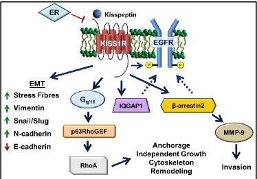
Studies from our laboratory have investigated the underlying molecular mechanisms
regulated by KP/KISS1R in breast cancer metastasis (Figure 1.7) [133,134]. Our first study
demonstrated that treatment of human TNBC cell lines, MDA-MB-231 and Hs578T, with
KP-10, stimulated migration and invasion [133]. Interestingly, KP-10 stimulation also
increased MMP-9 secretion and activity [133]. We found that activation of KISS1R
signaling promoted the activation of EGFR via a β-arrestin2 dependent pathway [133].
Lastly, fluorescence resonance energy transfer (FRET) analysis determined that KISS1R
interaction [133]. In our second study, we found that exogenous KISS1R expression or
KP-10 treatment induced EMT and cell invasion in ERα-negative non-malignant mammary
epithelial MCF10A cells [134]. This demonstrates that KP signaling stimulates malignant
transformation. KISS1R overexpression in ERα-negative SKBR3 breast cancer cells
stimulated extravasation in vivo using the chick chorioallantoic membrane assay [134]. Most
interestingly, KP-10 failed to stimulate invasion and EGFR activation in ERα-positive MCF7
and T47D breast cancer cells, leading us to conclude that ERα negatively regulates KISS1R
signaling in breast cancer [134]. This concept was validated by exogenously expressing ERα
in the ERα-negative MDA-MB-231 breast cancer cells, and upon estradiol stimulation
KP-10-induced invasion and EGFR activation was blocked, and KISS1R protein expression was
decreased [134]. This demonstrates that ERα negatively regulates KISS1R expression and
signaling. Finally, we discovered that KISS1R tranactivation of EGFR is dependent upon an
actin cytoskeletal binding protein, IQ-motif-containing GTPase activating protein 1
(IQGAP1), and that IQGAP1 is a novel binding partner of KISS1R [134]. However, the
molecular mechanism by which KP/KISS1R signaling stimulates TNBC cell invasion is still
1.5.4 β-arrestins
β-arrestins are ubiquitously expressed proteins that function as signaling adaptors
and molecular scaffolds [135], and play a crucial role in the phosphorylation of intracellular
targets. The two β-arrestin isoforms (β-arrestin1 and β-arrestin2) display a high degree of
homology (~70% of similar sequence identity) and may function similarly to regulate GPCR
desensitization and internalization [136]. β-arrestins can also regulate the signaling and
trafficking of other types of receptors, such as EGFR and the TGF-β receptor [137].
β-arrestins are capable of scaffolding numerous cell signaling molecules, including many that
have oncogenic roles and regulate the cytoskeleton, such as proto-oncogene tyrosine-protein
kinase Src (Src), ERK1/2 and AKT [138].
β-arrestins are also emerging regulators of breast cancer progression and metastasis.
β-arrestin1 was shown to regulate phosphorylation of ERK1/2 in ERα-positive MCF7 breast
cancer cells, and arrestin1 depletion resulted in decreased tumor cell proliferation [139].
β-arrestin1 has also been shown to promote survival of breast cancer cells in hypoxic
conditions via increased expression of VEGF and positively regulate breast cancer metastatic
spread in nude mice [136]. Furthermore, depletion of both β-arrestin1 and β-arrestin2 in
MDA-MB-231 breast cancer cells decreased tumor growth and increased mouse survival rate
in experimental metastasis assays [136]. Another breast cancer study has linked amplified
stromal β-arrestin1 expression with poor patient prognosis and increases in HIF-1 expression,
tumor growth and metastases [140]. Clinical data from our lab has shown that the expression
of β-arrestin2 is elevated in invasive human breast cancer, and that β-arrestin2 regulates the
Rap1A [141,142]. Lastly, we have shown that KP-10-induced EGFR transactivation and cell
invasion is dependent on β-arrestin2 [133].
1.6 Rationale, Hypothesis and Objectives
1.6.1 Rationale
Fibulin-3 is overexpressed in breast cancer effusions [83], however, whether plasma
fibulin-3 concentrations change in TNBC patients is currently unknown. Despite evidence
that fibulin-3 is capable of binding and activating EGFR [79], as well as upregulating
MMP-9 [76], a role for fibulin-3 in TNBC cell invasion has not yet been determined. Previous
studies from our lab have demonstrated that in TNBC cells, KISS1R signaling stimulates cell
invasion by stimulating EGFR activation and MMP-9 secretion and activity [133,134]. Here
we will investigate whether fibulin-3 regulates KISS1R-induced TNBC cell invasion and the
underlying mechanism(s).
1.6.2 Hypothesis
KISS1R stimulates TNBC cell invasion via a fibulin-3 dependent mechanism.
1.6.3 Objectives
To determine if:
1) Plasma fibulin-3 levels are increased in TNBC patients compared to healthy
controls.
2) Fibulin-3 regulates KISS1R-induced TNBC cell invasion, and uncover the
potential underlying mechanism(s).
1.7 References
†
http://www.cbcf.org/ontario/AboutBreastCancerMain/FactsStats/Pages/Breast-Cancer-Canada.aspx
1. Singletary, S. E., & Connolly, J. L. (2006). Breast cancer staging: working with the sixth edition of the AJCC Cancer Staging Manual. CA Cancer J Clin, 56(1), 37-47; quiz 50-31.
2. Bland, K. I., Menck, H. R., Scott-Conner, C. E., Morrow, M., Winchester, D. J., & Winchester, D. P. (1998). The National Cancer Data Base 10-year survey of breast carcinoma treatment at hospitals in the United States. Cancer, 83(6), 1262-1273. 3. Sakorafas, G. H., & Tsiotou, A. G. (2000). Ductal carcinoma in situ (DCIS) of the breast:
evolving perspectives. Cancer Treat Rev, 26(2), 103-125. doi: 10.1053/ctrv.1999.0149
4. Fantozzi, A., & Christofori, G. (2006). Mouse models of breast cancer metastasis. Breast Cancer Res, 8(4), 212. doi: 10.1186/bcr1530
5. Bagaria, S. P., Ray, P. S., Sim, M. S., Ye, X., Shamonki, J. M., Cui, X., & Giuliano, A. E. (2014). Personalizing breast cancer staging by the inclusion of ER, PR, and HER2. JAMA Surg, 149(2), 125-129. doi: 10.1001/jamasurg.2013.3181
6. Zhang, M., Zhang, X., Zhao, S., Wang, Y., Di, W., Zhao, G., . . . Zhang, Q. (2013). Prognostic value of survivin and EGFR protein expression in triple-negative breast cancer (TNBC) patients. Target Oncol. doi: 10.1007/s11523-013-0300-y
7. Barbieri, V., Sanpaolo, P., & Genovesi, D. (2011). Prognostic impact of triple negative phenotype in conservatively treated breast cancer. Breast J, 17(4), 377-382. doi: 10.1111/j.1524-4741.2011.01100.x
8. Brady-West, D. C., & McGrowder, D. A. (2011). Triple negative breast cancer: therapeutic and prognostic implications. Asian Pac J Cancer Prev, 12(8), 2139-2143. 9. Elnashar, A. T., Ali el, S. M., & Gaber, A. (2012). The prognostic value of triple negative in stage II/III breast cancer. J Oncol Pharm Pract, 18(1), 68-75. doi: 10.1177/1078155211398299
10. Carey, L. A., Perou, C. M., Livasy, C. A., Dressler, L. G., Cowan, D., Conway, K., . . . Millikan, R. C. (2006). Race, breast cancer subtypes, and survival in the Carolina Breast Cancer Study. JAMA, 295(21), 2492-2502. doi: 10.1001/jama.295.21.2492 11. Dent, R., Trudeau, M., Pritchard, K. I., Hanna, W. M., Kahn, H. K., Sawka, C. A., . . .
Narod, S. A. (2007). Triple-negative breast cancer: clinical features and patterns of recurrence. Clin Cancer Res, 13(15 Pt 1), 4429-4434. doi: 10.1158/1078-0432.ccr-06-3045
12. Ihemelandu, C. U., Leffall, L. D., Jr., Dewitty, R. L., Naab, T. J., Mezghebe, H. M., Makambi, K. H., . . . Frederick, W. A. (2007). Molecular breast cancer subtypes in premenopausal African-American women, tumor biologic factors and clinical outcome. Ann Surg Oncol, 14(10), 2994-3003. doi: 10.1245/s10434-007-9477-6 13. Atchley, D. P., Albarracin, C. T., Lopez, A., Valero, V., Amos, C. I., Gonzalez-Angulo,
A. M., . . . Arun, B. K. (2008). Clinical and pathologic characteristics of patients with BRCA-positive and BRCA-negative breast cancer. J Clin Oncol, 26(26), 4282-4288. doi: 10.1200/jco.2008.16.6231
receptor-negative/HER-2-negative patient: a promising candidate for epidermal growth factor receptor-targeted therapy? Breast J, 12(4), 360-362. doi: 10.1111/j.1075-122X.2006.00276.x
15. Yarden, Y. (2001). The EGFR family and its ligands in human cancer. signalling mechanisms and therapeutic opportunities. Eur J Cancer, 37 Suppl 4, S3-8.
16. Pan, D., & Lin, X. (2013). Epithelial growth factor receptor-activated nuclear factor kappaB signaling and its role in epithelial growth factor receptor-associated tumors. Cancer J, 19(6), 461-467. doi: 10.1097/ppo.0000000000000001
17. Heitz, F., Harter, P., Lueck, H. J., Fissler-Eckhoff, A., Lorenz-Salehi, F., Scheil-Bertram, S., . . . du Bois, A. (2009). Triple-negative and HER2-overexpressing breast cancers exhibit an elevated risk and an earlier occurrence of cerebral metastases. Eur J Cancer, 45(16), 2792-2798. doi: 10.1016/j.ejca.2009.06.027
18. Peddi, P. F., Ellis, M. J., & Ma, C. (2012). Molecular basis of triple negative breast cancer and implications for therapy. Int J Breast Cancer, 2012, 217185. doi: 10.1155/2012/217185
19. Hanahan, D., & Weinberg, R. A. (2011). Hallmarks of cancer: the next generation. Cell, 144(5), 646-674. doi: 10.1016/j.cell.2011.02.013
20. Greene FL, P. D., Fleming ID, Fritz AG, Balch CM, Haller DG, Morrow M (Ed.). (2002) (6th ed.). New York: Springer.
21. Mehlen, P., & Puisieux, A. (2006). Metastasis: a question of life or death. Nat Rev Cancer, 6(6), 449-458. doi: 10.1038/nrc1886
22. Welch, D. R., Steeg, P. S., & Rinker-Schaeffer, C. W. (2000). Molecular biology of breast cancer metastasis. Genetic regulation of human breast carcinoma metastasis. Breast Cancer Res, 2(6), 408-416.
23. Solomayer, E. F., Diel, I. J., Meyberg, G. C., Gollan, C., & Bastert, G. (2000). Metastatic breast cancer: clinical course, prognosis and therapy related to the first site of metastasis. Breast Cancer Res Treat, 59(3), 271-278.
24. Fidler, I. J. (2003). The pathogenesis of cancer metastasis: the 'seed and soil' hypothesis revisited. Nat Rev Cancer, 3(6), 453-458. doi: 10.1038/nrc1098
25. Liotta, L. A., Steeg, P. S., & Stetler-Stevenson, W. G. (1991). Cancer metastasis and angiogenesis: an imbalance of positive and negative regulation. Cell, 64(2), 327-336. 26. Tse, J. C., & Kalluri, R. (2007). Mechanisms of metastasis: epithelial-to-mesenchymal
transition and contribution of tumor microenvironment. J Cell Biochem, 101(4), 816-829. doi: 10.1002/jcb.21215
27. Marchiando, A. M., Graham, W. V., & Turner, J. R. (2010). Epithelial barriers in homeostasis and disease. Annu Rev Pathol, 5, 119-144. doi: 10.1146/annurev.pathol.4.110807.092135
28. Meyer, T., & Hart, I. R. (1998). Mechanisms of tumour metastasis. Eur J Cancer, 34(2), 214-221.
29. Micalizzi, D. S., Farabaugh, S. M., & Ford, H. L. (2010). Epithelial-mesenchymal transition in cancer: parallels between normal development and tumor progression. J Mammary Gland Biol Neoplasia, 15(2), 117-134. doi: 10.1007/s10911-010-9178-9 30. Gupta, G. P., & Massague, J. (2006). Cancer metastasis: building a framework. Cell,
127(4), 679-695. doi: 10.1016/j.cell.2006.11.001
31. Hunter, K. W., Crawford, N. P., & Alsarraj, J. (2008). Mechanisms of metastasis. Breast Cancer Res, 10 Suppl 1, S2. doi: 10.1186/bcr1988