Clinical Ophthalmology
Preliminary results of an intravitreal
dexamethasone implant (Ozurdex
®
) in patients
with persistent diabetic macular edema
Elena Pacella1
Anna Rita Vestri2
Roberto Muscella1
Maria Rosaria Carbotti1
Massimo Castellucci1
Luigi Coi1 Paolo Turchetti3
Fernanda Pacella1
1Department of Sense Organs,
Faculty of Medicine and Dentistry, Sapienza University of Rome, Rome, Italy; 2Department of Public Health
and Infectious Diseases, Faculty of Pharmacy and Medicine, Sapienza University of Rome, Rome, Italy;
3National Institute for Health,
Migration and Poverty (INMP/ NIHMP), Rome, Italy
Correspondence: Elena Pacella Department of Sense Organs, Faculty of Medicine and Dentistry, “Sapienza” University of Rome, Viale del Policlinico Clinica Oculistica, 00161 Rome, Italy Tel +39 0649975303 or
+39 336783409
Email elena.pacella@uniroma1.it
Background: To evaluate the efficacy and safety of an intravitreal dexamethasone implant
(Ozurdex®; Allergan Inc, Irvine, CA, USA) in patients with persistent diabetic macular edema
(DME) over a 6-month follow-up period.
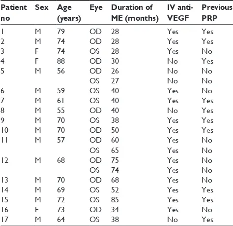
Methods: Seventeen patients (20 eyes) affected by DME were selected. The mean age was
67 ± 8 years, and the mean duration of DME was 46.3 ± 18.6 months. The eligibility criteria
were: age $18, a best-corrected visual acuity between 5 and 40 letters, and macular edema with
a thickness of $275 µm. Thirteen patients had also previously been treated with anti-vascular
endothelial growth factor medication.
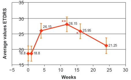
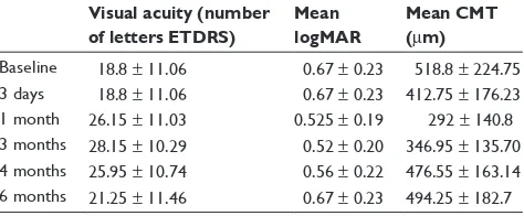
Results: The mean ETDRS (Early Treatment Diabetic Retinopathy Study) value went from
18.80 ± 11.06 (T0) to 26.15 ± 11.03 (P= 0.04), 28.15 ± 10.29 (P= 0.0087), 25.95 ± 10.74
(P= 0.045), 21.25 ± 11.46 (P= 0.5) in month 1, 3, 4, and 6, respectively. The mean logMAR
(logarithm of the minimum angle of resolution) value went from 0.67 ± 0.23 (at T0) to
0.525 ± 0.190 (P= 0.03), 0.53 ± 0.20 (P= 0.034), and 0.56 ± 0.22 (P= 0.12) in month 1, 3,
and 4, respectively, to finally reach 0.67 ± 0.23 in month 6. The mean central macular thickness
value improved from 518.80 ± 224.75 µm (at T0) to 412.75 ± 176.23 µm, 292.0 ± 140.8 µm
(P, 0.0001), and 346.95 ± 135.70 (P= 0.0018) on day 3 and in month 1 and 3, respectively,
to then increase to 476.55 ± 163.14 µm (P= 0.45) and 494.25 ± 182.70 µm (P= 0.67) in
month 4 and 6.
Conclusion: The slow-release intravitreal dexamethasone implant, Ozurdex, produced signifi-cant improvements in best-corrected visual acuity and central macular thickness from the third day of implant in DME sufferers, and this improvement was sustained until the third month.
Keywords: macular edema, diabetes, intravitreal implant, Ozurdex®
Introduction
Diabetic macular edema (DME) results from the exuding and accumulation of extracel-lular liquid and proteins in the macula1–3 following structural changes to the endothelium
of the retinal blood vessels that lead to the rupture of the hematoretinal barrier and thus to an increase in vascular permeability.4 The pathological neo-angiogenesis at the
basis of such alterations is provoked by the increase in cytokines (like interleukin-6 and -8), prostaglandins, and vascular endothelial growth factor (VEGF).4,5 Laser
pho-tocoagulation, considered for a long time as the main treatment option for DME, may lead to paracentral deficits of the visual field and reduced color vision and sensitivity to contrast.1,2 For these reasons, intravitreal therapies with anti-VEGF have been
con-sidered as an efficient treatment strategy for patients affected by DME,5,7 with drugs
such as pegaptanib6,9,10 ranibizumab,8 and bevacizumab11 being principally used.
Dove
press
M E T H O D O L O g y open access to scientific and medical research
Open Access Full Text Article
Clinical Ophthalmology downloaded from https://www.dovepress.com/ by 118.70.13.36 on 21-Aug-2020
For personal use only.
Number of times this article has been viewed
This article was published in the following Dove Press journal: Clinical Ophthalmology
However, not all patients respond favorably to intravitreal anti-VEGF treatment. Steroids reduce inflammatory media-tors through a more widespread action that blocks VEGFs, inflammatory cytokines, and prostaglandins.12
Our study investigates an intravitreal dexamethasone implant (Ozurdex®; Allergan Inc, Irvine, CA, USA) and its
efficacy as a treatment for DME.13 This implant was
devel-oped to guarantee sustained levels of dexamethasone in the posterior section of the eye for a period of 6 months.14,15
Ozurdex has recently been approved by the US Food and Drug Administration and by the European Union (EU), and is licensed in all EU countries for the treatment of macular edema (ME) following retinal vein occlusion.16,17
Nonethe-less, there is evidence of its efficacy in multiple clinical applications including DME, ME associated to uveitis or Irvine-Gass syndrome, DME in eyes having undergone vitrectomy,18 noninfectious vitritis, and as an adjuvant
therapy for age-related macular degeneration.19 In this study,
we evaluate the effects of a single intravitreal injection of Ozurdex, through a 6-month follow-up time period, in eyes affected with persistent DME.
Materials and methods
The study was conducted at the Policlinico Umberto I Hospital of “Sapienza” University of Rome. The eligibil-ity criteria were: age $18, a best-corrected visual acuity (BCVA) between 5 (corresponding to 1/10, logarithm of the minimum angle of resolution [logMAR] 1.0 or more) and 40 (corresponding to 5/10, logMAR 0.3 or less) let-ters, and macular edema with a thickness of $275 µm. The initial BCVA before the implant (at T0) was an average of 18.80 ± 11.06 letters (logMar 0.67 ± 0.23), and the mean central macular thickness (CMT) was 518.80 ± 224.75 µm. All patients had persistent DME although 13 of the patients recruited had previously undergone treatment with anti-VEGF: three with bevacizumab (Avastin®; Roche, Basel,
Switzerland), two with pegaptanib (Macugen®; Eyetech
Pharmaceuticals, Inc, New York, NY, USA), and eight with ranibizumab (Lucentis®; Genentech Inc, South San
Francisco, CA, USA) in the 3 months prior to investigation. The remaining patients presented counter recommendations to intravitreal injections of anti-VEGF (such as a certified diagnosis of vascular accidents).
Patients were excluded if: pregnant, had uncontrolled arterial hypertension, venous occlusions, evolved cataract, glaucoma, an epiretinal membrane visible by optical coher-ence tomography (OCT), age-related macular degeneration, uveitis, a history of vitreal surgery, cataract surgery (in
the previous 6 months), YAG laser capsulotomy (within 2 months prior to the trial), or had undergone recent panreti-nal laser photocoagulation or grid laser photocoagulation (in the 3 months prior to investigation).
The treatment was applied to only one eye of each participant: the eye selected for treatment was the one that showed inferior visual acuity (VA) and a greater macular thickness with respect to the other eye. The other eye was untreated and used as the control eye. The treatment protocol established that should the control eye have deteriorated to such an extent as to require intervention, then the treatment used would be applied to that eye also, if necessary.
All patients underwent: general preoperative anamnesis, cardiological examination, electrocardiogram, and blood tests that included glycosylated hemoglobin (HbA1c). All patients gave their informed consent to the injection treatment after they had been briefed regarding the benefits, risks, and pos-sible complications of the intervention.
At baseline, ocular exploration was carried out: fluores-cein angiography was performed to evaluate the presence of macular ischemia only at baseline, whilst BCVA was assessed through Early Treatment Diabetic Retinopathy Study (ETDRS) tables placed at a distance of 4 m, by slit-lamp biomicroscopy, ocular tonometry (using a Goldman applanation tonometer), fundus biomicroscopy, OCT (for the measurement of macular thickness and morphology using a Spectralis HRA-OCT produced by Heidelberg Engineering [Heidelberg, Germany] with a volumetric 512 × 49-scan), fluorescein angiography, and color fundus photography. These exams were carried out at day 3, and month 1, 3, 4, and 6 post-injection.
The controls carried out on the day after the injection were the following: examination of the anterior section of the eye using slit lamp, tonometry, and indirect fundus biomicroscopy.
Primary outcome measures included mean change from baseline in BCVA and central retinal thickness at all follow-up visits. We considered the efficacy of the implant as a mean improvement of visus (VA) of $10 letters (two lines) equivalent to a mean logMAR of $0.2.
Secondary outcomes included the analysis of the retinal layer structure using OCT. The outcomes expected were: a reduced mean CMT $ 250 µm, including a structural layer analysis of the retina with OCT. The evaluation of the integrity of the external membrane, and the inner and outer segments of the photoreceptor interface, was carried out at baseline and at 6 months after the implant.
Dovepress
Pacella et al
Clinical Ophthalmology downloaded from https://www.dovepress.com/ by 118.70.13.36 on 21-Aug-2020
All implants were performed under sterile operating room conditions by author EP, after preparation of the conjunctiva using 5% povidone–iodine solution, topical anesthetic with ropivacaine, and positioning of the blepharostat. A 700 µg slow-release intravitreal dexamethasone implant (Ozurdex) was placed in the vitreous cavity, behind the crystalline lens.11–14
Patients were treated with a topical ophthalmic antibiotic for 7 days after the treatment. All patients were monitored for local or systemic adverse effects relative to the implant for the duration of the study. Demographic data of the pooled patients, duration of DME, and previous treatments were recorded.
Statistical analysis
Wilcoxon tests were carried out to measure mean differences between pre- and post-implant values of all the parameters evaluated (ETDRS, logMAR, and CMT) and obtained at different temporal follow-up points (at day 3 to month 6). A P,0.05 was considered as a significant clinical result (Figures 1–3).
Safety criteria
The appearance of undesired side-effects correlated to the drug, such as inflammation of the anterior chamber, ocular pain, keratitis, or vitreous opacity, was monitored; those correlated to the surgical intervention itself, such as endophthalmitis, perforation of the eye, conjunctival hem-orrhage, and systemic effects related to the drug, were also monitored closely.
Reinjection criteria
Patients who showed a worsening of their clinical-functional condition at month 4 were recommended for a second
treat-ment cycle. Indicators of this worsening were considered as a reduced VA (a reduction of logMAR scores of at least 0.2 or 10 letters) and an increase of macular thickness (of at least 150 µm, as measured with OCT).
Results
The results are reported in terms of means ± standard deviation (SD). Seventeen patients were selected (and a total of 20 eyes): 14 males and three females, mean age 67 ± 8 years and affected with DME for an average 46.30 ± 18.64 months.
The response to treatment was evaluated independent of age, sex, and concurrent pathologies. The final analysis of the data allows us to compare VA and CMT from baseline to month 6. No patients had a worsening of their cataract dur-ing this (brief) period of study. In two patients, the recorded CMT values at month 6 were higher than those recorded at baseline, and they were thus reconsidered for treatment. An increment of intraocular pressure was seen in one patient only, and this happened 2 months after the implant (26 mmHg).
−5 0
18.8 18.8
28.15 25.95
21.25
15 20 25 30 35
5 10
Weeks
* **
Average values ETDRS
15 20 25 30
26.15
Figure 1 graphshowing trend in ETDRS values. Notes: *P # 0.05;**P# 0.01.
Abbreviation: ETDRS,Early Treatment Diabetic Retinopathy Study.
−5 0
0.675
0.56
0.53 0.525
0.67 0.675
0.45 0.5 0.55 0.6 0.65 0.7 0.75
5 10 Weeks
Average values logMAR
15 20 25 30
* *
Figure 2 graphshowing trend in logMAR values. Note: *P # 0.05.
Abbreviation: logMAR,logarithm of the minimum angle of resolution.
−5 0
494.2
346.95 476.5
292 518.8
412.7
200 250 300 400 350 450 500 550 600
5 10 Weeks
Average values CM
T
15 20 25 30
***
**
Figure 3 graphshowing trend in CMT values. Notes: **P # 0.01;***P# 0.001.
Abbreviation: CMT,central macular thickness.
Dovepress Intravitreal dexamethasone implant in patients with persistent DME
Clinical Ophthalmology downloaded from https://www.dovepress.com/ by 118.70.13.36 on 21-Aug-2020
This condition lasted 2 weeks but was successfully treated with a topical antiglaucomatous medication.
The evaluation of the integrity of the external membrane, and the inner and outer segments of the photoreceptor interface, performed at baseline and at 6 months after the implant was kept.
At day 3 after the intravitreal injection, the mean VA was 18.80 ± 11.06, mean logMAR 0.67 ± 0.23, and mean CMT 412.75 ± 176.23. At 1 month follow-up, patients showed a mean ETDRS of 26.15 ± 11.03 (P= 0.04), a mean logMAR 0.525 ± 0.190 (P= 0.03), and a mean CMT 292.0 ± 140.8 (P, 0.0001). At month 3, mean ETDRS was 28.15 ± 10.29 (P= 0.0087), mean logMAR was 0.52 ± 0.20 (P= 0.034), and mean CMT was 346.95 ± 135.70 (P = 0.0018). At month 4 follow-up, mean ETDRS was 25.95 ± 10.74 (P= 0.045), mean logMAR was 0.56 ± 0.22 (P= 0.12), and mean CMT was 476.55 ± 163.14 (P= 0.45). The last follow-up visual examination was carried out at month 6, and the evaluation of all parameters showed that mean ETDRS was 21.25 ± 11.46 (P= 0.5), mean logMAR was 0.67 ± 0.23 (P = 1) and mean CMT was 494.25 ± 182.7 (P= 0.67) (Figure 4).
Regarding the control eyes (14 eyes, because three patients had bilateral treatment at a later stage due to the worsening of DME), mean VA expressed as logMAR scores at month 4 follow-up with respect to baseline increased
0.0 200
Thickness
[
µ
m]
400 600 800 1000
200 0 Follow-up Follow-up thinner Follow-up thicker Reference
Thickness
[
µ
m]
400 600 800 1000
0.5 1.0 1.5 2.0 2.5 3.0 3.5 4.0 4.5
Position [mm]
808
5.0 5.5 6.0 6.5 7.0 7.5 8.0 8.5 0.0 0.5 1.0 1.5 2.0 2.5 3.0 308 (+46)
3.5 4.0 4.5
Position [mm]
5.0 5.5
200 0 Follow-up Follow-up thinner Follow-up thicker Reference
Thickness
[
µ
m
]
400 600 800 1000
0.0 0.5 1.0 1.5 2.0 2.5 3.0 260 (-163)
3.5 4.0 4.5
Position [mm]
5.0 5.5
200 0 Follow-up Follow-up thinner Follow-up thicker Reference
Thickness
[
µ
m
]
400 600 800 1000
0.0 0.5 1.0 1.5 2.0 2.5 3.0 260 (-163)
3.5 4.0 4.5
Position [mm]
5.0 5.5
A
C D
B
Figure 4 Opticalcoherence tomography images of a patient with persistent diabetic macular edema. OD (right eye) central macular thickness: (A), baseline; (B), 1 month after treatment; (C), 3 months after treatment; (D), 6 months after treatment.
Table 1 Demographic characteristics and medical history of the study population
Patient no
Sex Age (years)
Eye Duration of ME (months)
IV anti- VEGF
Previous PRP
1 M 79 OD 28 yes yes
2 M 74 OD 28 yes yes
3 F 74 OS 28 yes No
4 F 88 OD 30 No yes
5 M 56 OD 26 No No
OS 27 No No
6 M 59 OS 40 yes No
7 M 61 OS 40 yes yes
8 M 55 OD 40 No yes
9 M 70 OS 38 yes yes
10 M 70 OD 50 yes yes
11 M 57 OD 60 yes No
OS 65 yes No
12 M 68 OD 75 yes No
OS 74 yes No
13 M 70 OD 68 yes No
14 M 69 OS 52 yes yes
15 M 72 OS 85 yes yes
16 F 73 OD 34 yes No
17 M 64 OS 38 No yes
Abbreviations: F, female; IV, intravitreal; M, male; ME, macular edema; PRP, panretinal photocoagulation; VEgF, vascular endothelial growth factor; OS, left eye; OD, right eye.
from a mean value of 0.35 (0.2–0.4) to 0.4 (0.2–0.5), whilst the mean ETDRS reduced from 39 (35–48 letters) to 36.5 (28–44 letters). Mean CMT increased from an initial 325.5 µm (260–347) to 344 µm (285–440).
Dovepress
Pacella et al
Clinical Ophthalmology downloaded from https://www.dovepress.com/ by 118.70.13.36 on 21-Aug-2020
At month 6 of follow-up, two eyes had received a cycle of treatment due to the reinjection criteria, ie, a worsening of their condition had been established. In the two patients who received reinjection, levels of glucose in the blood were not balanced, in fact HbA1c was on average greater than 11%.
Discussion and conclusion
From the data at 6 months follow-up, we can see that the slow-release intravitreal dexamethasone implant, Ozurdex, shows efficacy for the treatment of DME, as both substantial improvements were registered in BCVA values, and signifi-cant reductions of CMT observed. In accordance with other literature, this significant improvement is seen from day 3 of the intravitreal implant.¹ The peak efficacy of the implant appears to be reached at month 1 through to month 3, and this then slowly decreases from month 4 to 6.¹¹ This result may be explained either by the reduced release of the drug, or by the worsening of the chronic diabetes.
The ETDRS, logMAR, and CMT values recorded at the end of the study, at month 6, were less than those recorded at baseline in all but two of the patients. In these two, a rebound effect was seen at month 6 after an initial improvement had been registered. However, these patients had not controlled their glycemic levels adequately, as testified by their high HbA1c levels (above 6%). In these patients, a second slow-release intravitreal dexamethasone implant was inserted.
Regarding the second aim of the study, ie, to evalu-ate the safety profile of the implant, we can say that our study is in accordance with others (Haller et al14 and
Kuppermann et al16,17) and that no particular complications
resulted from either the implant or the drug itself.
We have to acknowledge that this study has some limitations. In particular, few eyes were evaluated, with a very short follow-up period, and hence, it is difficult to reach robust conclusions. However, this study suggests that the slow-release intravitreal dexamethasone implant (Ozurdex)
is both efficient and safe for the treatment of secondary macular edema caused by diabetic retinopathy. The results that Ozurdex has a beneficial short-term effect on VA and retinal thickness are not surprising, and consistent with previ-ous works.13–17,22–24 Perhaps the association of this treatment
intervention and other therapeutic strategies may help better the outcomes for this pathology.20,21 Similar efficacy and
safety studies are certainly needed, with a greater number of patients and for a longer period of time.
Disclosure
The authors report no conflicts of interest in this work.
References
1. King H, Aubert RE, Herman WH. Global burden of diabetes,
1995–2025: prevalence, numerical estimates, and projections. Diabetes
Care. 1998;21:1414–1431.
2. Richter B, Kohner E. Medical interventions for diabetic retinopathy.
In: Wardnold R, Smeeth L, Henshaw K, editors. Evidence-Based
Ophthalmology. London; UK: BMJ Books; 2004:331–340.
3. Ciulla TA, Amador AG, Zinman B. Diabetic retinopathy and diabetic macular edema: pathophysiology, screening and novel therapies.
Diabetes Care. 2003;26(9):2653–2664.
4. Antcliff RJ, Marshall J. The pathogenesis of edema in diabetic
maculopathy. Semin Ophthalmol. 1999;14(4):223–232.
5. Rechtman E, Harris A, Garzozi HJ, Ciulla TA. Pharmacologic therapies
for diabetic retinopathy and diabetic macular edema. Clin Ophthalmol.
2007;1:383–391.
6. Pacella E, La Torre G, Impallara D, et al. Efficacy and safety of the intravitreal treatment of diabetic macular edema with pegaptanib:
a 12-month follow-up. Clin Ter. 2013;164(2):1–3.
7. Nguyen QD, Shah SM, Khwaja AA, et al; READ-2 Study Group. Two-year outcomes of the Ranibizumab for Edema of the
Mac-ula in Diabetes (READ-2) study. Ophthalmology. 2010;117:
2146–2151.
8. Katz B. A Phase II randomized double-masked trial of pegaptanib, an anti-vascular endothelial growth factor aptamer, for diabetic macular
edema. Ophthalmology. 2005;112(10):1747–1757.
9. Starita C, Patel M, Katz B, Adamis A. Vascular endothelial growth
factor and the potential therapeutic use of pegaptanib (Macugen®) in
diabetic retinopathy. Dev Ophthalmol. 2007;39:122–148.
10. Scott IU, Edwards AR, Beck RW, et al. A Phase II randomized clinical trial of intravitreal bevacizumab for diabetic macular edema.
Ophthalmology. 2007;114:1860–1867.
11. Zucchiatti I, Lattanzio R, Queques G, et al. Intravitreal dexam-ethasone implant in patients with persistent diabetic macular edema.
Ophthalmologica. 2012;228:117–122.
12. Funatsu H, Noma H, Mimura T, et al. Association of vitreous
inflammatory factors with diabetic macular edema. Ophthalmology.
2009;116:73–79.
13. Haller JA, Dugel P, Weinberg DV, et al. Evaluation of safety and performance of an applicator for a novel intra-vitreal dexamethasone
drug delivery system for the treatment of macular edema. Retina.
2009;29:46–51.
14. Haller JA, Kuppermann BD, Blumenkranz MS, et al. Dexamethasone DDS Phase II Study Group: randomized controlled trial of an intrav-itreous dexamethasone drug delivery system in patients with diabetic
macular edema. Arch Ophthalmol. 2010;128:289–296.
15. Rishi P, Rishi E, Kuniyal L, et al. Short-term results of intravitreal
dexamethasone implant (OZURDEX®) in the treatment of
recalci-trant diabetic macular edema: a case series. Oman J Ophthalmol.
2012;5(2):79–82.
Table 2 Mean changes from baseline visual acuity ETDRS, logMAR, and CMT
Visual acuity (number of letters ETDRS)
Mean logMAR
Mean CMT (µm)
Baseline 18.8 ± 11.06 0.67 ± 0.23 518.8 ± 224.75 3 days 18.8 ± 11.06 0.67 ± 0.23 412.75 ± 176.23 1 month 26.15 ± 11.03 0.525 ± 0.19 292 ± 140.8 3 months 28.15 ± 10.29 0.52 ± 0.20 346.95 ± 135.70 4 months 25.95 ± 10.74 0.56 ± 0.22 476.55 ± 163.14 6 months 21.25 ± 11.46 0.67 ± 0.23 494.25 ± 182.7 Abbreviations: CMT, central retinal thickness; ETDRS, Early Treatment Diabetic Retinopathy Study; logMAR, logarithm of the minimum angle of resolution.
Dovepress Intravitreal dexamethasone implant in patients with persistent DME
Clinical Ophthalmology downloaded from https://www.dovepress.com/ by 118.70.13.36 on 21-Aug-2020
Clinical Ophthalmology
Publish your work in this journal
Submit your manuscript here: http://www.dovepress.com/clinical-ophthalmology-journal
Clinical Ophthalmology is an international, peer-reviewed journal covering all subspecialties within ophthalmology. Key topics include: Optometry; Visual science; Pharmacology and drug therapy in eye diseases; Basic Sciences; Primary and Secondary eye care; Patient Safety and Quality of Care Improvements. This journal is indexed on
PubMed Central and CAS, and is the official journal of The Society of Clinical Ophthalmology (SCO). The manuscript management system is completely online and includes a very quick and fair peer-review system, which is all easy to use. Visit http://www.dovepress.com/ testimonials.php to read real quotes from published authors. 16. Kupper mann BD, Chou C, Weinberg DV, Whitcup SM,
Haller JA, Blumenkranz MS; Dexamethasone DDS Phase II Study Group. Intravitreous dexamethasone effects on different
pat-terns of diabetic macular edema. Arch Ophthalmol. 2010;128(5):
642–643.
17. Kuppermann BD, Blumenkranz MS, Haller JA, et al. Dexamethasone DDS Phase II Study Group: randomized controlled study of an intrav-itreous dexamethasone drug delivery system in patients with persistent
macular edema. Arch Ophthalmol. 2007;125:309–317.
18. Boyer DS, Faber D, Gupta S, et al; Ozurdex CHAMPLAIN Study Group. Dexamethasone intravitreal implant for treatment of diabetic macular
edema in vitrectomized patients. Retina. 2011;31(5):915–923.
19. Haller JA, Bandello F, Belfort R Jr, et al. Ozurdex GENEVA Study Group: randomized, sham-controlled trial of dexa-methasone intravitreal implant in patients with macular edema due to retinal vein occlusion.
Ophthalmology. 2010;117:1134–1146.
20. Sharma A, Madhusudhan RJ, Nadahalli V, et al. Change in macular thickness in a case of refractory diabetic macular edema with dexam-ethasone intravitreal implant in comparison to intravitreal bevacizumab:
a case report. Indian J Ophthalmol. 2012;60(3):234–235.
21. Callanan DG, Gupta S, Boyer DS, et al; Ozurdex PLACID Study Group. Dexamethasone intravitreal implant in combination with laser photocoagulation for the treatment of diffuse diabetic macular edema.
Ophthalmology. Epub May 22, 2013.
22. Bezatis A, Spital G, Höhn F, et al. Functional and anatomical results after a single intravitreal Ozurdex injection in retinal vein occlusion:
a 6-month follow-up – the SOLO study. Acta Ophthalmol. Epub
May 3, 2013.
23. Schmitz K, Maier M, Clemens CR, et al. [Reliability and safety of
intravitreal Ozurdex injections: the ZERO study]. Ophthalmologe. Epub
April 6, 2013. German.
24. Meyer CH, Klein A, Alten F, et al. Release and velocity of micron-ized dexamethasone implants with an intravitreal drug delivery
system: kinematic analysis with a high-speed camera. Retina. 2012;
32(10):2133–2140.
Dovepress
Dove
press
Pacella et al
Clinical Ophthalmology downloaded from https://www.dovepress.com/ by 118.70.13.36 on 21-Aug-2020