Marc Dorfman, Jose´-Eduardo Gomes,
1,2Sean O’Rourke
1and Bruce Bowerman
3Institute of Molecular Biology, University of Oregon, Eugene, Oregon 97403 Manuscript received May 7, 2009
Accepted for publication June 8, 2009
ABSTRACT
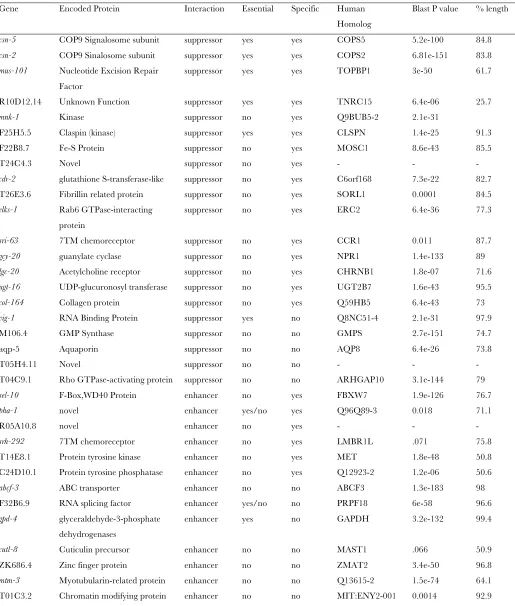
The essential Caenorhabditis elegans generfl-1 encodes one subunit of a heterodimeric E1-activating enzyme in the Nedd8 ubiquitin-like protein conjugation pathway. This pathway modifies the Cullin scaffolds of E3 ubiquitin ligases with a single Nedd8 moiety to promote ligase function. To identify genes that influence neddylation, we used a synthetic screen to identify genes that, when depleted with RNAi, enhance or suppress the embryonic lethality caused byor198ts, a temperature-sensitive (ts) mutation in rfl-1. We identified reproducible suppressor and enhancer genes and employed a systematic specificity analysis for each modifier using four unrelated ts embryonic lethal mutants. Results of this analysis highlight the importance of specificity controls in identifying genetic interactions relevant to a particular biological process because 8/14 enhancers and 7/21 suppressors modified lethality in other mutants. Depletion of the strongest specific suppressors rescued the early embryonic cell division defects in rfl-1(or198ts)mutants. RNAi knockdown of some specific suppressors partially restored Cullin neddylation in rfl-1(or198ts) mutants, consistent with their gene products normally opposing neddylation, and GFP fusions to several suppressors were detected in the cytoplasm or the nucleus, similar in pattern to Nedd8 conjugation pathway components in early embryonic cells. In contrast, depletion of the two strongest specific enhancers did not affect the early embryonic cell division defects observed in rfl-1(or198ts) mutants, suggesting that they may act at later times in other essential processes. Many of the specific modifiers are conserved in other organisms, and most are nonessential. Thus, when controlled properly for specificity, modifier screens using conditionally lethal C. elegans mutants can identify roles for nonessential but conserved genes in essential processes.
U
BIQUITIN-mediated proteolysis regulates many biological processes (Nandi et al.2006). In the early Caenorhabditis elegans embryo, these include oo-cyte maturation, cell cycle progression, cell polariza-tion, and cell fate patterning, all of which require the timely destruction of maternally expressed proteins (Bowerman and Kurz 2006; Greenstein and Lee 2006). One C. elegans protein targeted for proteolysis early in embryogenesis is MEI-1, the AAA-ATPase subunit of the microtubule-severing complex called katanin (Mains et al. 1990; Dow and Mains 1998; Srayko et al. 2000; Kurz et al. 2002; Pintard et al. 2003a; Xuet al.2003). Katanin is a heterodimer of two subunits called p60 and p80 in vertebrates andMEI-1andMEI-2inC. elegans. Katanin inC. elegansis required for proper assembly and function of the small, barrel-shaped meiotic spindles (Albertson and Thomson 1993; McNallyet al.2006) and must be degraded after meiotic divisions to permit assembly of the much larger first mitotic spindle in the one-cell zygote. In mutants that fail to degrade katanin after the completion of meiosis, the first mitotic spindle is fragmented and mis-oriented, cytokinesis is defective, and the embryos die without hatching (Dowand Mains1998; Sraykoet al. 2000; Kurzet al.2002).
The katanin subunit MEI-1 is targeted for poly-ubiquitylation and proteolytic destruction by a Cullin-based E3 ligase (Kurz et al. 2002). This complex includes the Cullin scaffolding protein CUL-3 and a substrate-specific adaptor called MEL-26that binds to CUL-3through a BTB domain and toMEI-1through a MATH domain (Pintardet al.2003b). Cullin 3-based E3 ligases in mammals also utilize substrate-specific adaptor proteins that, likeMEL-26, have both a Cullin-binding BTB/POZ domain and another protein– protein interaction domain that binds to the substrate
Supporting information is available online athttp://www.genetics.org/ cgi/content/full/genetics.109.104885/DC1.
1These authors contributed equally to this article.
2Present address: Institut Jacques Monod, CNRS, Universite´ Paris Diderot, 15 Rue Helene Brion, 75205, Paris Cedex 13, France.
3Corresponding author:Institute of Molecular Biology, 1229 University of Oregon, Eugene, OR 97403-1229.
E-mail: [email protected]
(Geyeret al.2003; Cullinanet al.2004; Angerset al. 2006). While MEI-1/Katanin downregulation by the CUL-3/MEL-26 E3 ligase is essential at most growth temperatures, amel-26null mutation is viable at the low growth temperature of 15°(Luand Mains2007). This bypass ofmel-26at 15°depends at least in part on the anaphase-promoting complex and its targeting ofMEI-1 for proteolytic degradation (Lu and Mains 2007). Phosphorylation by the kinase MBK-2 primes MEI-1 for proteolysis (Quintinet al.2003; Stitzelet al.2007) and also promotes the downregulation ofMEI-1by the anaphase-promoting complex (Luand Mains2007).
CUL-3is the onlyC. elegansCullin thus far identified that requires modification by the ubiquitin-like protein Nedd8 (Bowerman and Kurz 2006). In contrast, C. elegans CUL-2 is required for progression through meiosis and for the localized degradation of cell fate determinants in one-cell-stage embryos (Liuet al.2004; Sonneville and Gonczy 2004), but neddylation-defective mutants do not exhibit these early defects (Bowerman and Kurz 2006). Cullin neddylation is mediated by the Nedd8 protein conjugation pathway, which begins with a heterodimeric E1-activating enzyme consisting of ULA-1 and RFL-1 (Uba3p in budding yeast) and also includes the E2-conjugating enzyme UBC-12( Jonesand Candido2000; Sraykoet al.2000; Kurzet al.2002) and the E3 ligaseDCN-1(Kurzet al. 2005).
The downregulation ofMEI-1/katanin by theCUL-3/ MEL-26 E3 ligase requires a balance of both CUL-3 neddylation, which is mediated by the Nedd8 conjuga-tion pathway, and deneddylaconjuga-tion, which is mediated by the conserved COP-9 Signalosome (Pintard et al. 2003a). Other Cullin-based E3 ubiquitin ligases also require a balance of neddylation and deneddylation (Lyapina et al. 2001; Schwechheimer et al. 2001; Bornsteinet al.2006; Hetfeldet al.2008). Deneddy-lation may modulate activation of the E3 ligase and thereby prevent the premature degradation of sub-strate adaptor proteins that also can become poly-ubiquitylated and degraded as a result of E3 ligase function.
To identify additional factors that influence neddyla-tion, and the downregulation of MEI-1/katanin after the completion of meiosis inC. elegans, we report here our use of RNA interference (RNAi) to reduce gene functions in a temperature-sensitive (ts) neddylation-defective mutant,rfl-1(or198ts). The discovery of RNAi and its systemic properties inC. elegans have made it possible to systematically target C. elegans genes for depletion by feeding worms bacterial strains that ex-press double-strand RNAs corresponding toC. elegans gene sequences (Fireet al.1998; Timmonset al.2001; Feinbergand Hunter2003; Baughet al.2005; Lehner et al. 2006; van Haaften et al. 2006). Furthermore, chemical mutagenesis screens have identified tempera-ture-sensitive mutations in many essential C. elegans
genes, which can be used for synthetic screens by choos-ing intermediate-growth temperatures that sensitize the genetic background and also optimize visual scoring of embryonic viability. Recently, genomewide RNAi screens have been used to identify C. elegans genes that, when reduced in function, restore viability to temperature-sensitive, embryonic-lethal mutants (Labbeet al. 2006; O’Rourke et al. 2007). Because a loss of suppressor function restores mutant viability, the suppressors may negatively regulate either the wild-type gene product or the process that requires the wild-type gene product.
Here we report our identification ofC. elegansgenes that, when reduced in function by feeding RNAi, reproducibly suppressed or enhanced rfl-1(or198ts) embryonic lethality. Most suppressors were specific for rfl-1(or198ts), while specific enhancement was less com-mon. Many of the rfl-1-specific suppressors and en-hancers are conserved but appear nonessential. GFP fusions to several specific suppressors exhibit localiza-tion patterns that resemble those known for neddyla-tion pathway components, and depleneddyla-tion of some of these partially restored CUL-3 neddylation in rfl-1 (or198ts)mutants. In addition to identifying possible roles for conserved genes in cullin neddylation, we report the first quantitative analysis of specificity for both the en-hancement and the suppression of a conditionally lethal mutant inC. elegans.Our results highlight the importance of testing genetic modifiers of conditionally lethal mu-tants for locus specificity.
MATERIALS AND METHODS
C.elegansstrains and culture:Strains were cultured accord-ing to standard procedures (Brenner 1974).
Temperature-sensitive mutants were maintained at 15°, and GFP-expressing strains were maintained at room temperature. Isolation of transgenic worms was performed with the microparticle bombardment method as previously described (Praitis
2006; O’Rourkeet al.2007).
RNAi screening and quantification of embryonic viability: Methods used for RNAi screening and quantification of embryonic viability in this study were those described in detail in O’Rourkeet al.(2007), with the following modifications.
For scoring enhancement, genes that previously had been identified as lethal when targeted by RNAi, as reported in large-scale screens, were not given candidate enhancer status (Kamathand Ahringer2003; Simmeret al.2003) To quantify
embryonic viability of embryos at 23.5°, the broods of 7–10 gravid adult worms were analyzed. For all quantitative analysis, experiments were repeated at least four times and the average viability was determined. For enhancement, we report the percentage of dead embryos (using the average of the replicates). For suppression, we report the percentage of viable larvae, calculated again by taking the average of the replicates. The total number of progeny counted, the per-centage viability or lethality, and the standard deviations used to generate Figure 3 and data for wild-type (N2 strain) embryonic viability are included in thesupporting informa-tion,Table S1.
dures were used for SDS–PAGE and Western blotting. Rabbit anti-CUL-3 (Pintard et al. 2003a) was used at a 1:2000
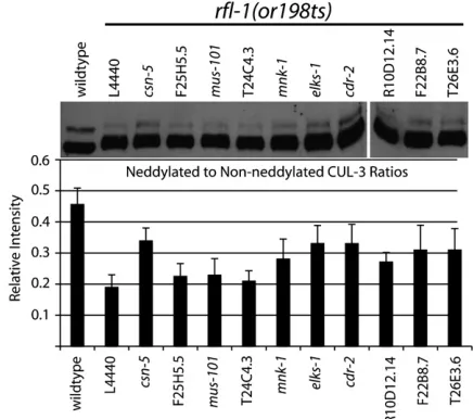
dilution in 4% milk and Tris-buffered saline plus 0.02% Tween-20. Anti-Rabbit secondary antibodies conjugated to peroxidase (Santa Cruz Biotechnology) were used at a concentration of 1:5000. Densitometry measurements of bands were carried out in Adobe Photoshop CS3. Integrated intensity was determined by multiplying the mean intensity (or average gray value) in each band, in a scanned and inverted image, by the number of pixels in the band. In-tegrated intensity of the neddylated band was divided by the integrated intensity of the non-neddylated band to determine relative intensity ratios. The graph in Figure 5 shows the average relative intensity of three separate exposures, and the error bars indicate6SD.
Molecular biology: For all pie-1 driven, N-terminal GFP constructs (rfl-1suppressors), genes were amplified using Pfu Turbo polymerase (Stratagene) from a cDNA library (Invitro-gen), with the exception ofR10D12.14, which was amplified from N2 genomic DNA. PCR products were subsequently ligated into pGEM-Tor pGEM-T-easy shuttle vectors (Promega). Inserted genes were sequenced at the University of Oregon sequencing facility prior to cleavage and ligation into pSO26 (pSO26 is described in O’Rourkeet al.(2007).
To construct N-terminal GFP and tdTomato (Shaneret al.
2004) fusions forsel-10,C24D10.1, andrfl-1, recombineering was used, as described in protocol #3 available at http:// recombineering.ncifcrf.gov/Protocol.asp (Warming et al.
2005). We used the following fosmid clones available from GeneService (http://www.geneservice.co.uk):WRM0610aC12
(sel-10), WRM0633dC (C24D10.1), andWRM066dF09(rfl-1). We used pSO26 as a template for amplifying GFP (O’Rourke
et al.2007). For the gap repair step, we used pPUB (Sarovet al.
2006) and designed primers to allow for inclusion of DNA sequence up to the next open reading frame (start or stop codon) from the gene of interest. To construct the N-terminal tdTomatoTRFL-1fusion, we used the pAA64 Vector to amplify the fluorescent protein-encoding gene. At least two indepen-dent transgenic lines for each construct were examined, and a representative line is shown in Figures 6 and 7. All fusion proteins are expressed from extrachromosomal arrays as determined by segregation analysis.
GFP imaging:Imaging of GFP and tdTomato fusion protein localization was done by mounting embryos or whole worms onM913% agarose pads on microscope slides and overlayed with a coverslip. Time-lapse videos were obtained on a spinning disk Leica DMI 4000B microscope using a Leica 63X/1.40-0.60 HCX Plan Apo oil objective, fitted with a Hamamatsu EM-CCD Digital Camera. Images in Figure 7 are projected stacks that include optical sections through the entire embryo or worm at a spacing of 0.5mmfor embryos and
of 1mmfor worms. Images of GFPTC24D10.1in Figure 7, D–F,
were taken with a Leica 40X/1.25-0.75 Plan Apo oil objective, and all other images were taken with the Leica 63X described above. Data were recorded using Velocity software and videos, and images were adjusted for contrast in ImageJ and adjusted for levels with Adobe Photoshop. ImageJ was used to obtain pixel intensity ratios of GFPTRFL-1in nuclei and cytoplasm of unenhanced images (Figure S1). Measurements were taken by
reached the L4 stage (60 hr at 20°). The incubation of rfl-1(or198ts) mutants at 20° through L4 stage allowed for slightly increased brood size and a decrease in the number of sterile adults. To image the embryos at a controlled temper-ature, we utilized a temperature-controlled microscope stage, equipped with an HEC-400 heat exchanger (20/20 Tech-nology) and a BC-110 bionomic controller (20/20 Technol-ogy). To calibrate the apparatus, we used an Omega HH12 temperature probe to obtain the temperature of aM913% agarose pad placed on a sapphire inset metallic microscope slide (20/20 Technology) while on the microscope stage with the light source on. We adjusted the Bionomic controller until a temperature of 23°was achieved. Use of the temperature-controlled stage prevented optimal focus of the condenser; DIC image quality was reduced but sufficient to score phenotypes.
Following incubation at 23° for 12-hr embryos were mounted on microscope slides and immediately transferred to the microscope stage. Images were recorded every 5 sec using a Dage MT1 VE1000 digital camera and Scion Image or ImageJ software. Contrast and levels were adjusted using Adobe Photoshop. Spindle angle measurements were done using the ImageJ angle tool. Spindle angles at cytokinesis were measured 1 min (12 frames) following the first appearance of cytokinetic furrow. The same procedure was used to image embryos for enhancement of cellular phenotypes except incubation of mutant worms were grown at a constant temperature throughout development (17°, 18°, or 20°). The microscope stage temperature was cali-brated to these lower temperatures for imaging as described above.
RESULTS
Quantifying embryonic viability to identify repro-ducible enhancers and suppressors: To screen for modifiers, we used an RNAi feeding library of 16,757 bacterial strains, each capable of inducibly expressing double-stranded RNA corresponding toC. elegansgene sequences, although some of the bacterial strains pro-duce dsRNAs that correspond to two genes due to updated gene annotations (Kamath and Ahringer 2003). We screened a total of 14,045 genes for suppres-sion and 8192 genes for enhancement ofrfl-1(or198ts) embryonic lethality after the mutant larvae matured into adults while feeding on the dsRNA-expressing bacterial strains at the semipermissive temperatures. Our initial qualitative screen yielded 248 candidate enhancers and 388 candidate suppressors. We then consolidated and systematically rescreened each candi-date enhancer and suppressor dsRNA using the same qualitative scoring method, reducing the number of candidate enhancers and suppressors to 101 and 104, respectively (see O’Rourkeet al.2007 for a description of the qualitative screening method).
To quantify suppression and enhancement, we com-pared the embryonic viability of broods from enhanced or suppressed young adults and from control young adults raised to adulthood on bacteria carrying the feeding RNAi vector without an insert (hereafter re-ferred to as empty-vector control broods). Thirty-two of the candidate enhancer genes, when reduced in func-tion by RNAi, increased embryonic lethality in the broods ofrfl-1(or198ts) mutants by at least 1.8-fold at 18°, compared to empty-vector control broods (Figure 2A). We chose this arbitrary and relatively nonstringent cutoff point because strong enhancers were less
com-mon than strong suppressors (see below), and we did not want to exclude potentially interesting genes entirely on the basis of strong enhancement of embry-onic lethality. To verify that depletion of these enhancer loci themselves did not cause embryonic lethality, we depleted each of these 32 genes using feeding RNAi and wild-type worms (Table S1). None of the enhancer genes were strongly required for embryonic viability under our conditions, with 5.7% embryonic lethality being the most penetrant essential requirement that we observed. We also tested each of the 32 enhancers with rfl-1(or198ts) mutants raised at 17°, at which tempera-ture 90% (SD64%) of unenhanced mutant embryos hatched. We found that 14 of the 32 enhancers still increased embryonic lethality by$2-fold at 17°(Figure 2B). We limited further analysis to these 14 enhancers that reproducibly acted at both semipermissive temper-atures (seeTable S2for modifier gene identities).
A similar quantitative brood analysis identified 21 suppressors that, when reduced in function by RNAi, consistently increased the viability of embryos from hermaphrodites raised at 23.5°by$1.8-fold, compared to worms raised on the empty-vector controlEscherichia coli(seeTable S2for gene identities). Depletion of 13 of the suppressors restored viability to.38% hatching (3-fold over the control); depletion of the strongest sup-pressor, csn-5, restored viability to 90% hatching. We chose an arbitrary and more restrictive cutoff of 3-fold suppression, compared to 1.8-fold and 2-fold for en-hancers at 17°and 18°, respectively, because suppressors tended to be both stronger and more specific.
Specificity of suppressors and enhancers: To test whether the modifiers that we identified specifically influenced rfl-1 function, or if depleting them can nonspecifically influence multiple conditionally mutant loci, we used four different temperature-sensitive, embryonic-lethal mutants that to our knowledge are not defective in functions related torfl-1or ubiquitin-mediated proteolysis: lit-1(or131ts), spn-4(or191ts), dhc-1(or195ts), andspd-5(or213ts). Thelit-1gene encodes a MAP Kinase that modulates Wnt signaling (Meneghini et al. 1999); spn-4 encodes a protein with an RNA-binding motif that regulates cell fate patterning in the early embryo (Gomes et al. 2001); dhc-1 encodes the heavy chain of the minus-end-directed microtubule motor dynein (Hamillet al.2002); andspd-5encodes a coiled-coil protein required for centrosome matura-tion (Hamillet al.2002). For each of these mutants, we used growth temperatures that gave nearly complete embryonic viability for testing enhancement or nearly complete embryonic lethality for testing suppression (O’Rourke et al. 2007). We quantified the effects of depleting modifier genes on embryonic viability with these four additional mutants and compared the results to those obtained withrfl-1(or198ts)(Figure 3).
We found that while enhancers were often nonspe-cific, most of the suppressors were specific for rfl-1. Figure 1.—Temperature vs. embryonic viability for
Reducing the function of 8 of the 14 enhancers in-creased embryonic lethality by at least 2-fold in one or more of the four other conditionally mutant strains. Depletions of the remaining six enhancer loci specifi-cally increased rfl-1(or198ts) embryonic lethality by between 2.1- and 9.6-fold (21% and 96.4% lethality), but reducing their function did not increase embryonic lethality by$2-fold in any of the four other conditional mutants that we tested (Figure 3 and Table S1). In contrast, of the 21 reproducible suppressor loci, 14 were specific forrfl-1(or198ts): reducing their function failed to suppress embryonic lethality by .2-fold in at least three of the four unrelated conditional mutants that we tested (Figure 3). All 14 specific suppressors failed to raise viability to $10% in any of the four unrelated mutants, while background viabilities on the empty-vector control ranged from 1.2% forlit-1(or131ts)as the lowest of the four to 3.7% forspn-4(or191ts) as the highest. Two of the specific and most penetrantrfl-1(or198ts) suppressors werecsn-2andcsn-5, which encode compo-nents of the COP-9 signalosome and have been shown previously to suppressrfl-1(or198ts)embryonic lethality when reduced in function (Pintardet al.2003a). These
were the only two signalosome components included in the 14,045 gene set that we tested for suppression, con-firming our ability to identify functionally important suppressors.
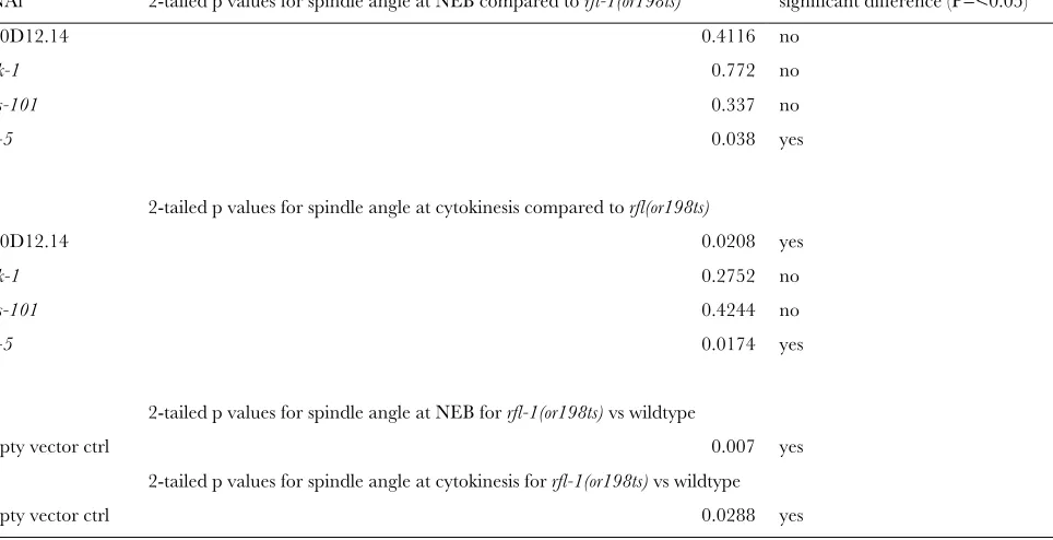
Modifier depletion and cell division defects in rfl-1(or198ts) mutant embryos: We next used DIC time-lapse videomicroscopy to examine live embryos at the first mitotic division in both suppressed and unsup-pressed rfl-1(or198ts) mutants. In unsuppressed em-bryos from rfl-1(or198ts) mutants grown at the fully restrictive temperature of 25°, the failure to degrade MEI-1/katanin leads to defects in mitotic spindle orientation, ectopic membrane furrows, and a failure to complete cytokinesis (Kurz et al. 2002). In unsup-pressed embryos, produced by rfl-1(or198ts) mutant worms raised at 23°and fed bacteria carrying the empty vector, 54% of the embryos failed to complete cytoki-nesis, 77% had ectopic cleavage furrow(s) following cytokinesis, and the first mitotic spindle was mis-oriented in comparison to the wild type (Figure 4 andTable S3). Depletion of the strongest suppressor,csn-5, almost com-pletely rescued the cytokinesis and ectopic furrowing defects: 0% and 10%, respectively, of the embryos ex-Figure 2.—Enhancement of rfl-1(or198ts)
hibited these defects, and the spindle orientation defect was also suppressed (Figure 4 andTable S3). Depletion of the specific suppressorsmnk-1,mus-101, andR10D12.14 produced less complete but still substantial suppression of these defects in early stage mutant embryos (Figure 4
andTable S3). We conclude that these specific suppres-sors influence the same early embryonic processes that require rfl-1 and that the suppression of these cell division defects may account at least in part for the increased viability of the suppressed mutant embryos.
Figure3.—Specificity of
The synthetic embryonic lethality caused by deple-tion of specific enhancers in rfl-1(or198ts) mutants raised at a semipermissive temperature could result from further compromising essential processes in the early embryo that require rfl-1 or from deleterious interactions that occur later in development. We there-fore used DIC time-lapse videomicroscopy to examine the first mitotic division in enhanced and unenhanced mutant embryos, focusing on the two most penetrant
and specific enhancer genes,sel-10andpha-1. At 17°on empty-vector RNAi-expressing bacteria, 0/15 unen-hancedrfl-1(or198ts)embryos exhibited cytokinesis fail-ures, while 2/15 had ectopic cleavage furrows. At 18°, 2/ 15 empty-vector RNAi-fed embryos exhibited cytokine-sis failure and 5/15 had ectopic furrows. While both sel-10(RNAi) and pha-1(RNAi) dramatically increased the rate of lethality at these temperatures, we observed no increase in cytokinesis failures, ectopic furrowing, or Figure4.—Suppression of early embryonic cell division defects inrfl-1(or198ts)mutants raised at 20°and then shifted to 23°for
7–12 hr prior to imaging (seematerials and methods). (A–P) Frames from time-lapse DIC videomicrographs at nuclear
enve-lope breakdown (A, E, I, M), during cytokinesis 1 min after first appearance of a cleavage furrow (B, F, J, N), and at the two-cell stage after the completion of cytokinesis (C, G, K, O). Black arrowheads indicate centrosome position; white arrowheads indicate ectopic membrane furrows; white arrow indicates nuclei of AB cell and P1 cell advancing toward each other after cytokinesis fail-ure; black arrow indicates regression of cytokinetic furrow. Temperature-controlled stage prevented use of optimal DIC optics (see
materials and methods). (Q) Quantification of ectopic cleavage furrows in suppressed and control embryos. (R) Quantitation
spindle orientation defects at either 17°or 18°(Table 1). This lack of enhancement of early cell division defects suggests that the synthetic lethal interaction(s) may occur at a later stage in embryonic development or were not detected by our methods if they occur in the early embryo.
To ask whetherrfl-1has functional requirements later in embryogenesis, beyond the first few cell divisions, we performed temperature upshift experiments after the completion of early cell divisions at the permissive temperature of 15°. After shifting 16- to 50-cell-stage rfl-1(or198ts) embryos to 26°, we found that 18/42 embryos (48.2%) failed to hatch, while in a control experiment with no temperature upshift, only 3/51 embryos (5.8%) failed to hatch, indicating that rfl-1 does have additional essential requirements later in embryonic development. We examined the terminal phenotypes of the unhatched, upshiftedrfl-1(or198ts) and found that 16/18 unenhanced and unhatched rfl-1(or198ts) embryos arrested after elongation to or beyond the threefold stage (data not shown). These results are consistent with a previous study that identified requirements for the neddylation pathway components ned-8,ubc-12, andula-1during postembryonic epidermal development and reported partially penetrant late-stage embryonic arrest after RNAi-mediated depletion of ubc-12andula-1( Jonesand Candido2000).
We next examined the terminal phenotypes of the unhatched synthetically lethal embryos using DIC mi-croscopy. Aftersel-10depletion at 18°, we found that 25/ 28 (89%) of the enhanced mutant embryos failed to hatch, and many of the unhatched embryos appeared to arrest after little if any elongation (data not shown), a more severe phenotype than we observed after upshifts of unenhancedrfl-1(or198ts)embryos to 26°(see above).
Other rfl-1(or198ts) embryos enhanced by sel-10 deple-tion arrested after a variable amount of elongadeple-tion (data not shown). We conclude that the synthetic-lethal inter-actions observed after enhancer depletion could result from interactions that occur after the early embryonic cell division processes known to requirerfl-1. However, we have not determined precisely where or when in embryo-genesis the synthetic-lethal interactions occur.
Suppressor depletions partially restore CUL-3 neddylation in rfl-1(or198ts) embryos: Neddylation of CUL-3 requires rfl-1 and other neddylation pathway components (Pintardet al.2003a). We therefore asked if the rfl-1 suppressors influence CUL-3 neddylation after isolating embryonic cell extracts from unsup-pressed and supunsup-pressed rfl-1(or198ts) mutants shifted to the nonpermissive temperature of 26°for 5 hr prior to sample collection (Figure 5). In comparison to wild type, we observed decreasedCUL-3neddylation in rfl-1(or198ts) mutant embryos, and partial restoration of neddylation after depletion of the signalosome compo-nent CSN-5, as expected (see Introduction). We ob-served partial restoration of CUL-3 neddylation reproducibly after depletion of six of the nine specific suppressors: most clearly with elks-1, C54D10.1/cdr-2, andF22B8.7, and more modestly but consistently with mnk-1,R10D12.14, andT26E3.6(Figure 5 and data not TABLE 1
Modifier depletion does not enhance early embryonic cell division defects inrfl-1(or198ts)mutants
Genotype
No. of embryos with ectopic
furrows
No. of embryos with cytokinesis
failure
rfl-1(or198); empty-vector RNAi 17°
2/15 0/15
rfl-(or198); sel-10(RNAi)17°
2/11 0/11
rfl-1(or198); pha-1(RNAi)17°
2/11 0/11
rfl-1(or198); empty-vector RNAi 18°
5/15 2/15
rfl-(or198); sel-10(RNAi)18°
5/13 0/13
rfl-1(or198); pha-1(RNAi)18°
3/13 1/13
Figure 5.—Neddylated CUL-3 levels in suppressed
rfl-1(or198ts) embryo extracts. Embryo extracts were prepared fromrfl-1(or198ts)embryos and wild-type embryos following a 5-hr shift to 26°and then loaded on an SDS–PAGE gel. After transferring the proteins to a membrane, the membrane was probed with an affinity-purified CUL-3 antibody. Lower band of82 kDa corresponds to the un-neddylated CUL-3, and up-per band of85 kDa corresponds to the neddylated CUL-3. Some enrichment of the neddylated CUL-3 band, relative to the un-neddylated band, is seen in seven of the samples. The bar graph shows the ratio of the integrated intensity of the neddylated CUL-3 band to the non-neddylated CUL-3 band (labeled as relative intensity) for each of the samples (see
shown). Thus these suppressor gene products may oppose, directly or indirectly, CUL-3 neddylation in wild-type embryos (seediscussion). We also attempted to detectMEI-1/katanin and RFL-1proteins but were unable to reproducibly analyze their levels in our ex-tracts with available antibodies.
Suppressor and enhancer protein localization over-lap with RFL-1: To gain further insight into how the specific modifiers might influencerfl-1-dependent pro-cesses, we constructed N-terminal GFP fusions to rfl-1 and to five of the specific suppressors, all driven by the maternalpie-1 promoter, and isolated transgenic lines for all six fusions (see materials and methods). We also constructed fusions to other suppressors but were unable to obtain germline-expressing transgenic strains for all constructs. Four of the five suppressors for which we did obtain germline expression (mnk-1,R10D12.14, F22B8.7, T26E3.6) are among the six that, when de-pleted by RNAi, partially restored CUL-3neddylation (see above). As observed for other Nedd8 pathway components (Kurzet al.2002; Pintardet al.2003a,b), we detected both cytoplasmic and enriched nuclear localization of the GFPTRFL-1 fusion protein in live embryos, using spinning disk confocal microscopy (Figure 6; see materials and methods). Fusion con-structs for both F25H5.5 (an ortholog of the human protein CLASPIN) and MNK-1 (a conserved kinase)
displayed nuclear enrichment in early embryonic cells. GFP fusions toF22B8.7(an uncharacterized conserved iron-sulfur domain-containing protein) and T26E3.6 (related to fibrillin) exhibited diffuse cytoplasmic and nuclear localization patterns. GFPTR10D12.14(a pro-tein of unknown function; seediscussion) localized to the cytoplasm and was largely excluded from nuclear and mitotic spindle regions. In addition, GFPTR10D12.14 was cortically enriched near cytoki-netic furrows and at cell boundaries in oocytes (Figure 6 and data not shown). While these are very general localization patterns, most of the suppressor proteins, including four that influenced CUL-3 neddylation, exhibited localization patterns similar at least in part to those observed for RFL-1,NED-8, andCSN-5, con-sistent with the possibility that some suppressor proteins could directly opposeCUL-3neddylation.
We also examined the expression and localization of GFP fusions to the proteins encoded by the two most specific enhancers,SEL-10(an F-box substrate adaptor for SCF-type E3 ligases) andC24D10.1(an uncharacter-ized protein tyrosine phosphatase), using recombineer-ing to construct GFP translational fusions regulated by native promoter and other noncoding sequences (see materials and methods). Embryonic expression of the other strong enhancer, pha-1, has been reported pre-viously on the basis of GFP fusion protein studies that Figure 6.—Expression
of pie-1 promoter-driven GFP fusions to suppressor-encoded proteins in early embryos from transgenic strains (seematerials and methods). Frames are from
detected cytoplasmic expression in most later-stage embryonic cells (Fayet al.2004). We did not detect any expression of GFPTSEL-10in early embryos, but we did detect cytoplasmic expression in some cells beginning at about the 50-cell stage (Figure 7A). In many cells throughout the embryo we detected higher levels of cytoplasmic expression beginning at about the bean stage (Figure 7B), after the completion of most embry-onic cell divisions. In larvae and adults, we observed expression in head and tail neurons and in unidentified cells along the entire length of the body (Figure 7, G and H). For the fusion GFPTC24D10.1, we again did not detect any expression in early embryos but first detected expression beginning at approximately the bean stage of embryogenesis, predominantly in nuclei and in many cells throughout the embryo (Figure 7, D and E). In larvae and adults, we observed GFPTC24D10.1 in the nuclei of many cells throughout the head.
The lack of early embryonic expression for the two most penetrant enhancers could be due to transgene silencing in the maternal germline, a frequent outcome for maternally expressed genes in transgenicC. elegans strains (Kellyand Fire1998). However, we did detect
presumably zygotic expression of the GFP fusions to these proteins in later-stage embryos, and we wanted to compare these later expression patterns to RFL-1. We therefore used recombineering to produce an N-terminal-tagged tdTomatoTRFL-1fusion driven by the rfl-1 promoter and generated a transgenic line that expresses this fusion (see materials and methods). We again did not observe any early embryonic expres-sion, presumably because of germline silencing. How-ever, we did detect strong cytoplasmic expression in larval stages and in adults in both head and tail neu-rons and in vulval epithelial cells and intestinal cells (Figure 7), consistent with previous studies of a rfl-1TGFP promoter fusion as a transcriptional reporter (Hunt-Newburyet al.2007). To summarize, although the two strongest specific rfl-1(or198ts) enhancers are expressed in later-stage embryos, we did not detect any RFL-1expression in later-stage embryos. For this reason, and because we did not detect any maternal expression of the two enhancer GFP fusions, we do not know in which cells RFL-1 might interact with SEL-10 or C24D10.1during embryogenesis, assuming such inter-actions are responsible for the enhanced embryonic Figure 7.—Spinning-disk confocal projected
Z-stack images of GFP and tdTomato fusion pro-teins driven by endogenous promoters (see
materials and methods). GFPTSEL-10
tions of other neddylation pathway components ( Jones and Candido2000).
To further investigate the requirements for theRFL-1 modifiers that we identified, we asked if depletion of the suppressor gene products can alter the localization of GFPTRFL-1and if depletion ofrfl-1or other neddyla-tion pathway components can change the localizaneddyla-tion of the GFP suppressor protein fusions in early embryonic cells. We did not detect changes in localization for any of the suppressor fusions followingrfl-1,nedd-8, andula-1 depletions (data not shown). Similarly, no change in GFPTRFL-1localization was observed upon depletion of the suppressor genes mus-101, R10D12.14, mnk-1, F25H5.5, T26E3.6, or F22B8.7 (data not shown). We also did not observe any change in early embryonic GFPTRFL-1localization upon depletion ofsel-10,pha-1, orC24D10.1, a result consistent with our finding that sel-10 andpha-1 depletion does not enhance the early embryonic phenotypes inrfl-1mutants.
Finally, we also examined GFPTRFL-1expression in transgenic embryos after depleting the neddylation pathway components ula-1,nedd-8, and ubc-12 (Figure S1). We did not observe changes inRFL-1localization after depletion of either nedd-8 or ubc-12, but we did observe a loss of GFPTRFL-1 nuclear enrichment following ula-1 depletion. ULA-1 and RFL-1/UBA-3 form the E1-activating enzyme complex for the Nedd8 conjugation pathway, with RFL-1 being the catalytic AAA-ATPase subunit ( Jones and Candido 2000). A comparison of pixel intensities showed a nuclear/ cytoplasmic ratio of 1.3 afterula-1depletion, compared to 5.7 in wild-type embryos (n ¼ 6 embryos for each analysis and then averaged). Using Western blots, we detected similar levels of GFPTRFL-1in extracts from ULA-1-depleted worms and wild-type worms (data not shown), suggesting that ULA-1 is required for RFL-1 nuclear enrichment independent of any changes in protein levels. To our knowledge, such a requirement for ULA-1 orthologs has not been reported in other organisms. Perhaps the heterodimerization of these two proteins allows for regulation of nuclear localization that is not observed when other neddylation pathway components are depleted.
DISCUSSION
To identify factors that influence neddylation and proteolytic regulation in C. elegans, we used a high-throughput RNAi screen to identify suppressors and
specific, with specific suppressors being more common. Depletion of the rfl-1-specific suppressors partially rescued the embryonic cell division defects associated with loss of rfl-1 function. Furthermore, we found similar subcellular distributions of several suppressor proteins andRFL-1, also consistent with the suppressors havingRFL-1-related roles in the early embryo. Impor-tantly, depletion of several suppressors partially restored CUL-3neddylation inrfl-1(or198ts)mutants, suggesting that these suppressors may normally oppose neddyla-tion. We also identified two highly penetrant and specific enhancers ofrfl-1(or198ts)embryonic lethality. While their depletion did not detectably enhance early embryonic cell division defects in mutant embryos, later requirements for rfl-1 may explain their synthetic le-thality. Finally, most of the modifiers do not themselves appear to be essential. Thus, when controlled for specificity, high-throughput synthetic screens that com-bine RNAi and conditionally lethalC. elegansmutations can be used to identify possible roles for nonessential genes in essential processes.
Synthetic screening with RNAi and temperature-sensitive, embryonic-lethalC.elegansmutants—controlling for specificity: While some large-scale synthetic screens using RNAi and temperature-sensitiveC. elegansmutants have addressed the issue of modifier specificity (Fraser 2004; Lehneret al.2006; O’Rourkeet al.2007), relatively few such studies have been reported thus far. We report here for the first time a systematic and quantitative analysis of specificity for both enhancers and suppressors of a conditional embryonic-lethalC. elegansmutant.
conditional embryonic lethality in more than one mutant background could represent functional links among the different mutant loci.
Synthetic lethal screens in budding yeast have been powerful tools for the discovery of new gene functions (Tonget al.2004; Ooiet al.2006), and it is interesting to compare the specificity observed in synthetic lethal C. elegansscreens with synthetic knockout screens done with budding yeast. For example, one yeast study analyzed 132 nonessential genes (Tong et al. 2004). Each of these 132 viable knockout mutants was mated with 4700 additional viable knockout mutants to produce double mutants. A total of 4000 synthetic lethal interactions were identified, with an average of 34 interactions/mutant. Of the interactions found, 27% were with pairs of genes known to be in the same or related genetic pathways. These findings suggest a substantial degree of specificity for synthetic lethal in-teractions in yeast, and thus the inin-teractions are relatively likely to reflect participation in a common process. By contrast, one study inC. elegansfound that over one-half of the genes on chromosomeIII, when reduced in function by feeding RNAi, significantly enhanced the embryonic lethality of a hypomorphic mutation in the transcription factor genedpl-l(Fraser 2004). Similarly, we found that many of the strong enhancers, when depleted by RNAi, enhanced embry-onic lethality with multiple conditional mutants.
This high degree of nonspecificity, compared to the synthetic-lethal screens in budding yeast, could reflect the added genetic complexity of the cellular and developmental processes in a multicellular organism. Alternatively, the different degrees of specificity ob-served in yeast and worms may simply reflect the different kinds of mutant alleles used for screening. The yeast synthetic-lethal screens used double mutants made with viable deletion alleles. In contrast, the synthetic screens inC. elegans have used partial inacti-vation of essential genes, usually by growing temperature-sensitive mutants at intermediate temperatures to sensitize genetic backgrounds, and RNAi to deplete the expression of other genes that may or may not themselves be essential. Perhaps temperature-sensitive mutants grown at just-viable temperatures are particularly vulnerable to non-specific synthetic lethality caused by the disruption of unrelated processes. It would be interesting to use genomewide feeding RNAi screens to detect synthetic lethality in fit, fertileC. elegansstrains that are homozygous for deletion mutations in nonessential genes. Conversely, it would be interesting to know if temperature-sensitive mutations in essential budding yeast genes are also prone to high levels of nonspecific enhancement and suppres-sion when other genes are reduced in function.
While nonspecific enhancement of embryonic lethal-ity is especially common, suppression of embryonic lethality by RNAi knockdown of other C. elegans loci also is frequently nonspecific. In a genomewide RNAi
screen for suppressors of a temperature-sensitive dynein heavy chain mutant, dhc-1(or195ts), 49 genes were identified that, when depleted by feeding RNA, restored dhc-1(or195ts)embryonic viability by threefold or more (O’Rourke et al. 2007). However, depletion of 57% of thedhc-1(or195ts)suppressors also significantly sup-pressed embryonic lethality for at least one of two other mutants with temperature-sensitive mutations in essen-tial loci unrelated in function to dhc-1. As we report here, 29% of therfl-1(or198ts)suppressors were similarly nonspecific. For at least some of the nonspecific dhc-1(or195ts) suppressor genes, their depletion has been reported by others to increase glycerol production, which may nonspecifically stabilize temperature-sensitive proteins and thereby increase embryonic viability at semipermissive temperatures (Lamitina et al. 2006; O’Rourkeet al.2007). Clearly, it is important to care-fully assess the specificity of both enhancer and suppres-sor interactions that influence the degree of lethality associated with temperature-sensitive mutations in es-sentialC. elegansgenes.
Known roles for specific modifier genes: The six suppressors that when depleted partially restored CUL-3neddylation inrfl-1mutants are all conserved, but how they might act is not apparent. Their influence on neddylation could be direct and of importance to other processes that involve neddylation, or it could be indi-rect. F22B8.7 encodes an uncharacterized conserved iron-sulfur domain-containing protein;T26E3.6, a fibrillin-related protein; cdr-2, a glutathione S-transferase-like protein (Donget al.2005);mnk-1, a conserved kinase; and elks-1, a Rab GTPase-interacting protein (Deken et al. 2005). The strong rfl-1suppressor R10D12.14is highly conserved only in nematodes and is one of six suppressors that, when depleted, partially restoredCUL-3 neddylation inrfl-1(or198ts)mutants. This gene is at least partially essential but its specific cellular requirements remain unknown (Pianoet al.2002). It encodes a pro-tein with a GYF domain (poly-proline interaction motif) and also was identified as a binding partner of dynein light chain (DLC-1) in a yeast two-hybrid screen of metazoan-specificC. elegansgenes (Liet al.2004). While the link to dynein might reflect roles in cell division, the GFPTR10D12.14fusion protein that we examined was largely excluded from nuclei and mitotic spindles and was mostly cytoplasmic, with some cortical enrichment in early embryonic cytokinesis furrows and in oocytes. Moreover, how binding to dynein light chain might re-late to neddylation is not obvious. Regardless of the functional significance of the dynein light chain inter-action, our analysis suggests that this protein of un-known function directly or indirectly opposes CUL-3 neddylation. It will be interesting to learn if any of these suppressors prove to influence neddylation in other model systems.
domain-containing proteins (Manke et al. 2003; Yu et al.2003). Some proteins with BRCT repeats, including BRCA1, have roles in mitosis as well as more established roles in DNA replication and repair. For example, BRCA1 binds to tubulin and localizes to centrosomes and spindle microtubules (Hsuand White1998), and high levels of BRCA1 are maintained throughout mitosis, while the protein is ubiquitinated and degraded during G1 and S phase (Choudhury et al. 2004). Furthermore, in both mammalian and Xenopus cells, BRCA1 is required for spindle pole assembly and the centrosomal accumulation of TPX2 ( Joukov et al. 2006). Perhaps the BRCA repeat-protein-encoded mus-101 influences cell division such that it can restore viability when depleted in rfl-1 mutants. Such a role might bypass requirements for neddylation, consistent with no influence of mus-101 depletion on CUL-3 neddylation.
The two strongest specific enhancer genes weresel-10 andpha-1. Thesel-10gene encodes an F-box protein and has been implicated as a substrate adaptor in E3 ligases that control the ubiquitin-mediated degradation of LIN-12/Notch,SEL-12/Presenilin, and the sex-determining proteinsFEM-1 andFEM-3(Hubbard et al. 1997; Wu et al.2001; Jageret al.2004). There are no identified requirements for Cullin neddylation in the ubiquitin-mediated degradation ofSEL-10targets, but our find-ings raise this possibility. Thepha-1gene encodes a novel protein that has been shown to function redundantly with class B SynMuv genes, such aslin-35/Rb andefl-1, to influence pharyngeal morphogenesis (Fayet al.2004). Furthermore, reducing the function of bothpha-1and any one of four ubiquitin ligases—ubc-18, C27A12.6, C27A12.7, and ari-1—results in pharyngeal morpho-genesis defects and partially penetrant early larval lethality (Qiuand Fay2006).ARI-1has been shown to interact withCSN-5by yeast two-hybrid screening, and inactivation ofcsn-5also enhances pharyngeal defects in some mutant backgrounds (D. Fay, personal commu-nication). Our results suggest that neddylation may be important for proper proteolytic regulation of pha-1 -and/orlin-35/Rb-dependent processes. While the F-box class of ubiquitin E3 ligases that may regulatepha-1and lin-35are not known to require neddylation, our results suggest that neddylation may promote their function.
Identifying requirements for nonessential genes: While mutational studies and genomewide RNAi screens inC. eleganshave identified thousands of genes that either are essential or have visible requirements,
that influence essential processes may be valuable drug targets: altering their function may allow for modifica-tion of an essential process without causing deleterious side effects that result from targeting more pleiotropic and essential disease genes. In our screen for modifiers of the essentialC. elegansgenerfl-1, 11 of the 16 specific suppressors and all 6 of the specific enhancers that we identified appear to be nonessential. Thus, when controlled for specificity, modifier screens that use RNAi and conditional mutations in essentialC. elegans genes may prove useful in identifying roles in essential processes for nonessential but conserved genes.
We thank I. Cruxent for help with RNAi screening; M. Price and J. Canman for microscopy assistance; L. Pintard for kindly providing theCUL-3antibody used in this study; and D. Greenstein, T. Herman, and a reviewer for helpful comments on the manuscript. This work was supported by a National Institutes of Health (NIH) Training Grant (to M.D.), by the Fundacxa˜o para a Cieˆncia e a Tecnologia (to J.-E.G.), and by NIH grant GM058017 (to B.B.).
LITERATURE CITED
Albertson, D. G., and J. N. Thomson, 1993 Segregation of
holo-centric chromosomes at meiosis in the nematode,Caenorhabditis elegans.Chromosome Res.1:15–26.
Angers, S., C. J. Thorpe, T. L. Biechele, S. J. Goldenberg, N. Zheng
et al., 2006 The KLHL12-Cullin-3 ubiquitin ligase negatively regulates the Wnt-beta-catenin pathway by targeting Dishevelled for degradation. Nat. Cell Biol.8:348–357.
Baugh, L. R., J. C. Wen, A. A. Hill, D. K. Slonim, E. L. Brownet al.,
2005 Synthetic lethal analysis ofCaenorhabditis elegansposterior embryonic patterning genes identifies conserved genetic interac-tions. Genome Biol.6:R45.
Bornstein, G., D. Ganothand A. Hershko, 2006 Regulation of
neddylation and deneddylation of cullin1 in SCFSkp2 ubiquitin ligase by F-box protein and substrate. Proc. Natl. Acad. Sci. USA 103:11515–11520.
Bowerman, B., and T. Kurz, 2006 Degrade to create:
developmen-tal requirements for ubiquitin-mediated proteolysis during early
C. elegansembryogenesis. Development133:773–784.
Brenner, S., 1974 The genetics ofCaenorhabditis elegans.Genetics
77:71–94.
Choudhury, A. D., H. Xuand R. Baer, 2004 Ubiquitination and
proteasomal degradation of the BRCA1 tumor suppressor is reg-ulated during cell cycle progression. J. Biol. Chem.279:33909– 33918.
Cullinan, S. B., J. D. Gordan, J. Jin, J. W. Harperand J. A. Diehl,
2004 The Keap1-BTB protein is an adaptor that bridges Nrf2 to a Cul3-based E3 ligase: oxidative stress sensing by a Cul3-Keap1 ligase. Mol. Cell. Biol.24:8477–8486.
Deken, S. L., R. Vincent, G. Hadwiger, Q. Liu, Z. W. Wanget al.,
2005 Redundant localization mechanisms of RIM and ELKS inCaenorhabditis elegans.J. Neurosci.25:5975–5983.
Dong, J., M. O. Songand J. H. Freedman, 2005 Identification and
Dow, M. R., and P. E. Mains, 1998 Genetic and molecular
charac-terization of theCaenorhabditis elegansgene,mel-26, a postmeiotic negative regulator of mei-1, a meiotic-specific spindle compo-nent. Genetics150:119–128.
Fay, D. S., X. Qiu, E. Large, C. P. Smith, S. Mangoet al., 2004 The
coordinate regulation of pharyngeal development inC. elegansby
lin-35/Rb,pha-1, andubc-18.Dev. Biol.271:11–25.
Feinberg, E. H., and C. P. Hunter, 2003 Transport of dsRNA into
cells by the transmembrane protein SID-1. Science 301:1545– 1547.
Fire, A., S. Xu, M. K. Montgomery, S. A. Kostas, S. E. Driveret al.,
1998 Potent and specific genetic interference by double-stranded RNA inCaenorhabditis elegans.Nature391:806–811. Fraser, A., 2004 Towards full employment: using RNAi to find roles
for the redundant. Oncogene23:8346–8352.
Geyer, R., S. Wee, S. Anderson, J. Yates and D. A. Wolf,
2003 BTB/POZ domain proteins are putative substrate adap-tors for cullin 3 ubiquitin ligases. Mol. Cell12:783–790. Gomes, J. E., S. E. Encalada, K. A. Swan, C. A. Shelton, J. C. Carter
et al., 2001 The maternal genespn-4encodes a predicted RRM protein required for mitotic spindle orientation and cell fate pat-terning in early C. elegans embryos. Development128: 4301– 4314.
Greenstein, D., and L. A. Lee, 2006 Oocyte-to-embryo transition:
kinase cabal plots regime change. Curr. Biol.16:R93–R95. Hamill, D. R., A. F. Severson, J. C. Carter and B. Bowerman,
2002 Centrosome maturation and mitotic spindle assembly in
C. elegansrequire SPD-5, a protein with multiple coiled-coil do-mains. Dev. Cell3:673–684.
Hetfeld, B. K., A. Peth, X. M. Sun, P. Henklein, G. M. Cohenet al.,
2008 The COP9 signalosome-mediated deneddylation is stimu-lated by caspases during apoptosis. Apoptosis13:187–195. Holway, A. H., C. Hungand W. M. Michael, 2005 Systematic,
RNA-interference-mediated identification of mus-101 modifier genes inCaenorhabditis elegans.Genetics169:1451–1460. Hsu, L. C., and R. L. White, 1998 BRCA1 is associated with the
cen-trosome during mitosis. Proc. Natl. Acad. Sci. USA95:12983– 12988.
Hubbard, E. J., G. Wu, J. Kitajewskiand I. Greenwald, 1997
sel-10, a negative regulator oflin-12activity inCaenorhabditis elegans, encodes a member of the CDC4 family of proteins. Genes Dev. 11:3182–3193.
Hunt-Newbury, R., R. Viveiros, R. Johnsen, A. Mah, D. Anastas
et al., 2007 High-throughput in vivo analysis of gene expression inCaenorhabditis elegans.PLoS Biol.5:e237.
Jager, S., H. T. Schwartz, H. R. Horvitz and B. Conradt,
2004 TheCaenorhabditis elegansF-box protein SEL-10 promotes female development and may target FEM-1 and FEM-3 for deg-radation by the proteasome. Proc. Natl. Acad. Sci. USA 101: 12549–12554.
Jones, D., and E. P. Candido, 2000 The NED-8 conjugating system
inCaenorhabditis elegansis required for embryogenesis and termi-nal differentiation of the hypodermis. Dev. Biol.226:152–165. Joukov, V., A. C. Groen, T. Prokhorova, R. Gerson, E. Whiteet al.,
2006 The BRCA1/BARD1 heterodimer modulates ran-dependent mitotic spindle assembly. Cell127:539–552.
Kamath, R. S., and J. Ahringer, 2003 Genome-wide RNAi
screen-ing inCaenorhabditis elegans.Methods30:313–321.
Kelly, W. G., and A. Fire, 1998 Chromatin silencing and the
main-tenance of a functional germline inCaenorhabditis elegans. Devel-opment125:2451–2456.
Kemphues, K., 2005 Essential genes. WormBook Dec24:1–7.
Kurz, T., L. Pintard, J. H. Willis, D. R. Hamill, P. Gonczyet al.,
2002 Cytoskeletal regulation by the Nedd8 ubiquitin-like pro-tein modification pathway. Science295:1294–1298.
Kurz, T., N. Ozlu, F. Rudolf, S. M. O’Rourke, B. Luke et al.,
2005 The conserved protein DCN-1/Dcn1p is required for cull-in neddylation cull-inC. elegansandS. cerevisiae.Nature435:1257– 1261.
Labbe, J. C., A. Pacquelet, T. Martyand M. Gotta, 2006 A
ge-nomewide screen for suppressors ofpar-2uncovers potential reg-ulators of PAR protein-dependent cell polarity inCaenorhabditis elegans.Genetics174:285–295.
Lamitina, T., C. G. Huangand K. Strange, 2006 Genome-wide RNAi
screening identifies protein damage as a regulator of
osmopro-tective gene expression. Proc. Natl. Acad. Sci. USA103:12173– 12178.
Lander, E. S., L. M. Linton, B. Birren, C. Nusbaum, M. C. Zody
et al., 2001 Initial sequencing and analysis of the human ge-nome. Nature409:860–921.
Lehner, B., C. Crombie, J. Tischler, A. Fortunato and A. G.
Fraser, 2006 Systematic mapping of genetic interactions in
Caenorhabditis elegansidentifies common modifiers of diverse sig-naling pathways. Nat. Genet.38:896–903.
Li, S., C. M. Armstrong, N. Bertin, H. Ge, S. Milstein et al.,
2004 A map of the interactome network of the metazoan
C. elegans.Science303:540–543.
Liu, J., S. Vasudevanand E. T. Kipreos, 2004 CUL-2 and ZYG-11
promote meiotic anaphase II and the proper placement of the anterior-posterior axis in C. elegans. Development131:3513– 3525.
Lu, C., and P. E. Mains, 2007 TheC. elegansanaphase promoting
complex and MBK-2/DYRK kinase act redundantly with CUL-3/MEL-26 ubiquitin ligase to degrade MEI-1 microtubule-sever-ing activity after meiosis. Dev. Biol.302:438–447.
Lyapina, S., G. Cope, A. Shevchenko, G. Serino, T. Tsugeet al.,
2001 Promotion of NEDD-CUL1 conjugate cleavage by COP9 signalosome. Science292:1382–1385.
Maeda, I., Y. Kohara, M. Yamamotoand A. Sugimoto, 2001
Large-scale analysis of gene function inCaenorhabditis elegansby high-throughput RNAi. Curr. Biol.11:171–176.
Mains, P. E., K. J. Kemphues, S. A. Sprunger, I. A. Sulstonand W. B.
Wood, 1990 Mutations affecting the meiotic and mitotic
divi-sions of the earlyCaenorhabditis elegansembryo. Genetics 126: 593–605.
Manke, I. A., D. M. Lowery, A. Nguyen and M. B. Yaffe,
2003 BRCT repeats as phosphopeptide-binding modules in-volved in protein targeting. Science302:636–639.
McNally, K., A. Audhya, K. Oegema and F. J. McNally,
2006 Katanin controls mitotic and meiotic spindle length. J. Cell Biol.175:881–891.
Meneghini, M. D., T. Ishitani, J. C. Carter, N. Hisamoto,
J. Ninomiya-Tsujiet al., 1999 MAP kinase and Wnt pathways
converge to downregulate an HMG-domain repressor in Caeno-rhabditis elegans.Nature399:793–797.
Nandi, D., P. Tahiliani, A. Kumarand D. Chandu, 2006 The
ubiquitin-proteasome system. J. Biosci.31:137–155.
Ooi, S. L., X. Pan, B. D. Peyser, P. Ye, P. B. Meluhet al., 2006 Global
synthetic-lethality analysis and yeast functional profiling. Trends Genet.22:56–63.
O’Rourke, S. M., M. D. Dorfman, J. C. Carterand B. Bowerman,
2007 Dynein modifiers inC. elegans: light chains suppress con-ditional heavy chain mutants. PLoS Genet.3:e128.
Piano, F., A. J. Schetter, D. G. Morton, K. C. Gunsalus, V. Reinke
et al., 2002 Gene clustering based on RNAi phenotypes of ovary-enriched genes inC. elegans.Curr. Biol.12:1959–1964. Pintard, L., T. Kurz, S. Glaser, J. H. Willis, M. Peter et al.,
2003a Neddylation and deneddylation of CUL-3 is required to target MEI-1/Katanin for degradation at the meiosis-to-mitosis transition inC. elegans.Curr. Biol.13:911–921.
Pintard, L., J. H. Willis, A. Willems, J. L. Johnson, M. Sraykoet al.,
2003b The BTB protein MEL-26 is a substrate-specific adaptor of the CUL-3 ubiquitin-ligase. Nature425:311–316.
Praitis, V., 2006 Creation of transgenic lines using microparticle
bombardment methods. Methods Mol. Biol.351:93–107. Qiu, X., and D. S. Fay, 2006 ARI-1, an RBR family ubiquitin-ligase,
functions with UBC-18 to regulate pharyngeal development in
C. elegans.Dev. Biol.291:239–252.
Quintin, S., P. E. Mains, A. Zinkeand A. A. Hyman, 2003 Thembk-2
kinase is required for inactivation of MEI-1/katanin in the one-cellCaenorhabditis elegansembryo. EMBO Rep.4:1175–1181. Sarov, M., S. Schneider, A. Pozniakovski, A. Roguev, S. Ernst
et al., 2006 A recombineering pipeline for functional genomics applied toCaenorhabditis elegans.Nat. Methods3:839–844. Schwechheimer, C., G. Serino, J. Callis, W. L. Crosby, S. Lyapina
et al., 2001 Interactions of the COP9 signalosome with the E3 ubiquitin ligase SCFTIRI in mediating auxin response. Science 292:1379–1382.
Shaner, N. C., R. E. Campbell, P. A. Steinbach, B. N. Giepmans,
Mains, 2000 MEI-1/MEI-2 katanin-like microtubule severing
activity is required for Caenorhabditis elegans meiosis. Genes Dev.14:1072–1084.
Stitzel, M. L., K. C. Chengand G. Seydoux, 2007 Regulation of
MBK-2/Dyrk kinase by dynamic cortical anchoring during the oocyte-to-zygote transition. Curr. Biol.17:1545–1554.
Timmons, L., D. L. Courtand A. Fire, 2001 Ingestion of bacterially
expressed dsRNAs can produce specific and potent genetic inter-ference inCaenorhabditis elegans.Gene263:103–112.
Tong, A. H., G. Lesage, G. D. Bader, H. Ding, H. Xu et al.,
2004 Global mapping of the yeast genetic interaction network. Science303:808–813.
vanHaaften, G., R. Romeijn, J. Pothof, W. Koole, L. H. M ullend-erset al., 2006 Identification of conserved pathways of
DNA-teins are substrate-specific adaptors in an SCF-like modular ubiq-uitin ligase containing CUL-3. Nature425:316–321.
Yamamoto, R. R., J. M. Axton, Y. Yamamoto, R. D. Saunders, D. M.
Gloveret al., 2000 The Drosophilamus101gene, which links
DNA repair, replication and condensation of heterochromatin in mitosis, encodes a protein with seven BRCA1 C-terminus do-mains. Genetics156:711–721.
Yu, X., C. C. Chini, M. He, G. Merand J. Chen, 2003 The BRCT
do-main is a phospho-protein binding dodo-main. Science302:639–642.
Supporting Information
http://www.genetics.org/cgi/content/full/genetics.109.104885/DC1
Using RNA Interference to Identify Specific Modifiers of a
Temperature-Sensitive, Embryonic-Lethal Mutation in the
Caenorhabditis elegans
Ubiquitin-Like Nedd8 Protein Modification Pathway E1-Activating Gene
rfl-1
Marc Dorfman, José-Eduardo Gomes, Sean O’Rourke and Bruce Bowerman
L4440(RNAi) ula-4(RNAi) ned-8(RNAi) ubc-12(RNAi)
GFP::RFL-1 SUPPLEMENTAL FIGURE 1
M. D. Dorfman et al. 3 SI
TABLE S1
Summary of modifier effects on embryonic lethality
Homolog
csn-5 COP9 Signalosome subunit suppressor yes yes COPS5 5.2e-100 84.8
csn-2 COP9 Sinalosome subunit suppressor yes yes COPS2 6.81e-151 83.8
mus-101 Nucleotide Excision Repair Factor
suppressor yes yes TOPBP1 3e-50 61.7
R10D12.14 Unknown Function suppressor yes yes TNRC15 6.4e-06 25.7
mnk-1 Kinase suppressor no yes Q9BUB5-2 2.1e-31
F25H5.5 Claspin (kinase) suppressor yes yes CLSPN 1.4e-25 91.3
F22B8.7 Fe-S Protein suppressor no yes MOSC1 8.6e-43 85.5
T24C4.3 Novel suppressor no yes - - -
cdr-2 glutathione S-transferase-like suppressor no yes C6orf168 7.3e-22 82.7
T26E3.6 Fibrillin related protein suppressor no yes SORL1 0.0001 84.5
elks-1 Rab6 GTPase-interacting protein
suppressor no yes ERC2 6.4e-36 77.3
sri-63 7TM chemoreceptor suppressor no yes CCR1 0.011 87.7
gcy-20 guanylate cyclase suppressor no yes NPR1 1.4e-133 89
lgc-20 Acetylcholine receptor suppressor no yes CHRNB1 1.8e-07 71.6
ugt-16 UDP-glucuronosyl transferase suppressor no yes UGT2B7 1.6e-43 95.5
col-164 Collagen protein suppressor no yes Q59HB5 6.4e-43 73
vig-1 RNA Binding Protein suppressor yes no Q8NC51-4 2.1e-31 97.9
M106.4 GMP Synthase suppressor no no GMPS 2.7e-151 74.7
aqp-5 Aquaporin suppressor no no AQP8 6.4e-26 73.8
T05H4.11 Novel suppressor no no - - -
T04C9.1 Rho GTPase-activating protein suppressor no no ARHGAP10 3.1e-144 79
sel-10 F-Box,WD40 Protein enhancer no yes FBXW7 1.9e-126 76.7
pha-1 novel enhancer yes/no yes Q96Q89-3 0.018 71.1
R05A10.8 novel enhancer no yes - - -
srh-292 7TM chemoreceptor enhancer no yes LMBR1L .071 75.8
T14E8.1 Protein tyrosine kinase enhancer no yes MET 1.8e-48 50.8
C24D10.1 Protein tyrosine phosphatase enhancer no yes Q12923-2 1.2e-06 50.6
abcf-3 ABC transporter enhancer no no ABCF3 1.3e-183 98
F32B6.9 RNA splicing factor enhancer yes/no no PRPF18 6e-58 96.6
gpd-4 glyceraldehyde-3-phosphate dehydrogenases
enhancer yes no GAPDH 3.2e-132 99.4
cutl-8 Cuticulin precursor enhancer no no MAST1 .066 50.9
ZK686.4 Zinc finger protein enhancer no no ZMAT2 3.4e-50 96.8
mtm-3 Myotubularin-related protein enhancer no no Q13615-2 1.5e-74 64.1
M. D. Dorfman et al. 5 SI
TABLE S3
Statistical analysis of suppressor effects on spindle
Student T-test
RNAi 2-tailed p values for spindle angle at NEB compared to rfl-1(or198ts) significant difference (P=<0.05)
R10D12.14 0.4116 no
mnk-1 0.772 no
mus-101 0.337 no
csn-5 0.038 yes
2-tailed p values for spindle angle at cytokinesis compared to rfl(or198ts)
R10D12.14 0.0208 yes
mnk-1 0.2752 no
mus-101 0.4244 no
csn-5 0.0174 yes
2-tailed p values for spindle angle at NEB for rfl-1(or198ts) vs wildtype
empty vector ctrl 0.007 yes
2-tailed p values for spindle angle at cytokinesis for rfl-1(or198ts) vs wildtype