kangaroo
, a Mobile Element From
Volvox carteri
, Is a Member of a Newly
Recognized Third Class of Retrotransposons
Leonard Duncan,
1Kristine Bouckaert, Fay Yeh
2and David L. Kirk
Department of Biology, Washington University, Saint Louis, Missouri 63130
Manuscript received February 28, 2002 Accepted for publication August 30, 2002
ABSTRACT
Retrotransposons play an important role in the evolution of genomic structure and function. Here we report on the characterization of a novel retrotransposon calledkangaroofrom the multicellular green alga,Volvox carteri.kangarooelements are highly mobile and their expression is developmentally regulated. They probably integrate via double-stranded, closed-circle DNA intermediates through the action of an encoded recombinase related to the-site-specific integrase. Phylogenetic analysis indicates thatkangaroo
elements are closely related to other unorthodox retrotransposons includingPAT(from a nematode),
DIRS -1(from Dictyostelium), andDrDIRS1(from zebrafish).PATandkangarooboth contain split direct repeat (SDR) termini, and here we show thatDIRS -1and DrDIRS1elements contain terminal features structurally related to SDRs. Thus,these mobile elements appear to define a third class of retrotransposons (the DIRS1 group) that are unified by common structural features, genes, and integration mechanisms, all of which differ from those of LTR and conventional non-LTR retrotransposons.
R
ETROTRANSPOSONS are mobile genetic ele- Boeke1993). LTR retrotransposons are closely related ments that are found in a wide range of eukaryotes to retroviruses and are bounded by direct repeats that (Xiongand Eickbush1990; Gabrieland Boeke1993). contain transcription-initiation and polyadenylation sig-They use reverse transcriptase (RT) to convert RNA nals. They typically contain one open reading frame intermediates into DNA copies that can then be inte- (ORF) that encodes a nucleic acid binding protein grated in new locations. Such replicative transposition (Gag) and a second ORF that encodes protease, RT, means that retrotransposons can greatly influence ge- RNAse H, and integrase domains (Figure 1A). They nome size: it is estimated that ⵑ40% of mammalian sometimes contain a third ORF encoding an envelope genomes andⵑ50% of the maize genome is composed protein. Non-LTR elements are simpler than (and prob-of retroelements (SanMiguel et al. 1996; Smit1999). ably ancestral to) the LTR class (Xiongand EickbushIn addition to increasing genome size, retroelements 1990): they lack terminal repeats, typically contain a can affect genome structure and function in other ways. poly(A)-rich sequence near their 3⬘ ends, and usually Although they often inactivate the gene into which they contain one ORF encoding a Gag protein and a second insert, there are now many examples in which novelcis- ORF encoding endonuclease, RT, and RNAse H func-regulatory sequences or protein domains are believed tions (Figure 1A).
to have been acquired from retroelements (Kumarand LTR and non-LTR elements use distinct transposition Bennetzen1999; Smit1999). For example, telomerase mechanisms (Craig 1997). LTR elements use RT to may have obtained its RT function this way (Eickbush generate a free, linear cDNA copy of the element that 1997). Retroelements can also transduce flanking se- is inserted into a target site by the action of an integrase quences, alter splicing of chimeric pre-mRNAs, create related to DNA transposases. In contrast, non-LTR ele-pseudogenes, and promote unequal crossing over and ments transpose by “target-primed reverse transcrip-other genomic rearrangements (Finnegan 1989; Cof- tion,” in which an RNA copy of the element is reverse
fin1993; Kumarand Bennetzen1999). transcribed only after an encoded endonuclease has
Most retrotransposons can readily be placed in either cleaved the target DNA to generate a primer for reverse the long terminal repeat (LTR) or the non-LTR classes transcription.
(Figure 1A; Xiong and Eickbush1990; Gabriel and RT-based molecular phylogenies generally identify ret-roelement clades whose individual members share other important features (Doolittleet al. 1989; Xiongand
Sequence data from this article have been deposited with the Eickbush1990; McClure1993). For example, the
ele-EMBL/GenBank Data Libraries under accession no. AY137241. ments placed in the gypsy, copia, BEL, and retrovirus
1Corresponding author:Cumbre, Inc., 1502 Viceroy Dr., Dallas, TX
clades on the basis of their RT sequence all contain
75235. E-mail: [email protected]
LTRs. Furthermore, elements in thecopiaclade all have 2Present address:University of Connecticut School of Medicine, 263
Farmington Ave., Farmington, CT 06030. an integrase gene upstream of the RT gene, whereas in
mid DNA,DNA, and hybridization probes were isolated using other LTR lineages (such asgypsy) the integrase gene
purification kits from QIAGEN (Valencia, CA). Volvox RNA is downstream of the RT gene.
was purified by minor modifications of a previously described A notable exception to this rule is the basis of this method (Kirkand Kirk1985). DNA was sequenced with Ap-report. An RT-based phylogeny places two unorthodox plied Biosystem’s (Foster City, CA) BigDye v. 2.0 premix using standard methods. Sequence data were collected on a depart-retrotransposons—PAT, from the nematodePanagrellus
mental MJ Research (Waltham, MA) Basestation and analyzed redivivus(deChastonay et al. 1992) andDIRS-1from
using PHRED/PHRAP/CONSED (http://www.phrap.org). Dictyostelium discoideum(Cappelloet al. 1985)—as
near-Cloningkangaroo-1:Two novelHindIII fragments that re-est neighbors on a branch located near the copia and sulted from the insertion ofkangaroo-1into thenitA(nitrate gypsyclades (Malikand Eickbush2001). However, nei- reductase-encoding) gene of CRH7 were separately cloned from subgenomic libraries generated from size-selected
Hin-ther PAT nor DIRS-1 encode either an LTR type of
dIII fragments of CRH7 genomic DNA ligated to Hin dIII-integrase or a non-LTR type of endonuclease. Moreover,
digested pBluescript II KS. The resulting 3.8-kb (pLD41) and PATandDIRS-1possess termini that appear to be very
7.5-kb (pLD40) inserts containing two segments ofkangaroo-1
different from one another and from the termini of were sequenced using a combination of primer walking and any other retroelement group.PATcontains split direct the Genome Priming System (NEB) to a final estimated error rate of ⬍0.01/10,000 bp. Next, we used OLV32 (5⬘-TTGTT repeat (SDR) termini, in which one copy of anⵑ300-bp
GGGCTGCTTTCCTC-3⬘) and OLV46 (5⬘-GGAAGCACACGAA direct repeat is found in the interior of the element
GTTGG-3⬘; Figure 2A) as PCR primers to amplify from CRH7 ( juxtaposed open and solid triangles, Figure 1B), while
genomic DNA a 2.7-kb DNA fragment that spans the internal the second copy is bifurcated, with about one-half of it HindIII site withinkangaroo-1.The sequence of this fragment present at each terminus (solo solid and open triangles, confirmed that these twoHindIII fragments that contain
kan-garoo-1sequences are juxtaposed as shown in Figure 2A. Figure 1B), such that the half-repeats alternate in the
Nucleic acid hybridization:Southern and Northern blotting order A, BA, B. DIRS-1, on the other hand, contains
experiments followed standard procedures (Sambrooket al. inverted terminal repeats (Figure 1B).
1989). [␣-32P]dCTP-labeled probes were prepared using the Our efforts to develop transposon-tagging tools for oligolabeling kit (Pharmacia, Piscataway, NJ) and purified us-use in studying the developmental genetics of the multi- ing NucTrap columns (Stratagene, La Jolla, CA).
cellular green alga,Volvox carteri(Kirk1998), led us to DNA fragments used as hybridization probes in the figures were as follows: Probe 2 was a 1.3-kbSmaI fragment isolated the discovery of a highly mobile retroelement called
from pLD41. Probe 3 was anⵑ600-bp fragment generated by kangaroo that contains SDR termini like those of PAT
PCR amplification from pLD40 using oligonucleotides OLV25 and that is closely related to PATandDIRS-1in terms (5⬘-GCACTTACGACCGTGAAACC-3⬘) and OLV67 (5⬘-AAAACG of its RT sequence. Here we report on the characteriza- GACGCTCCACGA-3⬘). Probe 4 was anⵑ4-kbXmnI-HindIII frag-tion ofkangaroo,its developmentally regulated expres- ment isolated from pLD40. Probe 5 was an ⵑ1-kb fragment generated by PCR amplification from pLD40 using oligonucleo-sion, and its probable method of integration.
tides OLV22 (5⬘-ATCCATCTTCGTATTTGCTG-3⬘) and OLV23 The unusual features shared byPAT,DIRS-1, and
kan-(5⬘-ACGAACGGGAGCACACTTAT-3⬘). Probe 6 was anⵑ1.4-kb garoo-1suggest that these elements must transpose by a
HindIII-DraI fragment isolated from pLD40. Probe 7 was a mechanism distinct from that used by either LTR or 325-bp fragment generated by PCR amplification from pLD41 non-LTR retroelements, despite the similarity of their using oligonucleotides OLV50 (5⬘-AATAGCGGGAAAGGG ATG-3⬘) and OLV63 (5⬘-GAAGTGTGAAGCCGACGA-3⬘). The RT proteins to those of well-known LTR elements.
Be-C38 probe has been described previously (Tam and Kirk
cause similar unorthodox elements are also found in the
1991). genomes of zebrafish and other metazoans, we conclude
Isolation ofkangaroo-2termini and preinsertion site:
kanga-that this DIRS1 group represents a widespread third roo-2was identified by Southern blot analysis (using probe 2,
class of retrotransposons. Figure 2A) as a 3.5-kb BamHI restriction fragment length
polymorphism (RFLP) present in LDV45 but absent from EVE. The DNA fragment corresponding to this RFLP was cloned, generating pLD35. An ⵑ400-bp fragment of DNA MATERIALS AND METHODS
(probe 8) derived from the nonretrotransposon sequence that flanks the left side ofkangaroo-2was amplified from pLD35 Volvox strains and cultivation conditions: Strains HK 9
by PCR using oligonucleotides OLV9 (5⬘-ATGGATGGGACTT (male) and HK 10 (female) of V. carteri f. nagariensis were
GCTGCTGAC-3⬘) and OLV10 (5⬘-CACCAATTTACCCGCCA isolated and described by Starr(1969) and later provided
GGATG-3⬘). Southern blotting experiments demonstrated that to us by the University of Texas Culture Collection of Algae.
probe 8 hybridized with a single copy sequence in LDV45 and EVE is our standard subclone of HK 10 (Harperet al. 1987)
and CRH7 (Miller et al. 1993) and LDV45 (L. Duncan, EVE genomic DNA and hybridized to the same 3.5-kbBamHI RFLP present in LDV45 that is recognized by probe 2. unpublished data) are first- and second-generation subclones
of EVE. The NIES male and female strains were isolated in To isolate thekangaroo-2preinsertion site, probe 8 was used to screen an EVE genomic library constructed inDASH II 1983 from a slightly different area of Japan than HK 9 and
HK 10 had been and were provided to us by the National (Kirket al. 1999). The preinsertion site was sequenced directly from bacteriophage DNA isolated from three independent Institute for Environmental Studies in Ibaraki, Japan. Volvox
cultures were maintained in standard Volvox medium under probe 8-hybridizing clones.
Next, the sequence of the preinsertion site was used to standardized culture conditions (Kirkand Kirk 1983) on a
16-hr-light/8-hr-dark cycle. design an oligonucleotide, OLV58 (5⬘-CACAGGGCGGGCAG
TTAT-3⬘), whose sequence was expected to be present within Nucleic acid purification and sequencing:Volvox genomic
kangaroo-2. Twokangaroospecific oligonucleotides, OLV42 (5⬘- element (Figure 2A),which we callkangaroo-1. We then
AGATTTGAGGCAGAGTAGG-3⬘) and OLV43 (5⬘-AGAAGA confirmed (as described inmaterials and methods)
CACAGTCGGATGAG-3⬘), were separately used in
conjunc-that the twoHindIII fragments are juxtaposed as shown. tion with OLV58 to PCR amplify anⵑ1-kb fragment from
kangaroo-1is an unorthodox retrotransposon:
kanga-LDV45 genomic DNA containing the junction between the
right side ofkangaroo-2and flanking DNA. Both independent roo-1possesses termini (solo solid and open triangles,
PCR products were sequenced. Figure 2A) that are distinct from those of DNA
transpo-Sequencing of retrotransposon:flanking DNA junctions sons and most retrotransposons and that are similar in fromkangaroo-3 through kangaroo-13: Plasmids pLD33 and
structure (but not in sequence) to the SDRs found in pLD34 were obtained by screening a partial LDV45 genomic
thePAT retroelement (Figure 1B). The terminal half-library withkangarooprobe 2, and they contain distinct inserts
that include the left termini ofkangaroo-5andkangaroo-4, re- repeats of kangaroo-1 are identical in sequence (data spectively.kangaroo-3andkangaroo-6throughkangaroo-13were not shown) to their counterparts within the full-length isolated by screening an EVEgenomic library (Kirk et al. internal repeat ( juxtaposed open and solid triangles, 1999) with a labeled 2.8-kbXmnI DNA fragment ofkangaroo-1
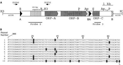
Figure 2A). Near its left end,kangaroo-1 also contains derived from pLD40. Thekangaroo:flanking DNA boundary
ⵑ12 contiguous copies of an 89-bp sequence (Figure sequences for several of these clones were determined by
directly sequencing the corresponding purified bacteriophage 2A). The first 10 copies of the 89-bp repeat are extremely DNA with oligonucleotides that hybridize to the left or right similar, although many of them can be distinguished terminus of kangarooelements. Forkangaroo-3 andkangaroo-6, by a small number of nucleotide deletions and/or poly-however,SalI DNA fragments containing the right side of the
morphisms (Figure 2B). The last two repeats (not shown) retroelement plus associated flanking DNA were first
sub-are less well conserved and more difficult to align. cloned into pBluescript II SK to generate pLD43 and pLD42.
PCR amplification of a portion of the putative circular form The two largest uninterrupted ORFs predicted by the ofkangaroo:PCR reactions containing 0.5mOLV2 (5⬘-AAG nucleotide sequence ofkangaroo-1are shown in Figure ACACAGTCGGATGAGGAG-3⬘), 0.5 m OLV93 (5⬘-CATTCT 2A. ORF-A and ORF-B potentially encode proteins 417 GGTGTCCTCCTT-3⬘; Figure 7B), and 0.5g of EVE DNA were
and 829 amino acids long, respectively. Importantly, a carried out using standard methods (Sambrooket al. 1989).
reverse position-specific BLAST search revealed that a The predominant PCR product, which was the expected size
(850 bp) to have been generated from a circular form of portion of the deduced amino acid sequence of ORF-B
kangaroo, was cloned into pGEM-T Easy (Promega, Madison, is strikingly similar (E ⫽ 6e-13) to the RT family of WI) to generate pLD53, which was then sequenced. proteins in the Pfam database (Bateman et al. 2002).
Isolation ofkangaroo-hybridizing cDNA clones:We purified
This similarity extends over and includes the conserved several clones that hybridized withkangarooprobe 4 (Figure
RT regions 2–7 as defined by Xiong and Eickbush
6A) from EVE cDNA libraries constructed ingt10 (Tamand
Kirk 1991; clones 5-11 and 5-13) or -Uni-ZAP XR (B. (1990) and the key conserved residues of RNAse H
Taillonand D.Kirk, unpublished data; clones5-2, 5-4, (Doolittleet al. 1989; McClure1993; Figure 3). Re-and5-5). The inserts in5-2,5-4, and5-5 were converted gion 5 of RT includes a highly conserved (Y/F)XDD to phagemids using the Rapid Excision kit (Stratagene) to
box that is thought to be essential for binding divalent generate pLD48, pLD49, and pLD50, respectively. The inserts
metal ions (Kohlstaedtet al. 1993). Although region from5-11 and5-13 were PCR amplified from purified
bacte-riophage DNA usinggt10 forward and reverse oligonucleo- 5 of RT from kangaroo-1 and two other independent tides and cloned into pGEM-T Easy to generate pLD56 and kangarooclones (data not shown) encodes a less
com-pLD57, respectively. mon LIDD (solid triangle, Figure 3), such divergence
has been observed previously in other RT and related proteins (Doolittleet al. 1989; Xiongand Eickbush
RESULTS
1990). We therefore conclude thatkangaroo-1 encodes RT/RNAse H and is likely to be a retrotransposon. We
Isolation ofkangaroo-1:Milleret al. (1993) previously
used a selection for chlorate-resistant individuals to en- note that in terms of nucleotide sequence, predicted amino acid sequences, overall organization, and phylo-rich forV. carterimutants carrying transposon insertions
in the nitrate-reductase-encoding gene,nitA (Gruber genetic position,kangaroo-1is clearly distinct from mem-bers of thecopia class of retrotransposons (e.g.,Osser) et al. 1992). One such mutant, CRH7, was found to
contain a large insertion withinnitAthat was unrelated that are found inV. carteri(Lindaueret al. 1993). Interestingly, the RT protein fromkangaroo-1is most to the transposon that became the major focus of that
study (Milleret al. 1993). We confirmed the existence closely related to the RT proteins encoded by PAT (BLASTP score:E⫽4e-24) andDIRS-1(BLASTP score: of such an insertion element by probing a Southern
blot containingHindIII-restricted genomic DNAs with E ⫽ 6e-16). Furthermore, as we discovered through TBLASTN searches of the databases and as was reported a probe derived from the 5⬘ end of the nitA coding
region: Theⵑ2.5-kbHindIII fragment derived from the by Goodwinand Poulter(2001), theDanio rerio (zebra-fish) genome contains several copies of a 6.1-kb retro-wild-typenitA gene was replaced in CRH7 by 3.8- and
7.5-kb restriction fragments (data not shown), a result transposon (DrDIRS1) that is strikingly similar in structure toDIRS-1(Figure 1B) and encodes a deduced RT protein consistent with the insertion of anⵑ9-kb DNA element.
Figure 1.—Three major groups of retrotransposons. (A) The generic structures of LTR and conventional non-LTR retroelements. The figures are not to scale and are not intended to repre-sent specific retrotranspo-sons. Large black arrows, long terminal repeats; PR, protease domain; RT, re-verse transcriptase domain; H, RNAse H domain; INT, integrase domain; UTR, un-translated region; EN, en-donuclease; An, poly(A)-rich sequence. Major ORFs are indicated by shading. (B) Diagrams of four members of the DIRS1 group of ret-roelements. PAT andTOC1
contain SDR termini (solid and open triangles desig-nated A and B), while the ter-mini ofDIRS-1andDrDIRS1
contain inverted repeats (dot-ted triangles). DIRS-1 and
DrDIRS1 also contain short terminal sequences (solid and open rectangles labeled A and B) that overlap with part of and, in some cases, extend beyond the inverted repeats and that are re-peated in an internal com-plementary region (ICR), where they are present in a juxtaposed and inverted ar-rangement (A⬘B⬘). Inver-sion of the ICR would create a structure very similar to that found in the SDR ele-ments. When comparing any two DIRS1-group elements, the A and B repeat units are similar in structure but unlated in sequence. REC, re-combinase. Accession num-bers for PAT, DIRS-1, and
TOC1 are X60774, M11339, and X56231, respectively. The copy ofDrDIRS1shown here is found in accession no. AL590134. The specificPATandDrDIRS1elements shown here contain nonsense codons (small solid triangles) in their rec genes. A second copy ofDrDIRS1(not shown) and, presumably, other copies of PATcontain an uninterrupted ORF-C.
domains from these four related retroelements is shown that the RT protein sequences from kangaroo-1, PAT, DIRS-1, and DrDIRS1 constitute a clade—the DIRS1 in Figure 3.
When we used PAUP* 4.0 to perform a neighbor- group—that apparently diverged after thecopia group and at about the same time as the retrovirus andgypsy joining phylogenetic analysis of the RT domains of
kan-garoo-1 and 22 other RT proteins, a tree (not shown) groups. This finding suggests that although the DIRS1 group of retrotransposons includes members with two was produced that was very similar in overall topology
to those previously published (Xiong and Eickbush different types of unusual termini, these subfamilies may nevertheless share a common mode of replication. In 1990; McClure1993; Malikand Eickbush2001). This
similar-Figure 2.—kangaroo-1 is an unorthodox retrotransposon containing SDR termini. (A) Diagram of kangaroo-1 (rectangle) inserted innitA(solid line). The solid and open triangles (labeled A and B) at opposite ends of the element represent the two halves of the SDR. The juxtaposed open and solid triangles (BA) represent the full-length interior direct repeat. The arrows above the diagram indicate the locations of PCR primers that were used to demonstrate that the twoHindIII fragments containingkangaroo-1are juxtaposed as shown here. The dashed vertical lines within the 89-bp repeat region represent the last two copies of the repeat, which are less well conserved. ORFs A, B, and C are shaded. Abbreviations for RT, H, and REC are as in Figure 1. The positions of relevant restriction enzyme sites are shown: H3,HindIII; S,SacI; R,RsaI; P,PstI; and Ap,ApaI. (B) Alignment of the first 10 89-bp repeats. Solid squares indicate positions that differ from the consensus. A dash indicates a gap. The numbers linked to the first and last nucleotides in the alignment correspond to the sequence ofkangaroo-1.
ity between the RT proteins from the four DIRS1-group kangaroo-1is a member of a dispersed, repetitive
fam-ily of mobile elements:We compared thekangaroo
ele-members extends⬎100 amino acids beyond the RNAse
H domain (Figure 3). This conserved C-terminal exten- ments of five closely relatedV. carterif.nagariensisstrains on DNA blots (Figure 4). Both probe 2 from the left sion is apparently not found in other RT proteins and
may indicate that DIRS1 members share some unknown, side and probe 3 from the right side ofkangaroo-1(see Figure 2A) recognized numerous, discrete bands in all conserved function, possibly related to their unusual
mode of transposition (see below). strains (Figure 4, A and B). Many bands appeared to
be present in all strains, but others were present in only Thekangaroo-1sequence includes nothing suggestive
of protease or envelope functions but, like most other one or a few of the strains examined. Most strikingly, many polymorphisms were visible between CRH7 and retrotransposons, it does contain a large ORF upstream
of the RT gene (ORF-A, Figure 2A). An ORF in this its clonal progenitor, EVE, which have been separated in culture for only a few years. Strains that have been location often encodes a Gag protein with one or more
C2HC “zinc-finger” motifs thought to bind nucleic acids isolated from one another for longer periods—such as EVE and HK9, which have been separate for at least (Reinet al. 1998). However, ORF-A fromkangaroo-1lacks
any discernible zinc-finger motif and BLASTP searches 35 years—showed a correspondingly greater number of RFLPs. Because the restriction enzyme/probe combina-fail to identify any other protein with significant
se-quence similarity. However, our observation that ORF- tions used were chosen to produce and reveal fragments with one end derived fromkangaroo-1and the other end A-specific transcripts are developmentally regulated
(see below) suggests that ORF-A may play a role in derived from flanking DNA, these results are consistent with kangaroo-1 being a member of a large family of retrotransposition. Finally, as discussed below,kangaroo-1
also encodes a recombinase that is distinct from the dispersed mobile elements.
Most members of the kangaroo family have similar
integrases and endonucleases normally associated with
ele-Figure3.—The RT fromkangaroo-1is closely related to the RT present in other members of the DIRS1 group. A portion of the ORF-B predicted amino acid sequence fromkangaroo-1(kan) is aligned with the deduced RT proteins ofDIRS-1,DrDIRS1, andPAT. The RT and RNAse H domains are outlined. Conserved regions 2–7 within the RT superfamily (Xiongand Eickbush
1990) are indicated by bars above the alignment. The solid triangle in region 5 indicates the first residue of the (Y/F)XDD box (see text). The alignment begins slightly upstream of region 2 and continues to the termination codon of each ORF (asterisk). The solid circles within the RNAse H domain indicate residues known to be present in the enzyme’s active site and believed to be important for catalysis. The alignment was created using CLUSTALX (with manual refinement of the RNAse H domain) and MacBoxshade. Identical and similar amino acids are indicated by black and gray shading, respectively, using a 75% consensus threshold. A dot represents a gap.
ments in the genome possessed the same general struc- zymes and probe 3, which is derived from the opposite side ofkangaroo-1(Figure 5B). We interpret these results ture as kangaroo-1, we probed a blot of RsaI-digested
EVE DNA with probe 2, which covers the region of to mean that a large fraction of the kangarooelements within V. carteri possess the same general structure as kangaroo-1containing the 89-bp repeats (see Figure 2A).
The predominant hybridizing band that was detected kangaroo-1.
To determine whether otherkangarooelements have (Figure 5A, lane 2) was identical in size (ⵑ2 kb) to
the hybridizing fragment produced byRsaI digestion of the same SDR termini askangaroo-1, we cloned several distinctkangaroo-hybridizing DNA fragments that con-clonedkangaroo-1(Figure 5A, lane 1), although
en-kangarooinsertions shown are bordered by the nucleo-tide dT on both their 5⬘and 3⬘ends (solid circles, Figure 5C). By comparing pre- and postintegration sites, we found that kangaroo-1 had inserted into the sequence 5⬘-CTG-3⬘, and kangaroo-2 had inserted into the se-quence 5⬘-CTT-3⬘ (Figure 5C and data not shown). These findings lead us to conclude that the dT residue at onekangaroo–flanking DNA junction is derived from the target site, while the other is derived from the retro-transposon. However, there is presently no way to be certain which dT is derived from which source. Thus (as noted in the caption to Figure 5C) there is a one-nucleotide uncertainty regarding the boundaries of the retrotransposon and its target site.
kangarooexpression is developmentally regulated:We
isolated fromV. cartericDNA libraries several clones that hybridized with kangaroo probe 4 (Figure 6A). These clones fall into two classes. Members of the first class (clones 2, 4, 5, and 13) encode all or part of ORF-A (Figure 6A) and terminate 13–16 bp downstream of a volvocalean polyadenylation signal sequence (5⬘ -TGTAA-3⬘; Kirk1998) that is located just upstream of ORF-B. cDNA 4 contains the longest insert of this class
Figure4.—kangaroo-1is a member of a dispersed repetitive (ⵑ2.1 kb) and corresponds to a transcript containing family. Autoradiograms of Southern blots containing 2g of
two exons with canonical splice sites. Interestingly, the restricted genomic DNA from five closely relatedV. carterif.
intron includes the region of 89-bp repeats. Because
nagariensisstrains: CRH7, EVE, HK9, NIES female, and NIES
cDNA 4 begins near the left end ofkangaroo-1, it proba-male (seematerials and methods). (A)HindIII-restricted
DNA hybridized with probe 2 (see Figure 2A). (B) SacI- bly represents a full-length or nearly full-length cDNA restricted DNA hybridized with probe 3 (see Figure 2A). clone. This suggests that a promoter may reside within the left half-repeat. The second cDNA class has only a single member, cDNA 11 (Figure 6A). This apparently designed to prime just inside each terminus and read partial cDNA clone corresponds to a transcript that en-into the DNA flanking the insertion site. One of these codes a portion of ORF-B and whose processed 3⬘end clones, kangaroo-2, corresponds to a recent insertion terminates midway through the internal full-length re-that is present in strain LDV45, but absent in its progeni- peat.
tor, EVE (data not shown), providing additional evi- We next analyzed the accumulation ofkangaroo tran-dence ofkangaroomobility. The other clones contained scripts by Northern blot analysis using developmentally randomly selectedkangaroo-hybridizing fragments from staged RNAs harvested at various points during the asex-EVE (kangaroo-3 and kangaroo-6 through kangaroo-13) ual life cycle ofV. carteri, which is outlined in Figure or from LDV45 (kangaroo-4 and kangaroo-5) genomic 6B. Using probe 4 (Figure 6A), we found that four major libraries. Figure 5C shows an alignment of these flanking kangaroo-hybridizing transcripts (2.0, 3.8, 7.1, and 9.0 DNA:kangaroojunctions, with the left and right ends of kb; a–d in Figure 6, C–E) are produced during develop-kangaroooriented as in Figure 2A. We found that these ment. Such transcripts were virtually undetectable in kangaroo clones are identical in sequence on the left precleavage gonidia, began to accumulate during cleav-side of the alignment beginning with the sequence 5⬘- age, reached a maximum level shortly after inversion, TGCATGTTGATTAA-3⬘ and, with one exception, are and then declined dramatically by 6 hr later (Figure also identical on the right side of the alignment until 6C, lanes 1–7). These transcripts then remained at low they all diverge after the sequence 5⬘-GACGTTTAAG levels throughout the rest of the asexual life cycle (time CAAT-3⬘(Figure 5C). We conclude that the majority of points 8–10; Figure 6E, lane 8 and data not shown). kangarooelements contain SDR termini similar to those Because the transcripts reached maximum abundance
ofkangaroo-1. during the dark period at the end of embryogenesis,
Analysis of kangarooinsertion sites:As shown above but are present at much lower levels during the dark
the alignment in Figure 5C, we observed some conserva- period 24 hr later (time points 9 and 10, Figure 6B; data tion in the DNA sequences that flank the variouskanga- not shown), we conclude that transcript accumulation rooelements that we have characterized, which suggests is controlled by developmental rather than circadian thatkangaroo integration may exhibit some degree of factors.
Figure5.—Mostkangarooelements have the same general structure askangaroo-1. (A) Auto-radiogram of a Southern blot hybridized with probe 2 (see Figure 2A). Lane 1, 6 ng ofRsa I-digested pLD41 (which contains the left half ofkangaroo-1); lane 2, 1.5g of EVE genomic DNA digested withRsaI. The 1.2-kb hybridiz-ing band flanked by dots in lane 1 is a fragment of pLD41 that contains only a short segment ofkangaroo-1 and whose size is unrelated to the true structure of the retrotransposon. (B) Autoradiogram of a Southern blot hybridized with probe 3 (see Figure 2A). Lane 1, 6 ng of
ApaI-digested pLD40 (which contains the right half of kangaroo-1); lane 2, 1.5 g of Apa I-digested EVE genomic DNA; lane 3, 6 ng of pLD40 digested with PstI; lane 4, 1.5 g of
PstI-digested EVE genomic DNA. (C) Aligned sequences of the retrotransposon:flanking DNA junctions of several independent kanga-roo clones (kangaroo-1 through kangaroo-13). Thekangaroosequences are enclosed by a rect-angle and shown in uppercase. Only a short segment of each terminus of thekangaroo ele-ments is shown. Flanking DNA is in lowercase and delineated by arrows. Note that (with one exception) thekangarooelements are flanked by a dT residue on both sides (solid circles), and it is unclear which residue is derived from the retroelement and which is derived from the target DNA; possibly, the rectangle deline-ating the boundaries of kangaroo should be shifted 1 bp to the left. HDR, half direct repeat; CON, conserved target DNA sequences; Y, py-rimidine; R, purine.
6A), because it is of the appropriate size (ⵑ2.1 kb), c are identical to those of transcript d (Figure 6, C and D, and data not shown) except that c fails to hybridize and it hybridizes with ORF-A-specific probe 6 (data not
shown) but not with intron-specific probe 7 (Figure 6E, to the intron-specific probe 7 (Figure 6E, lane 6). These results lead us to propose that transcripts b and c have lane 6), or with ORF-B-specific probe 5 (Figure 6D, lane
6), or with probe 3, which is derived from the right side the structures shown in Figure 6A. The sizes of tran-scripts b and c are consistent with this interpretation. ofkangaroo(data not shown).
Transcript d is about the right size to be a full-length, Finally, because cDNA 11 (Figure 6A) apparently does not correspond to any of the majorkangaroo-hybridizing unsplicedkangarooRNA species (Figure 6A). Consistent
with this view, transcript d hybridizes with all of the transcripts visualized by Northern blot analysis, it seems likely that it represents a relatively low-abundance mes-kangarooprobes that we have used in Northern blotting
experiments (Figure 6, C–E and data not shown). Pre- sage.
kangaroo may integrate as a closed-circle,
double-sumably, transcript d is the template used during reverse
transcription. stranded DNA copy: A common feature of LTR and
non-LTR retrotransposon integration is the generation The discovery of an intron withinkangaroo-1prompted
us to also examine the nature of transcripts b and c. of element-specific target-site duplications (Gabriel
and Boeke1993). Thus, one interpretation of the find-We found that the hybridization patterns for transcript
b are identical to those of transcript a (Figure 6, C and ing that kangaroo elements are bounded by single dT residues (Figure 5C) is thatkangaroomay integrate using D and data not shown) except that b also hybridizes
Figure 7.—A model for
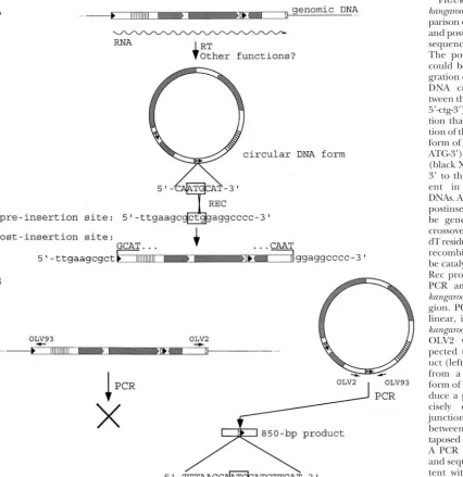
kangarooinsertion. (A) Com-parison of the observed pre-and postinsertion target-site sequences for kangaroo-1. The postinsertion product could be generated if inte-gration occurred by a single DNA crossover event be-tween the target site (boxed 5⬘-ctg-3⬘) and the circle junc-tion that is created by liga-tion of the ends of the linear form ofkangaroo(boxed 5⬘ -ATG-3⬘). Here, the crossover (black X) is shown as being 3⬘ to the dT residues pres-ent in the recombining DNAs. Alternatively, the same postinsertion product could be generated if the DNA crossover occurred 5⬘to the dT residues (not shown). This recombination event could be catalyzed by thekangaroo
Rec protein (see text). (B) PCR amplification of the
kangaroocircle junction re-gion. PCR amplification of linear, integrated forms of
kangaroo with OLV93 and OLV2 would not be ex-pected to produce a prod-uct (left), but amplification from a closed-circle DNA form ofkangaroowould pro-duce a product with a pre-cisely defined size and junction (boxed 5⬘-ATG-3⬘) between the two newly jux-taposed half-repeats (right). A PCR product whose size and sequence is fully consis-tent with the existence of such a circular form was ob-tained (see text).
our knowledge, no LTR or non-LTR retrotransposon is Figure 7A, the integration product that is observed for kangaroo-1 (Figure 5C) could be produced by a DNA known to create a single-base-pair target-site
duplica-tion. More importantly,kangarooand other members of crossover event between the newly formed circle junc-tion of kangaroo and the target site. The integration the DIRS1 group do not encode a conventional
DDE-integrase or a non-LTR-like endonuclease. These results product produced by insertion of kangaroo-2 (Figure 5C) can be explained in a similar manner (not shown). and the asymmetric nature of kangaroo’s termini lead
us to propose the insertional model presented in Figure In accord with this model, we were able to use the PCR primers shown in Figure 7B to amplify a fragment 7A, which involves an extrachromosomal DNA circle
very similar to the one that was proposed as an interme- from EVE DNA whose size and sequence is fully consis-tent with the product that would be produced from diate in the replication ofDIRS-1(Cappelloet al. 1985)
and a second, related element calledTOC1(Dayet al. the postulated closed-circle DNA intermediate––but not from a linear—DNA form of kangaroo. It is formally 1988; see below). Specifically, it is postulated that a
critical intermediate formed during retrotransposition possible that such a product could have been produced by amplification from two copies ofkangaroointegrated ofkangaroois an extrachromosomal, closed-circle,
double-stranded DNA copy in which the ends of the retroele- in the genome in a head-to-tail manner. However, we believe that even if such a tandem juxtaposition of linear ment are fused to generate an identical second copy of
be unlikely to have coincidentally generated a sequence those of the LTR and the conventional non-LTR retro-elements. The first members of this group to be recog-identical to that which would be created by ligation of
the half-repeats in a circularkangarooform. nized wereDIRS-1, from Dictyostelium (Cappelloet al. 1985), andPAT, fromP. redivivus(deChastonayet al.
kangaroo encodes a protein related to-site-specific
integrase:Goodwinand Poulter(2001) reported that 1992), which were united solely by the similarity of their
RT proteins and appeared to differ greatly in the struc-members of the DIRS1 group of retrotransposons
po-tentially encode a protein (ORF-C, Figure 1B) related ture of their termini. A less-well-characterized, but clearly related, retroelement calledPrt1was also found to the Int family of recombinases (Nunes-Duby et al.
1998). The quintessential member of this protein family in the fungusPhycomyces blakesleeanus(Ruiz-Perezet al. 1996). Here we have characterized the first member of is the integrase protein from bacteriophage , which
catalyzes the integration and excision of the-genome the group to be identified in a photoautotroph, namely kangaroo-1ofV. carteri, a green alga, and we have shown at a specific site within theEscherichia colichromosome
(Landy1989). Int family members also catalyze the non- thatkangaroonot only is closely related toDIRS-1andPAT in terms of RT amino acid sequence, but also has split site-specific integration of circular forms of conjugative
(DNA) transposons (Scottand Churchward1995). direct repeat termini very similar in structure to the termini of PAT. Meanwhile, Goodwin and Poulter
It is important to note that the Int recombinases are
distinct in structure and catalytic mechanism from the (2001) have reported discovering several additional metazoan members of the DIRS1 group by screening DDE-type integrases normally associated with
retrovi-ruses and LTR retrotransposons (Haren et al. 1999) public databases. These includedDrDIRS1of the zebra-fish Danio rerio,TnDIRS1of the pufferfishTetraodon ni-and from the endonucleases associated with non-LTR
elements (Furano 2000). Members of the Int family groviridis, andCbPAT1ofCaenorhabditis briggsae, as well as several fragmentary DIRS1-like sequences from two are quite divergent in sequence. Indeed, only four
amino acids, which comprise the “RHRY” tetrad essen- species of Xenopus and the sea urchin,Strongylocentrotus purpuratus (Goodwinand Poulter2001; L. Duncan, tial for catalysis, are present in all members of this group
(Nunes-Dubyet al. 1998). In support of the results of unpublished data). DIRS1-group retroelements have not yet been found in insects or vascular plants. Goodwinand Poulter(2001) we have found that
kan-garooalso potentially encodes a 225-amino-acid protein However, earlier studies (Day et al. 1988; Day and Rochaix 1991) had revealed that Chlamydomonas rein-related to-integrase (ORF-C, Figure 2A). Figure 8 shows
part of the deduced ORF-C protein from kangaroo-1 hardtii, the closest unicellular relative ofV. carteri, con-tains a mobile element called TOC1with SDRs similar aligned with portions of -integrase and several other
members of the Int family, including the deduced re- in structure (but not sequence) to those ofkangaroo-1 and PAT(Figure 1B). TOC1also contains a stretch of combinase proteins from PAT, DIRS-1, and DrDIRS1.
The alignment identifies the conserved box I and box 76-bp repeats that is strikingly similar in position and length to the stretch of 89-bp repeats inkangaroo-1 (com-II regions containing the catalytic RHRY tetrad (Nunes
-Duby et al. 1998) and, in extension of the results of pare Figures 1B and 2A), although the nucleotide se-quences of their repetitive units are wholly unrelated. Goodwin and Poulter (2001), we have found that
the recombinase proteins from the DIRS1 family also Because TOC1does not encode RT or other proteins that play a role in retrotransposition, it appears to be contain the “Patch II” and “Patch III” regions identified
by Nunes-Duby et al. (1998; Figure 8). Although the a nonautonomous element and could not be included in the phylogenetic analysis of retroelement RT se-ORF-C protein fromkangaroo-1is predicted to contain
several insertions and deletions of amino acids relative quences. Nevertheless, the studies reported here lead us to predict that TOC1probably replicates and trans-to other proteins shown in Figure 8 (e.g., immediately
following box I), these insertions/deletions are likely poses by a mechanism similar to the one that we have proposed for kangaroo in Volvox, using functions that to fall within loop regions of the folded protein (Nunes
-Duby et al. 1998) and presumably do not prevent its are encoded by an autonomous kangaroo-like element elsewhere in the Chlamydomonas genome. Consistent adopting a structure similar to-integrase. These results
lead us to conclude that ORF-C encodes a member of with this hypothesis, we have found that several Chlamy-domonas expressed sequence tags are present in the the Int family and to speculate that this enzyme catalyzes
the integration ofkangaroovia the mechanism we have public databases that encode peptides with significant similarity to regions of thekangarooRT protein, includ-proposed in Figure 7A.
ing the C-terminal extension that we believe is diagnos-tic for the DIRS1 family of transposons (L. Duncan, DISCUSSION
unpublished data). This, in turn, leads us to suspect that kangaroo-like elements may be widely distributed kangaroois a member of a newly recognized but
rap-idly growing class of retrotransposons—the DIRS1 within the order Volvocales. Indeed, with representa-tives now known to be present in slime molds, fungi, group—whose members share structures, genes, and
Figure8.—DIRS1-group members encode a recombinase related to-site-specific integrase. The predicted ORF-C proteins from kangaroo-1, DIRS-1, DrDIRS1, and PAT are aligned with a number of recombinase proteins from the Int family. The conserved residues forming the RHRY catalytic tetrad are indicated by asterisks. For clarity, the sequences FGPDYSHVI and RGGGPFRGFPLPLPDPFGA were removed from theDrDIRS1andkangarooproteins, respectively, at the positions indicated just to the right of box I. The other aligned proteins are: Cb (Coxiella burnetiiintegrase, CAA75853), Psp (Pseudomonas sp. integrase, CAA67462), Ll (Lactobacillus leichmanniiXerC, CAA59018), Asp (Anabaena sp. integrase, BAB77331), and(integrase, P03700). The alignment was created using CLUSTALX (with manual refinement) and MacBoxshade. Identical and similar residues are shaded black and gray, respectively, using a 50% consensus threshold. A dot indicates a gap.
seems reasonable to postulate that DIRS1-group ele- can be symbolized as: A⬎ . . .⬍ A⬍B . . . B⬎, and we propose that it may represent an altered form of the ments may be almost as widely distributed among the
eukaryotes as the LTR and conventional non-LTR ret- type of SDR seen in PAT,TOC1, and kangaroo, which can be symbolized as: A ⬎ . . . B⬎ A ⬎ . . . B ⬎. It roelements are now known to be.
The two DIRS1-group subfamilies contain related ter- is tempting to speculate that one of the DIRS1-group
subfamilies was derived from the other subfamily by
minal structures:As discussed in the Introduction, most
RT-based phylogenies identify clades of retroelements inversion of the full interior repeat (Figure 1B).
DIRS1 elements appear to integrate by a novel
mecha-that share other important structural and genetic
fea-tures. Thus, we were initially surprised to find that the nism: Here we have proposed that members of the DIRS1 group may transpose by a mechanism similar to DIRS1 clade grouped elements that apparently contain
dissimilar termini: namely, those containing inverted the one proposed initially by Cappelloet al. (1985) and Dayet al. (1988), in which RT generates a closed-circle, repeats and those containing SDRs. However, we
subse-quently realized that the elements with inverted-repeat double-stranded DNA intermediate that is then inserted into a target site by a single crossover event. If this model termini have other features that are structurally similar
to the SDRs found inkangaroo,PAT, andTOC1. Specifi- is correct, then the integration mechanism used by the DIRS1-group members would be clearly distinct from cally, both DIRS-1 and DrDIRS1 have short sequences
(labeled A and B in Figure 1B) that extend past the that used during insertion of known LTR and conven-tional non-LTR retrotransposons.
end of, or are part of, the terminal inverted repeats,
and these same sequences are also found juxtaposed This model was initially based on: (1) our observation that members of the DIRS1 group contain unorthodox, internally in an inverted orientation (A⬘B⬘) in a
struc-ture called the internal complementary region (ICR; but related, terminal structures and do not encode a conventional retrotransposon integrase or endonucle-Cappelloet al. 1985).TnDIRS1also has a similar
postinte-gration sites; and (3) our demonstration that we could (Mut9 andMut11) that are involved in transcriptional gene silencing (TGS). Furthermore, the levels ofTOC1 amplify a PCR product from Volvox DNA whose
struc-ture is consistent with the existence of the postulated RNA are also controlled, at least in part, by degradation that is dependent on theMut6RNA helicase (Wu-Scharf
closed-circular DNA intermediate. These latter two
re-sults constitute the first pieces of experimental evidence et al. 2000), which is a component of the post-transcrip-tional gene silencing (PTGS) machinery ofC. reinhardtii. in support of this kind of model.
Meanwhile,Goodwinand Poulter(2001) proposed It is conceivable thatkangarooexpression inV. carteriis controlled in an analogous manner by uncharacterized, a very similar model, after discovering that members of
the DIRS1 group encode proteins of the Int family of developmentally regulated TGS or PTGS mechanisms.
kangarooas a molecular genetic tool:At present, the
recombinases, some of which (such as-site-specific
in-tegrase) are known to mediate this type of integration only method available for cloning genes by forward ge-netics inV. carteri has involved tagging with the DNA process. We have now shown thatkangarooalso encodes
such a recombinase. Thus, our study and that ofGood- transposon, Jordan(Milleret al. 1993). Although this approach has been used successfully to clone several
winand Poulter(2001) are mutually reinforcing.
However, whereas Goodwin and Poulter (2001) developmentally important genes (Kirk et al. 1999; Millerand Kirk1999; I.Nishii, personal communica-consider the DIRS1 elements as a “group of LTR
retro-transposons,” we believe that because of their very sub- tion), we have encountered several cases in which inter-esting mutations that have the earmarks of transposon-stantial differences from LTR elements in termini, gene
content, and probable integration mechanisms, the induced mutations cannot be correlated with Jordan RFLPs (our unpublished observations). Evidence pre-DIRS1 group should be considered to be a third class
of retrotransposons, distinct from both the LTR and sented here identifieskangarooas a second, highly mo-bile element within theV. carterigenome that is capable the traditional non-LTR classes.
Developmental regulation ofkangarooexpression:We of causing gene disruptions. Whetherkangaroowill turn
out to have other properties required to make it a sec-have shown that the accumulation of four discrete
tran-scripts produced bykangaroo is developmentally regu- ond useful transposon-tagging tool forV. carteri develop-mental biologists, only time will tell.
lated, which is a property shared with numerous other
retrotransposons. The expression of at least 19 different We thank I. Nishii for staged RNAs and the Northern blot (with Drosophila retroelements is controlled both temporally associated C38 loading control data) used in Figure 6C; A. Horton and D. Berg for helpful advice; and J. Umen, S. Miller, I. Nishii, and
and spatially during development (Dingand Lipshitz
C. Shaffer for comments on the manuscript. L.D. was a postdoctoral
1994; Frommer et al. 1994; Mozer and Benzer 1994;
fellow of, and this investigation has been aided by, a grant from the
Bronner et al. 1995; Awasaki et al. 1996; Kerber et
Jane Coffin Childs Memorial Fund for Medical Research. This work
al. 1996; Marsano et al. 2000), as is expression of the was also supported by a National Science Foundation grant (IBN-zebrafish retroelement bhikhara (Vogel and Gerster 9904739) to D.K.
1999) and the Xenopus 1A11 element (Greene et al. 1993). Similarly, transcription ofTyelements in
Saccha-romyces cerevisiae(Erredeet al. 1987), LINE-1 in mam- LITERATURE CITED
mals (Ostertag and Kazazian 2001), and DIRS-1 in
Awasaki, T., N. JuniandK. M. Yoshida, 1996 An eye imaginal
disc-D. discoideum(Cohenet al. 1984) all appear to be under specific transcriptional enhancer in the long terminal repeat
of the tomretrotransposon is responsible for eye morphology
cell-type or stage-specific controls. As with expression
mutations ofDrosophila ananassae.Mol. Gen. Genet.251:161–166.
of more conventional kinds of genes, developmentally
Bateman, A., E. Birney, L. Cerruti, R. Durbin, L. Etwilleret al.,
regulated expression of retrotransposons has been 2002 The Pfam protein families database. Nucleic Acids Res.
30:276–280.
shown to involve both cis-regulatory elements located
Bronner, G., H. TaubertandH. Jackle, 1995 Mesoderm-specific
within the transposons themselves andtrans-acting
fac-B104expression in the Drosophila embryo is mediated by internal
tors encoded elsewhere (Erredeet al. 1987; Dingand cis-acting elements of the transposon. Chromosoma103:669–675.
Cappello, J., K. HandelsmanandH. F. Lodish, 1985 Sequence of
Lipshitz1994; Mozer and Benzer1994; Bronner et
DictyosteliumDIRS-1: an apparent retrotransposon with inverted
al. 1995; Awasakiet al. 1996; Kerberet al. 1996; Vogel
terminal repeats and an internal circle junction sequence. Cell
and Gerster1999). It has been argued that the develop- 43:105–115.
Coffin, J., 1993 Reverse transcriptase and evolution, pp. 445–479
mentally regulated expression of such retroelements
inReverse Transcriptase, edited by A. M.Skalkaand S. P.Goff.
may have been selected for by virtue of its benefit to
Cold Spring Harbor Laboratory Press, Cold Spring Harbor, NY.
the host (Dingand Lipshitz1994; Bronneret al. 1995), Cohen, S. M., J.Cappelloand H. F.Lodish, 1984 Transcription
ofDictyostelium discoideumtransposable elementDIRS-1. Mol. Cell.
but convincing evidence in favor of this hypothesis
re-Biol.4:2332–2340.
mains lacking.
Craig, N. L., 1997 Target site selection in transposition. Annu. Rev.
Interestingly, observations made in the related unicell Biochem.66:437–474.
Day, A., andJ. D. Rochaix, 1991 A transposon with an unusual
C. reinhardtii raise the possibility that developmental
LTR arrangement from Chlamydomonas reinhardtiicontains an
accumulation ofkangarootranscripts could be regulated
internal tandem array of 76 bp repeats. Nucleic Acids Res.19:
epigenetically.Jeonget al. (2002) have shown thatTOC1 1259–1266.
Day, A., M. Schirmer-Rahire, M. R. Kuchka, S. P. Mayfieldand
J. D. Rochaix, 1988 A transposon with an unusual arrangement S. P.Goff. Cold Spring Harbor Laboratory Press, Cold Spring Harbor, NY.
of long terminal repeats in the green algaChlamydomonas
rein-Kumar, A., andJ. L. Bennetzen, 1999 Plant retrotransposons. Annu.
hardtii.EMBO J.7:1917–1927.
Rev. Genet.33:479–532.
de Chastonay, Y., H. Felder, C. Link, P. Aeby, H. Tobleret al.,
Landy, A., 1989 Dynamic, structural, and regulatory aspects of 1992 Unusual features of the retroid elementPAT from the
site-specific recombination. Annu. Rev. Biochem.58:913–949. nematodePanagrellus redivivus.Nucleic Acids Res.20:1623–1628.
Lindauer, A., D. Fraser, M. BruderleinandR. Schmitt, 1993
Re-Ding, D., andH. D. Lipshitz, 1994 Spatially regulated expression
verse transcriptase families and acopia-like retrotransposon,Osser, of retrovirus-like transposons duringDrosophila melanogaster
em-in the green algaVolvox carteri.FEBS Lett.319:261–266. bryogenesis. Genet. Res.64:167–181.
Malik, H. S., andT. H. Eickbush, 2001 Phylogenetic analysis of
Doolittle, R. F., D. F. Feng, M. S. JohnsonandM. A. McClure,
ribonuclease H domains suggests a late, chimeric origin of LTR 1989 Origins and evolutionary relationships of retroviruses. Q.
retrotransposable elements and retroviruses. Genome Res.11:
Rev. Biol.64:1–30.
1187–1197.
Eickbush, T. H., 1997 Telomerase and retrotransposons: Which
Marsano, R. M., R. Moschetti, C. Caggese, C. Lanave, P. Barsanti
came first? Science277:911–912.
et al., 2000 The completeTiranttransposable element in
Dro-Errede, B., M.Companyand C. A.Hutchison, 1987 Ty1sequence
sophila melanogastershows a structural relationship with retrovirus-with enhancer and mating-type-dependent regulatory activities.
like retrotransposons. Gene247:87–95. Mol. Cell. Biol.7:258–265.
McClure, M. A., 1993 Evolutionary history of reverse transcriptase,
Finnegan, D. J., 1989 Eukaryotic transposable elements and genome
pp. 425–444 inReverse Transcriptase, edited by A. M.Skalkaand evolution. Trends Genet.5:103–107.
S. P.Goff. Cold Spring Harbor Laboratory Press, Cold Spring
Frommer, G., R. SchuhandH. Jackle, 1994 Localized expression
Harbor, NY. of a novelmicropia-like element in the blastoderm ofDrosophila
Miller, S. M., andD. L. Kirk, 1999 glsA, a Volvox gene required
melanogaster is dependent on the anterior morphogen bicoid.
for asymmetric division and germ cell specification, encodes a Chromosoma103:82–89.
chaperone-like protein. Development126:649–658.
Furano, A. V., 2000 The biological properties and evolutionary dy- Miller, S. M., R. SchmittandD. L. Kirk, 1993 Jordan, an active namics of mammalian LINE-1 retrotransposons. Prog. Nucleic Volvox transposable element similar to higher plant transposons. Acid Res. Mol. Biol.64:255–294. Plant Cell5:1125–1138.
Gabriel, A., and J. D.Boeke, 1993 Retrotransposon reverse tran- Mozer, B. A., andS. Benzer, 1994 Ingrowth by photoreceptor axons scription, pp. 275–328 inReverse Transcriptase, edited by A. M. induces transcription of a retrotransposon in the developing
Dro-Skalkaand S. P.Goff. Cold Spring Harbor Laboratory Press, sophila brain. Development120:1049–1058.
Cold Spring Harbor, NY. Nunes-Duby, S. E., H. J. Kwon, R. S. Tirumalai, T. Ellenberger
Goodwin, T. J., and R. T. Poulter, 2001 The DIRS1 group of andA. Landy, 1998 Similarities and differences among 105 retrotransposons. Mol. Biol. Evol.18:2067–2082. members of the Int family of site-specific recombinases. Nucleic
Greene, J. M., H. Otani, P. J. GoodandI. B. Dawid, 1993 A novel Acids Res.26:391–406.
family of retrotransposon-like elements inXenopus laeviswith a Ostertag, E. M., and H. H.Kazazian, Jr., 2001 Biology of mamma-transcript inducible by two growth factors. Nucleic Acids Res.21: lianL1retrotransposons. Annu. Rev. Genet.35:501–538.
2375–2381. Rein, A., L. E. HendersonandJ. G. Levin, 1998
Nucleic-acid-chap-Gruber, H., S. D. Goetinck, D. L. KirkandR. Schmitt, 1992 The erone activity of retroviral nucleocapsid proteins: significance for nitrate reductase-encoding gene ofVolvox carteri: map location, viral replication. Trends Biochem. Sci.23:297–301.
sequence and induction kinetics. Gene120:75–83. Ruiz-Perez, V. L., F. J. Murillo and S. Torres-Martinez, 1996
Haren, L., N. Ton-Hoang andM. Chandler, 1999 Integrating Prt1, an unusual retrotransposon-like sequence in the fungus DNA: transposases and retroviral integrases. Annu. Rev. Micro- Phycomyces blakesleeanus. Mol. Gen. Genet.253:324–333. biol.53:245–281. Sambrook, J., E. F.Fritschand T.Maniatis, 1989 Molecular
Clon-Harper, J. F., K. S. HusonandD. L. Kirk, 1987 Use of repetitive ing: A Laboratory Manual. Cold Spring Harbor Laboratory Press, sequences to identify DNA polymorphisms linked toregA, a devel- Cold Spring Harbor, NY.
SanMiguel, P., A. Tikhonov, Y. K. Jin, N. Motchoulskaia, D.
Zakh-opmentally important locus in Volvox. Genes Dev.1:573–584.
arovet al., 1996 Nested retrotransposons in the intergenic
re-Jeong, B., D.Wu-Scharf, C.Zhangand H.Cerutti, 2002
Suppres-gions of the maize genome. Science274:765–768. sors of transcriptional transgenic silencing in Chlamydomonas
Scott, J. R., andG. G. Churchward, 1995 Conjugative transposi-are sensitive to DNA-damaging agents and reactivate transposable
tion. Annu. Rev. Microbiol.49:367–397. elements. Proc. Natl. Acad. Sci. USA99:1076–1081.
Smit, A. F., 1999 Interspersed repeats and other mementos of
trans-Kerber, B., S. Fellert, H. TaubertandM. Hoch, 1996 Germ line
posable elements in mammalian genomes. Curr. Opin. Genet. and embryonic expression ofFex, a member of the Drosophila
Dev.9:657–663.
F-element retrotransposon family, is mediated by an internalcis
-Starr, R. C., 1969 Structure, reproduction and differentiation in regulatory control region. Mol. Cell. Biol.16:2998–3007.
Volvox carterif.nagariensisIyengar, strains HK9 and HK10. Arch.
Kirk, D., 1998 Volvox: Molecular-Genetic Origins of Multicellularity and
Protistenkd.111:204–222.
Cellular Differentiation. Cambridge University Press, Cambridge,
Tam, L. W., andD. L. Kirk, 1991 Identification of cell-type-specific UK.
genes ofVolvox carteriand characterization of their expression
Kirk, D. L., andM. M. Kirk, 1983 Protein synthetic patterns during
during the asexual life cycle. Dev. Biol.145:51–66. the asexual life cycle ofVolvox carteri.Dev. Biol.96:493–506.
Vogel, A. M., andT. Gerster, 1999 Promoter activity of the
zebra-Kirk, M. M., andD. L. Kirk, 1985 Translational regulation of protein
fishbhikhariretroelement requires an intact activin signaling path-synthesis, in response to light, at a critical stage of Volvox develop- way. Mech. Dev.85:133–146.
ment. Cell41:419–428. Wu-Scharf, D., B. Jeong, C. ZhangandH. Cerutti, 2000
Trans-Kirk, M. M., K. Stark, S. M. Miller, W. Muller, B. E. Taillonet gene and transposon silencing inChlamydomonas reinhardtiiby a al., 1999 regA, a Volvox gene that plays a central role in germ- DEAH-box RNA helicase. Science290:1159–1162.
soma differentiation, encodes a novel regulatory protein. Devel- Xiong, Y., andT. H. Eickbush, 1990 Origin and evolution of ret-opment126:639–647. roelements based upon their reverse transcriptase sequences.
Kohlstaedt, L. A., J.Wang, P. A.Rice, J. M.Friedmanand T. A. EMBO J.9:3353–3362.
Steitz, 1993 The structure of HIV-1 reverse transcriptase, pp.