O R I G I N A L R E S E A R C H
Lithium chloride with immunomodulatory function
for regulating titanium nanoparticle-stimulated
in
fl
ammatory response and accelerating osteogenesis
through suppression of MAPK signaling pathway
This article was published in the following Dove Press journal: International Journal of Nanomedicine
Chao Yang* Wei Wang* Kechao Zhu Wei Liu Yao Luo Xiangwei Yuan Jiaxing Wang Tao Cheng Xianlong Zhang
Department of Orthopedics, Shanghai Jiao Tong University Affiliated Sixth People’s Hospital, Shanghai 200233, People’s Republic of China
*These authors contributed equally to this work
Background: Wear particle-induced inflammatory osteolysis and the consequent aseptic loosening constitute the leading reasons for prosthesis failure and revision surgery. Several studies have demonstrated that the macrophage polarization state and immune response play critical roles in periprosthetic osteolysis and tissue repair, but the immunomodulatory role of lithium chloride (LiCl), which has a protective effect on wear particle-induced osteolysis by
suppressing osteoclasts and attenuating inflammatory responses, has never been investigated.
Methods: In this work, the immunomodulatory capability of LiCl on titanium (Ti) nano-particle-stimulated transformation of macrophage phenotypes and the subsequent effect on
osteogenic differentiation were investigated. We first speculated that LiCl attenuated Ti
nanoparticle-stimulated inflammation responses by driving macrophage polarization and
generating an immune micro-environment to improve osteogenesis. Furthermore, a metal
nanoparticle-stimulated murine air pouch inflammatory model was applied to confirm this
protective effect in vivo.
Results:The results revealed that metal nanoparticles significantly activate M1 phenotype
(proinflammatory macrophage) expression and increase proinflammatory cytokines
secre-tions in vitro and in vivo, whereas LiCl drives macrophages to the M2 phenotype
(anti-inflammatory macrophage) and increases the release of anti-inflammatory and bone-related
cytokines. This improved the osteogenic differentiation capability of rat bone marrow mesenchymal stem cells (rBMSCs). In addition, we also provided evidence that LiCl inhibits the phosphorylation of the p38 mitogen-activated protein kinase (p38) and extracellular signal-regulated kinase (ERK) pathways in wear particle-treated macrophages.
Conclusion: LiCl has the immunomodulatory effects to alleviate Ti nanoparticle-mediated
inflammatory reactions and enhance the osteogenic differentiation of rBMSCs by driving
macrophage polarization. Thus, LiCl may be an effective therapeutic alternative for
prevent-ing and treatprevent-ing wear debris-induced inflammatory osteolysis.
Keywords: lithium chloride, Ti nanoparticle, macrophage polarization, osteoimmunology, osteogenesis, immunomodulatory
Introduction
Total joint arthroplasty is one of the most successful surgical treatments for various
end-stage joint diseases.1 However, wear particle-induced aseptic loosening remains the
leading reason for limiting the long-term survival of total joint arthroplasty.2,3 Wear particles, which come from the interface between the bone and the implant materials,
Correspondence: Xianlong Zhang; Tao Cheng
Department of Orthopedics, Shanghai Jiao Tong University Affiliated Sixth People’s Hospital, Shanghai 200233, People’s Republic of China Tel +86 216 436 9183 Fax +86 216 470 1363
Email [email protected]; [email protected]
International Journal of Nanomedicine
Dove
press
open access to scientific and medical research
Open Access Full Text Article
International Journal of Nanomedicine downloaded from https://www.dovepress.com/ by 118.70.13.36 on 22-Aug-2020
play a crucial role in the particle-induced inflammatory cas-cade and consequent aseptic loosening.4–6 Previous studies have shown that wear debris are responsible for the stimula-tion of macrophages, which subsequently increase the produc-tion of proinflammatory factors, such as interleukin (IL)-6 and tumor necrosis factor alpha (TNF-α).7,8These proinfl
amma-tory cytokines can promote local inflammation by inducing
the activation and differentiation of macrophages to a
proin-flammatory phenotype (M1 macrophages). Recently, the
con-cept of macrophage polarization was supposed to play a crucial role in the process of wear particle-induced infl amma-tory osteolysis.9M1 macrophage regulation of proinfl
amma-tory cytokine (IL-6, TNF-α) secretion is well known, and
recognized to enhance osteoclastic activities and exert immu-nostimulatory effects.10–12 By contrast, anti-inflammatory phenotypes, M2 macrophages, enhance tissue repair by secret-ing relevant cytokines (IL-4, IL-10, vascular endothelial growth factor [VEGF], and bone morphogenetic protein [BMP]-2) which contribute to the process of osteogenesis.13,14 Some studies have already indicated that the local micro-environmental conditions induced by M2 macrophages is
beneficial for osseointegration and angiogenesis; this is
known as osteoimmunology.15–17 Thus, the regulation of
macrophage polarization is deemed an effective strategy to alleviate wear particle-induced inflammatory osteolysis and enhance osseointegration of implants.
Lithium chloride (LiCl) is one of the currently prescribed drugs for treating patients with bipolar disorder and epilepsy. Even though many studies have indicated that LiCl can
regulate some biological processes, such as inflammation,
apoptosis, and glycogen synthesis, the mechanisms involved in these processes are not well understood.18–20Furthermore, it has been reported that LiCl enhances osteogenesis in wear particle-induced osteolysis via Glycogen synthase kinase-3β (GSK-3β) signaling pathway inhibition and attenuates osteo-clastogenesis by suppressing the nuclear factor-kappa B
(NF-κB) pathway.19,21In addition to these biological processes, the effects of LiCl in regulating inflammation in different immune cell models, especially macrophages, in the context
of inflammatory diseases, have also been explored.22–24
However, until now, not much was known about the immu-nomodulatory effect of LiCl on macrophage polarization and
its subsequent influence on the osteogenic differentiation
potential of mesenchymal stem cells.
Therefore, the purpose of this study was to investigate the potential immunomodulatory capacity of LiCl on wear par-ticle-treated macrophages and its subsequent impact on osteogenic differentiation in vitro and in vivo. Interestingly,
LiCl alleviated titanium (Ti) particle-induced inflammation and promoted osteogenesis in a conditioned medium. Furthermore, the results of this study suggest that these immunomodulatory effects may be attributed to the regula-tion of macrophage polarizaregula-tion in the local microenviron-ment through the inhibition of the p38 mitogen-activated protein kinase (p38) and extracellular signal-regulated kinase (ERK) phosphorylation.
Materials and methods
Ti particles
Ti particles (99.99% purity) were obtained from Johnson Matthey Chemical (MA, USA). Ti particles were endotoxin-negative, as verified by a limulus amebocyte lysate assay.25 The Ti particles characteristics were observed by a transmis-sion electron microscope (TEM). The particle diameters were determined by ImageJ software as previously described.26
Cell culture
RAW264.7 macrophages were obtained from the Type Culture Collection of the Chinese Academy of Sciences (Shanghai, China), whereas rat bone marrow mesenchymal stem cells (rBMSCs) were isolated and cultured as
pre-viously described.27RAW264.7 macrophages and rBMSCs
were cultured in Dulbecco’s Modified Eagle’s Medium
(DMEM, HyClone), containing 10% fetal bovine serum (Gibco) and 1% penicillin/streptomycin (HyClone) at 37°C in 5% CO2. The cells were seeded onto plates before stimu-lation with or without Ti particles (0.1 mg/mL) or treatment with a combination of wear particles and different concentra-tions of LiCl (0, 1 or 5 mM). The medium was replaced every 2 days. Images of RAW264.7 macrophages were photo-graphed via a light microscope (Leica).
Cell proliferation assay
Cell proliferation was analyzed using a Cell Counting Kit-8 (CCK-Kit-8) Assay. RAW264.7 cells were seeded onto a
plate at 5×10⁴per well, whereas rBMSCs were seeded at
2.5×10⁴ per well. The cells were washed thrice and 10%
CCK-8 containing medium was added for 4 h at 37°C. Then a microplate reader was used to measure the absor-bance at 450 nm.
Immuno
fl
uorescent staining of
macrophage polarization
Immunofluorescence staining was performed to assess the
activation of the C-C chemokine receptor type 7 (CCR7,
International Journal of Nanomedicine downloaded from https://www.dovepress.com/ by 118.70.13.36 on 22-Aug-2020
M1 phenotype) and arginase-1 (Arg-1, M2 phenotype).
After culturing for 4 days, the cells were fixed in
paraf-ormaldehyde (4%), then blocked with 1% bovine serum albumin (BSA) for 30 min and cultured with CCR7 (1:100, Abcam) and Arg-1 (1:100, Abcam) antibodies overnight at 4°C. The next day, the cells were rinsed with PBS, treated with the secondary antibodies, donkey anti-mouse Alexa Fluor 594 (1:200, Abcam) and donkey anti-rabbit Alexa Fluor 488 (1:200, Abcam), and incubated
for 1 h in the dark. Then, 4ʹ,6-diamidino-2-phenylindole
(DAPI) was used to stain cell nuclei for 10 min.
Flow cytometry
The expression of the M1 marker, CCR7, and M2 marker, cluster of differentiation 206 (CD206) were analyzed by
flow cytometry. After culturing for 4 days, cells were
scraped, washed, and resuspended in 1% BSA for 30 min. Then, they were treated with allophycocyanin-conjugated CCR7 (eBioscience) and phycoerythrin-conju-gated CD206 (eBioscience) antibodies for 1 h. Moreover, allophycocyanin-conjugated Armenian hamster immuno-globulin G and phycoerythrin-conjugated rat
immunoglo-bulin G2a, κ, were used as isotype control. Guava flow
cytometer and software (Millipore, USA) were used to analyze the results.
Enzyme-linked immunosorbent assay
(ELISA)
After culturing for 4 days, the cell medium was collected
and centrifuged. The concentrations of TNF-α, IL-4, IL-6,
and IL-10 in the supernatants were determined using ELISA kits (Anogen, Canada) according to the manufac-turer’s instructions.
Real-time polymerase chain reaction
(RT-PCR)
Gene expression of the M1 macrophage marker CD86, the M2 macrophage marker CD163, and the bone-related gene
BMP-2 and VEGF were quantified via RT-PCR. After 4 days
of culture, TRIzol reagent (Invitrogen) was used to extract
total RNA. Complementary DNA was synthesized from 1μg
of total RNA using a RevertAid First Strand cDNA Synthesis kit (Thermo). Quantitative gene analysis was performed using FastStart Universal SYBR Green Master (Rox, Roche) and a thermocycler instrument (ABI). The primers used in this section are shown inTable 1.
Western blot analysis
The cells were pretreated with 5 mM LiCl for 4 h. Ti
particles were then added to the medium at specific
times. Afterwards, the cells were lysed for protein extrac-tion in radioimmunoprecipitaextrac-tion assay (RIPA) lysis
buf-fer, containing protease and phosphatase inhibitors
(Yeasen, Shanghai), for 30 min on ice. Total cell protein was separated by sodium dodecyl sulfate-polyacrylamide gel electrophoresis and subsequently transferred to
poly-vinylidene fluoride membranes. The membranes were
blocked in 5% skim milk for 1 h, and probed with the
relevant antibodies: ERK, p-ERK, p38, p-p38, and β
-actin. After washing thrice with Tris-buffered saline and Tween, the membranes were incubated with secondary antibodies for 1 h. Finally, the protein bands were
devel-oped with an enhanced chemiluminescence agent
(Millipore, China). The relative gray levels of p-ERK
and p-p38 were quantified and normalized to β-actin
using ImageJ software.
Osteogenic differentiation effect of
rBMSCs in conditioned medium
Preparation of conditioned medium
After 4 days of culture, conditioned medium was collected and the supernatants from the culture media were mixed
with complete medium at a 1:2 ratio.10 Next, rBMSCs
were seeded onto a plate at a density of 2×10⁴ cells per
well. After incubating for 12 h, the conditioned medium was used for further incubation.
Table 1Primers for RT-PCR used to quantify the expression of RAW264.7
Gene Primer sequences (F: forward; R: reverse; 5′−3′)
Length (bp)
CD86 F: TGGGCGCAGAGAAACTTGAT 127
R: AAGCCCGTGTCCTTGATCTG
CD163 F: GTGGTCAACTCCGCTTGGTA 126
R: CTTGGGGCACCATCTGTGAT
BMP-2 F: AACGAGAAAAGCGTCAAGCC 135
R: AGGTGCCACGATCCAGTCAT
VEGF F: GCAAGAGAAGACACGGTGGT 138
R: CAGGAGGTGGGGTAAGGAG
GAPDH F: AAATGGTGAAGGTCGGTGTG 142
R: AGGTCAATGAAGGGGTCGTT
International Journal of Nanomedicine downloaded from https://www.dovepress.com/ by 118.70.13.36 on 22-Aug-2020
Alkaline phosphatase (ALP) and alizarin red staining (ARS)
After culturing for 2 weeks, the cells were fixed and
subjected to ALP or ARS dye (Beyotime). ALP activity was measured using the ALP kit (Beyotime) after 14 days and the values were normalized against the protein con-centration, which was determined using a bicinchoninic acid protein assay kit (Beyotime). In addition, quantitative analysis of ARS was initiated by the addition of 10% cetylpyridinium chloride and the optical density was mea-sured using a microplate reader at 600 nm.
Immunofluorescent staining
Immunofluorescent staining of ALP was also used to
assess osteogenic differentiation. After culturing in
condi-tioned medium for a specific period, rBMSCs were fixed
and permeabilized with 0.1% Triton-X for 15 min, then blocked with 1% BSA and incubated with ALP (1:100, Abcam) antibodies overnight at 4°C. The next day, the cells were rinsed with PBS and incubated with the sec-ondary antibody for 1h. Then, phalloidin and DAPI were used to stain the cytoskeleton and nuclei, respectively, for 10 min.
In vivo mouse air pouch model
The animal experiment was approved by the Animal Care
and Experiment Committee of Sixth People’s Hospital
affiliated with Shanghai Jiao Tong University. Animal
care and use were conducted according to the policies of the Institutional Animal Care and Use Committee of Shanghai Jiao Tong University, the regulations for the
Administration of Affairs Concerning Experimental
Animals (China, 2014), and the National Institutes of Health Guide for the Care and Use of Laboratory Animals (GB14925-2010). C57BL/6 mice were used for
the experiment as previously described.28After air pouch
formation, pouches were injected with 0.5 mL PBS alone or 0.5 mL PBS containing Ti particles with or without different concentrations of LiCl. After 4 days of injection,
the mice were sacrificed and the air pouches were washed
with 2 mL PBS. The exudates were collected, centrifuged
and stored at −80°C for ELISA as previously described.9
Finally, the air pouch was harvested and collected for further histological analysis. In addition, the heart, kidney,
liver, and lung of mice were also harvested and fixed to
evaluate the bio-safety of LiCl on major organs. Sections of major organs were embedded, cut and subjected to hematoxylin and eosin staining.
The air pouch membranes were embedded and cut into
4-μm sections using a microtome. Then, the tissue was
subjected to hematoxylin and eosin and Masson trichrome
staining to assess the inflammatory reaction of the pouch
membranes. The thickness and cell infiltration of the
pouch membranes were measured via the Image-Pro Plus software, and other tissue sections were subjected to
immunofluorescence staining to assess the expression of
CCR7 and Arg-1-positive cells as described above.14,29
Statistical analysis
All the data were analyzed by SPSS 17.0 software and expressed as the mean ± standard deviation (SD). Differences among groups were analyzed with one-way
ANOVA followed by the Student's t-test were used to
evaluate the significance of differences. Differences with
P<0.05 were considered statistically significant.
Results
Particles characterization
The morphology of the Ti particles was observed by TEM and the characteristics of Ti particles are shown inFigure 1AandB. Most Ti particles had irregular morphology, and almost 80% of the particles ranged from 30 to 90 nm with a mean diameter of 52.59±20.48 nm (Figure 1C). These characteristics were simi-lar to those of previously described wear particles.30,31
Cell morphology and proliferation
Light microscopy and CCK-8 assay were used to assess cell morphology and proliferation, and the results are
depicted in Figure 2. In the control group, RAW cells,
unstimulated by wear particles, were round and small.
However, the cells stimulated by Ti particles were flat
with many synaptic structures. Polygonal cells decreased in groups treated with LiCl, and cone-shaped cells increased in the group treated with high LiCl concentration (Figure 2A). The results of cell proliferation are presented
in Figure 2B and C. After incubation for 1 and 4 days,
LiCl and Ti particles were not obviously cytotoxic to RAW264.7 and rBMSCs.
In vitro macrophage polarization
After culturing for 4 days, the expression of CCR7 (green) and Arg-1 (red) in RAW264.7 cells were assessed by immu-nofluorescence staining. As shown inFigure 3, the trend of Arg-1 expression was: Ti < Control < Ti+LiCl1< Ti+LiCl5. The result of immunofluorescence staining indicated that the
International Journal of Nanomedicine downloaded from https://www.dovepress.com/ by 118.70.13.36 on 22-Aug-2020
groups treated with LiCl featured a higher expression of the M2 phenotype than the control and Ti particle groups. CCR7,
however, indicated a contrary trend: more CCR7-positive cells were detected in the Ti particle group.
Figure 1Characterization of Ti particles.
Notes:(A) and (B) Representative TEM image of Ti particles. (C) Ti particles size distribution, particles with sizes of 52.59±20.48 nm (mean ± SD). Scale bar =100μm. Abbreviations:Ti, titanium; TEM, transmission electron microscopy; SD, standard deviation.
Figure 2Cell morphology and proliferation.
Notes:(A) Morphology of RAW macrophages obtained by a light microscope. Scale bar: 50μm. (B) and (C) Cell proliferation was evaluated by CCK-8 after 1 and 4 days of culture.
Abbreviation:CCK-8, cell counting kit-8.
International Journal of Nanomedicine downloaded from https://www.dovepress.com/ by 118.70.13.36 on 22-Aug-2020
Flow cytometry was used to quantify the percentage of CCR7 and CD206-positive cells simultaneously. Representative histograms of CCR7 and CD206 are
shown in Figure 4A and B and the dot plot of RAW
cells is depicted in Figure 4C. The percentage of CCR7
and CD206 are presented inFigure 4D andE. The results
shown that the expression of M1-positive cells reduced from 79.27% in the Ti group to 41.99% in the Ti+LiCl5 group. In addition, the expression of M2-positive cells was higher in the Ti+LiCl5 group (47.19%) than in the Ti
(6.87%) and control (14.19%,Figure 4BandE) groups.
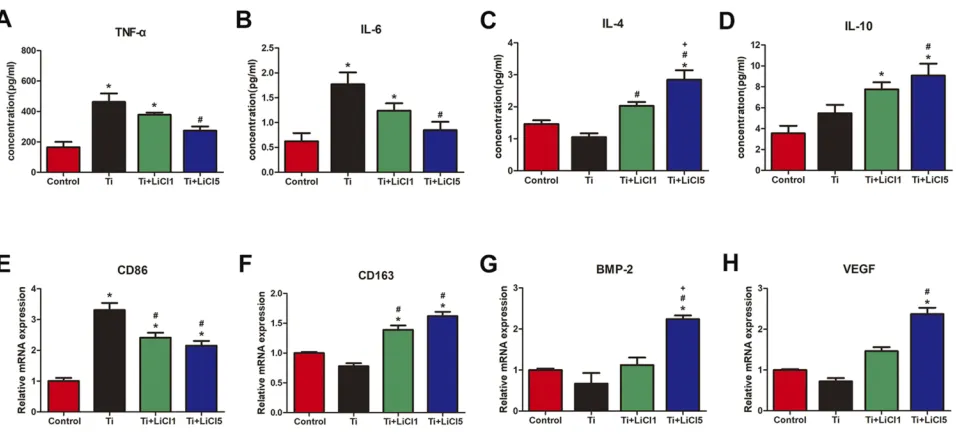
ELISA was performed to detect cytokine secretion levels and RT-PCR was performed to determine the
expression of related genes. As depicted inFigure 5A-D,
macrophages in the Ti+LiCl5 group released the highest amounts of IL-4 and IL-10, which are mainly generated by
anti-inflammatory macrophages. However, the secretion of
inflammatory cytokines, TNF-αand IL-6, were the highest
in the Ti group. The M1 phenotype marker gene, CD86, was downregulated in the LiCl-treated groups compared to
the control and Ti particle groups (Figure 5E). However,
the expression of marker gene, CD163, representing M2 macrophages, was elevated in the LiCl-treated groups and
not in the other groups (Figure 5F). In addition, the expres-sion levels of BMP-2 and VEGF (bone-related gene) were
also upregulated in the Ti+LiCl5 group (Figure 5GandH),
suggesting a potential osteogenic capacity in the LiCl-treated groups.
LiCl attenuates Ti particle-induced activation
of ERK and p38 phosphorylation in vitro
Previous studies have reported that poly (methyl metha-crylate) and hydroxyapatite particles stimulate the activa-tion of the ERK and p38 pathways, and drive M1
macrophage polarization.32However, whether Ti particles
and LiCl are involved in the mitogen-activated protein kinase (MAPK) pathway, or not, is unknown. In this study, RAW cells were stimulated with different concen-tration of Ti particles over the course of 30 min, then, ERK and p38 activation, as indicated by phosphorylation, was
measured by Western blot analysis (Figure 6), which
revealed that ERK and p38 phosphorylation were signifi
-cantly increased by Ti particles stimulation (Figure 6A–C).
As presented in Figure 6D–F, the activation of ERK and
p38 was reduced by LiCl treatment compared to Ti treat-ment, indicating that LiCl attenuated wear particle-induced
Figure 3Immunofluorescence staining of RAW cells.
Notes:CCR7 (greenfluorophore) indicates M1 macrophages; Arg-1 (redfluorophore) indicates M2 macrophages; nuclei are stained with DAPI (bluefluorophore), scale bar: 50μm.
Abbreviations:CCR7, C-C chemokine receptor type 7; Arg-1, arginase-1; DAPI, 4ʹ,6-diamidino-2-phenylindole.
International Journal of Nanomedicine downloaded from https://www.dovepress.com/ by 118.70.13.36 on 22-Aug-2020
Figure 4In vitro polarization of macrophage.
Notes:(A) and (B) Representative histograms offlow cytometry results after cultured for 4 days, percentage of CCR7 and CD206 positive cells, representing M1 or M2 macrophages, respectively. (C) Flow cytometry analysis of RAW264.7 cells. (D) and (E) Percentage of CCR7 and CD206 positive cells respectively. (*# and + represent P<0.05 when compared with Control, Ti, and Ti+LiCl1 respectively).
Abbreviations:CCR7, C-C chemokine receptor type 7; CD206, cluster of differentiation 206.
Figure 5ELISA determination of cytokine, RT-PCR analysis of genes from RAW cells.
Notes:(A) TNF-α, (B) IL-6, (C) IL-4, (D) IL-10, (E) Gene expression of M1 marker CD86, (F) Gene expression of M2 marker CD163, (G) BMP-2 and (H) VEGF. (*# and + representP<0.05 when compared with Control, Ti, and Ti+LiCl1 respectively).
Abbreviations:ELISA, enzyme-linked immunosorbent assay; RT-PCR, real-time polymerase chain reaction; TNF-α, tumor necrosis factor alpha; IL, interleukin; CD, cluster of differentiation; BMP-2, bone morphogenetic protein-2; VEGF, vascular endothelial growth factor.
International Journal of Nanomedicine downloaded from https://www.dovepress.com/ by 118.70.13.36 on 22-Aug-2020
inflammation via the suppression of ERK and p38 phos-phorylation in vitro.
The evaluation of osteogenic
differentiation capacity in conditioned
medium
RAW cell culture supernatant was used as conditioned medium to evaluate its effect on osteogenic differentiation of rBMSCs. The results of ALP and ARS staining demon-strated a higher level of ALP expression and extracellular matrix mineralization (ECM) in the Ti+LiCl5 group,
fol-lowed by the Ti+LiCl1 group (Figure 7AandB). The ALP
activity and optical density values of ECM showed similar
results (Figure 7CandD). Consistent with ALP and ARS
staining, a similar trend was detected in immunofl
uores-cence staining: the Ti+LiCl5 group had a higher ALP
fluorescence intensity than other groups, and the lowest
ALP fluorescence intensity was observed in the Ti group
(Figure 7E).
In vivo air pouch model
To assess the inflammatory response and different
macro-phages phenotypes infiltrate the skin of the air pouch, we
carried out histological and immunofluorescent staining of
the skin sections. The hematoxylin and eosin- and Masson
trichrome- staining sections are shown inFigure 8AandD.
The blue arrows represent thefibrous layer. The results of
histological staining suggested that air pouches treated with
wear particles provoked a pronounced inflammatory
reac-tion, presenting as increased thickness and cell infiltration of the pouch membrane compared to other groups. In con-trast to Ti groups, LiCl attenuated the inflammatory reaction
by decreasing the thickness of the fibrous layer and cell
infiltration. The general observation was consistent with the quantitative results (Figure 8BandC). As shown inFigure
8E–H, the concentration of proinflammatory cytokines
(TNF-α and IL-6) in the LiCl-treated groups were lower
than those in the Ti group. However, the anti-inflammatory
cytokines, IL-4 and IL-10, were upregulated in the exudates injected with LiCl. The ELISA results from in vivo experi-ments, were in accordance with those in vitro. Additionally,
an in vivo bio-safety assay of major organs confirmed that
LiCl attenuated the inflammatory responses by regulating
the ratio of M1/M2 macrophages that were non-toxic to key organs (Figure S1).
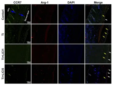
The expression of CCR7 (green, yellow arrows) and Arg-1 (red, white arrows) in RAW264.7 cells were also
Figure 6LiCl attenuates Ti particle-induced the activation of ERK and p38 phosphorylation in RAW cells.
Notes:(A) Western blots performed after macrophages were treated with various concentrations (0, 10, 50, 100μg/mL) of Ti particles for the indicated times. (B) and (C) The density of Western blot bands shown in (A) was quantified using ImageJ software. (D) Western blots performed after macrophages were treated with PBS (control), Ti particles or Ti particles+LiCl. (E) and (F) The density of Western blot bands shown in (D) was quantified using ImageJ software. (*# and + representP<0.05 when compared with Control, Ti, and Ti+LiCl1 respectively).
Abbreviations:LiCl, lithium chloride; Ti, titanium; ERK, extracellular signal-regulated kinase.
International Journal of Nanomedicine downloaded from https://www.dovepress.com/ by 118.70.13.36 on 22-Aug-2020
assessed by immunofluorescence staining in vivo. The
blue arrows show the fibrous layer and skin (Figure 9).
As shown in Figure 9, more CCR7 positive cells were
observed in the Ti particle group, but not in other groups; in contrast, the Ti+LiCl5 group featured a higher propor-tion of Arg-1 positive cells. Therefore, the in vivo experi-mental results were highly consistent with the results from the in vitro experiments, indicating that LiCl can drive
macrophage polarization and induce an osteogenic
immune microenvironment.
Discussion
Total joint arthroplasty is one of the most successful treatment strategies to be developed in past decades;
however, wear debris, generated from orthopedic mate-rials, can stimulate host immune cells to enhance
inflammatory responses and induce osteolysis.33,34 The
wear particles are preponderantly phagocytosed by
macrophages, which produce various inflammatory
che-mokines and cytokines.31 Specifically, several studies
have reported that the state of macrophage polarization
may affect inflammatory response and generate an
osteogenic immune microenvironment.10,35,36 Recently,
LiCl has been studied in relation to various bone
loss-related diseases and inhibited inflammatory cytokine
expression, especially in wear debris-induced infl
amma-tory response in vivo.21,37 Moreover, the
immune-mod-ulatory properties of LiCl have also drawn much
Figure 7Osteogenic differentiation effect of macrophage-conditioned medium.
Notes:(A) ALP staining and (B) ARS staining of rBMSCs cultured in conditioned medium for 14 days. (C) ALP activity of rBMSCs cultured in conditioned medium for 14 days. (D) Quantitative analysis of Alizarin red staining. (E) ALP immunofluorescent staining of rBMSCs cultured in conditioned medium: ALP (green), actin (red), nuclear (blue). scale bar: 50μm. (*# and + representP<0.05 when compared with Control, Ti, and Ti+LiCl1 respectively).
Abbreviations:ALP, Alkaline phosphatase; ARS, alizarin red staining; rBMSCs, rat bone marrow mesenchymal stem cells.
International Journal of Nanomedicine downloaded from https://www.dovepress.com/ by 118.70.13.36 on 22-Aug-2020
attention. Therefore, we explored the immunomodula-tory ability of LiCl on Ti particle-treated macrophages and its osteogenic effects on rBMSCs.
Macrophages are versatile cells that play a crucial role in response to microenvironmental signals. Previous studies have demonstrated that macrophages can polarize into either the M1 or M2 phenotype, which represent extremes of a continuum of functional states.30,38 However, polarization
is not fixed, as macrophages are sufficiently plastic and
dynamic for integrating multiple signals, such as damaged tissues, microbes, and the normal tissue environment.11,39 The morphology of macrophages suggested that wear
particles stimulate obvious M1 phenotypes with many synap-tic structures, whereas LiCl enhanced the differentiation of M2 macrophages with cone-shaped ones and decreased the number of M1-like macrophages, especially in the high-LiCl concentration group (Figure 1A). According to the cell CCK-8 assay results, LiCl and Ti particles have no obvious
cyto-toxicity (Figure 1B). Then, we explored whether LiCl
reg-ulates macrophage polarization in vitro. The results of immunostaining showed that LiCl upregulated Arg-1
expres-sion and downregulated CCR7 expresexpres-sion (Figure 3). Flow
cytometry results were consistent with immunostaining
results, and changes in proinflammatory and
anti-Figure 8Images of hematoxylin eosin and Masson trichrome staining on the air pouch tissues at day 4.
Notes:(A) hematoxylin eosin stained images showing a whole structure and layers of the air-pouch tissues. (B) Thickness offibrous layer. (C) Numbers of infiltration cells. (D) Masson’s trichrome stained images obviously displaying thefibrous tissues and the infiltration cells. Cytokines in the air-pouch exudates were evaluated by ELISA: (E) TNF-α; (F) IL-6; (G) IL-4; and (H) IL-10. scale bar: 200μm. (*# and + representP<0.05 when compared with Control, Ti, and Ti+LiCl1 respectively).
Abbreviations:ELISA, enzyme-linked immunosorbent assay; TNF-α, tumor necrosis factor alpha; IL, interleukin.
International Journal of Nanomedicine downloaded from https://www.dovepress.com/ by 118.70.13.36 on 22-Aug-2020
inflammatory cytokines, as detected by ELISA, further
reflected the functional differences between M1 and M2
phenotypes. At the genetic level, the mRNA expression of
CD86 and CD163 were used by RT-PCR to further confirm
the effect of LiCl on macrophage polarization. LiCl decreased CD86 expression and increased CD163 expres-sion, which is characteristic of a downregulation of infl
am-matory activation and an upregulation of anti-inflammatory
expression. In addition, the expression of BMP-2 and VEGF, both of which enhance tissue healing and bone formation, were also elevated in the LiCl-treated group. To further investigate the osteoimmunology of LiCl, cell and
immuno-fluorescence staining were performed to investigate the
osteogenic differentiation of rBMSCs cultured in condi-tioned medium. The results obtained were consistent with the results of in vitro macrophage polarization. It has been demonstrated that osseous repair requires the coordination of diverse osteogenic and angiogenic factors during the healing
cascade.40 In summary, the results of the immune
experi-ments indicated that LiCl can promote M2 macrophage polarization and enhance the secretion of anti-inflammatory and osteogenic cytokines. Furthermore, previous studies
have already demonstrated that the immunomodulatory effects between macrophage polarization and osteogenesis may be one of the mechanisms accounting for bone regeneration.41–44
In this study, we also investigated the wear
particle-stimulated inflammatory response and macrophage
polari-zation using an air pouch model.9 The injection of Ti
particles into the air pouch on the back of mice resulted in
increased thickness of thefibrous layers and inflammatory
cell infiltration compared to other groups. However, mice
treated with LiCl had fewer infiltrating cells and thinner
fibrous layers, suggesting a comparatively anti-infl
amma-tory effect (Figure 8BandC). ELISA results also revealed that the LiCl treated group induced the production of higher
levels of anti-inflammatory cytokines and lower levels of
proinflammatory cytokines. Furthermore, immunofl
uores-cence images indicated that an air pouch treated with wear particles and 5 mM LiCl presented a lower percentage of CCR7-positive cells and a higher percentage of Arg-1-posi-tive cells than those treated with wear debris alone. The results of the in vivo air pouch model were consistent with the results obtained in vitro.
Figure 9Images of immunofluorescence staining on air-pouch tissues at day 4.
Notes:CCR7 (M1 marker) positive cells were stained in green and Arg-1 (M2 marker) positive cells were stained in red; nuclei were stained in blue; scale bar: 100μm. Abbreviations:CCR7, C-C chemokine receptor type 7; Arg-1, arginase-1.
International Journal of Nanomedicine downloaded from https://www.dovepress.com/ by 118.70.13.36 on 22-Aug-2020
We also provided evidence that LiCl inhibits the activation of ERK and p38 in Ti particle-treated macrophages. Previous studies have demonstrated that wear debris immediately sti-mulates the M1 phenotype bringing about a sustained infl am-matory response via the MAPK pathway.32,45Moreover, wear debris activate the MAPK pathway in bone marrow-derived macrophages, promoting the differentiation of osteoclast pre-cursor cells.46 Furthermore, it has been reported that LiCl
significantly suppressed LPS-induced activation of ERK
phosphorylation in macrophages.47,48Thus, MAPK pathway
targeting has the capacity to regulate the two key processes resulting in aseptic loosening: wear particle-induced infl am-mation and osteoclast foram-mation. Given the close relationship between LiCl and the MAPK pathway, we reasoned that LiCl might drive wear particle-treated macrophages through sup-pression of the ERK or p38 pathway. First, we confirmed that Ti particles activated ERK and p38 MAPKs in RAW264.7. Then, we detected that LiCl reduced wear particle-induced ERK and p38 phosphorylation in macrophages. These results suggested that wear particles could modulate M1 macrophage
polarization and inflammatory responses in RAW264.7. Our
results also revealed that LiCl was able to alleviate the phos-phorylation of p38 and ERK, which have been shown to be
concerned with wear particle-induced inflammatory
responses, suggesting that LiCl induced macrophage
polariza-tion and inflammatory responses via the MAPK pathway
(Figure 10). However, the exact molecular mechanisms by
which LiCl influences inflammatory responses and
macro-phage polarization need to be subsequently investigated. In addition, this phenomenon might be one of the mechanisms underlying the immunomodulatory effects of LiCl on the osteogenic differentiation of rBMSCs.
Conclusion
In this study, we indicated that LiCl played an immuno-modulatory role in macrophage polarization and Ti parti-cle-stimulated inflammatory responses in vitro and in vivo, and promoted the differentiation of RAW264.7 cells to
osteo-specific phenotypes by secreting osteogenic and
angiogenic factors to enhance the osteogenic differentia-tion of rBMSCs. This immunomodulatory capacity of LiCl may be attributed to the inhibition of the MAPK signaling pathway. Conclusively, these results suggest that the immunomodulatory properties of LiCl to macrophage polarization and its subsequent effect on the osteoimmune
Figure 10LiCl modulated macrophage polarization to promote the osteogenic differentiation of rBMSCs via the suppression of ERK and p38 phosphorylation. Notes:LiCl exerted an immunomodulatory effect on macrophage polarization and attenuated Ti particle-induced inflammatory responses in vitro and in vivo, and generated an immune microenvironment to improve the osteogenic differentiation capability of rBMSCs. This immunomodulatory capacity of LiCl may be attributed to the inhibition of ERK and p38 signaling pathway.
Abbreviations:LiCl, lithium chloride; rBMSCs, rat bone marrow mesenchymal stem cells; ERK, extracellular signal-regulated kinase; Ti, titanium.
International Journal of Nanomedicine downloaded from https://www.dovepress.com/ by 118.70.13.36 on 22-Aug-2020
environment may be of importance to the mitigation and treatment of wear particle-induced osteolysis.
Acknowledgment
This work was supported by the National Natural Science Foundation of China (81772309), Shanghai Pujiang Talent Program (18PJD035), and Shanghai Sailing Program (18YF1418800).
Author contributions
All authors contributed to data analysis, drafting and revising the article, gavefinal approval of the version to be published, and agree to be accountable for all aspects of the work.
Disclosure
The authors report no conflicts of interest in this work.
References
1. Bin Abd Razak HR, Tan CS, Chen YJ, et al. Age and preoperative knee society score are significant predictors of outcomes among Asians following total knee arthroplasty. J Bone Joint Surg Am. 2016;98(9):735–741. doi:10.2106/JBJS.15.00280
2. Rao AJ, Gibon E, Ma T, et al. Revision joint replacement, wear particles, and macrophage polarization. Acta Biomater. 2012;8 (7):2815–2823. doi:10.1016/j.actbio.2012.03.042
3. Goodman SB, Gibon E, Pajarinen J, et al. Novel biological strategies for treatment of wear particle-induced periprosthetic osteolysis of orthopaedic implants for joint replacement. J R Soc Interface. 2014;11(93):20130962. doi:10.1098/rsif.2013.0962
4. Gallo J, Goodman SB, Konttinen YT, Raska M. Particle disease: biolo-gic mechanisms of periprosthetic osteolysis in total hip arthroplasty.
Innate Immun.2013;19(2):213–224. doi:10.1177/1753425912451779 5. Purdue PE, Koulouvaris P, Potter HG, Nestor BJ, Sculco TP. The cellular
and molecular biology of periprosthetic osteolysis.Clin Orthop Relat Res. 2007;454:251–261. doi:10.1097/01.blo.0000238813.95035.1b 6. Nine MJ, Choudhury D, Hee AC, Mootanah R, Osman NAA. Wear debris
characterization and corresponding biological response: artificial hip and knee joints.Materials (Basel).2014;7(2):980–1016. doi:10.3390/ma7020 980
7. Yang H, Xu Y, Zhu M, et al. Inhibition of titanium-particle-induced inflammatory osteolysis after local administration of dopamine and sup-pression of osteoclastogenesis via D2-like receptor signaling pathway.
Biomaterials.2016;80:1–10. doi:10.1016/j.biomaterials.2015.11.046 8. Ping Z, Wang Z, Shi J, et al. Inhibitory effects of melatonin on
titanium particle-induced inflammatory bone resorption and osteo-clastogenesis via suppression of NF-kappaB signaling. Acta Biomater.2017;62:362–371. doi:10.1016/j.actbio.2017.08.046 9. Li B, Hu Y, Zhao Y, et al. Curcumin attenuates titanium
particle-induced inflammation by regulating macrophage polarization in vitro and in vivo.Front Immunol.2017;8:55.
10. Liu W, Li J, Cheng M, et al. Zinc-modified sulfonated polyetherether-ketone surface with immunomodulatory function for guiding cell fate and bone regeneration. Adv Sci (Weinh). 2018;5(10):1800749. doi:10.1002/advs.201800749
11. Murray PJ. Macrophage Polarization.Annu Rev Physiol. 2017;79 (541–566). doi:10.1146/annurev-physiol-022516-034339
12. Kang H, Kim S, Wong DSH, et al. Remote manipulation of ligand nano-oscillations regulates adhesion and polarization of macrophages in vivo.
Nano Lett.2017;17(10):6415–6427. doi:10.1021/acs.nanolett.7b03405 13. Li B, Cao H, Zhao Y, et al. In vitro and in vivo responses of
macrophages to magnesium-doped titanium.Sci Rep.2017;7:42707. doi:10.1038/srep42707
14. Yuan X, Cao H, Wang J, et al. Immunomodulatory effects of calcium and strontium co-doped titanium oxides on osteogenesis. Front Immunol.2017;8:1196. doi:10.3389/fimmu.2017.01196
15. Ono T, Takayanagi H. Osteoimmunology in bone fracture healing.Curr Osteoporos Rep.2017;15(4):367–375. doi:10.1007/s11914-017-0381-0 16. Takayanagi H. Osteoimmunology: shared mechanisms and crosstalk between the immune and bone systems.Nat Rev Immunol.2007;7 (4):292–304. doi:10.1038/nri2062
17. Takayanagi H. Osteoimmunology in 2014: two-faced immunology-from osteogenesis to bone resorption.Nat Rev Rheumatol.2015;11 (2):74–76. doi:10.1038/nrrheum.2014.219
18. Hao HP, Wen LB, Li JR, et al. LiCl inhibits PRRSV infection by enhancing Wnt/beta-catenin pathway and suppressing inflammatory responses. Antiviral Res. 2015;117:99–109. doi:10.1016/j.antiviral. 2015.02.010
19. Kim S, Bong N, Kim OS, et al. Lithium chloride suppresses LPS-mediated matrix metalloproteinase-9 expression in macrophages through phosphorylation of GSK-3beta. Cell Biol Int. 2015;39 (2):177–184. doi:10.1002/cbin.10352
20. Chen HC, Chien WC, Chang MY, et al. The iNOS/Src/FAK axis contributes to lithium chloride-mediated macrophage migration.
Nitric Oxide.2015;47:58–64. doi:10.1016/j.niox.2015.04.001 21. Gu Y, Wang Z, Shi J, et al. Titanium particle-induced osteogenic
inhibition and bone destruction are mediated by the GSK-3beta/beta-catenin signal pathway. Cell Death Dis. 2017;8(6):e2878. doi:10.1038/cddis.2017.518
22. Qian K, Cheng X, Zhang D, et al. Antiviral effect of lithium chloride on replication of avian leukosis virus subgroup J in cell culture.Arch Virol.2018;163(4):987–995. doi:10.1007/s00705-017-3692-7 23. Khan MS, Ali T, Abid MN, et al. Lithium ameliorates
lipopolysac-charide-induced neurotoxicity in the cortex and hippocampus of the adult rat brain. Neurochem Int. 2017;108:343–354. doi:10.1016/j. neuint.2017.05.008
24. Leu SJ, Yang YY, Liu HC, et al. Valproic acid and lithium meditate anti-inflammatory effects by differentially modulating dendritic cell differentiation and function.J Cell Physiol.2017;232(5):1176–1186. doi:10.1002/jcp.25604
25. Jiang C, Xiao F, Gu X, et al. Inhibitory effects of ursolic acid on osteoclastogenesis and titanium particle-induced osteolysis are mediated primarily via suppression of NF-kappaB signaling.
Biochimie.2015;111:107–118. doi:10.1016/j.biochi.2015.02.002 26. Frellsen AF, Hansen AE, Jolck RI, et al. Mouse positron emission
tomography study of the biodistribution of gold nanoparticles with different surface coatings using embedded copper-64. ACS Nano. 2016;10(11):9887–9898. doi:10.1021/acsnano.6b03144
27. Li B, Jha RK, Qi YJ, et al. Early cellular responses of BMSCs genetically modified with bFGF/BMP2 co-cultured with ligament fibroblasts in a three-dimensional model in vitro. Int J Mol Med. 2016;38(5):1578–1586. doi:10.3892/ijmm.2016.2752
28. Sedgwick AD, Sin YM, Edwards JC, Willoughby DA. Increased inflammatory reactivity in newly formed lining tisssue. J Pathol. 1983;141(4):483–95. doi:10.1002/path.1711410406.
29. Cheng T, Zhang GY, Guo CJ, Zhang X. Effects of NF-kappaB inhibitor on titanium particulate-induced inflammation in a murine model.J Surg Res.2010;162(2):225–230. doi:10.1016/j.jss.2009.03. 034
30. Wang Z, Xue K, Bai M, et al. Probiotics protect mice from CoCrMo particles-induced osteolysis. Int J Nanomedicine. 2017;12:5387– 5397. doi:10.2147/IJN.S130485
International Journal of Nanomedicine downloaded from https://www.dovepress.com/ by 118.70.13.36 on 22-Aug-2020
31. Deng Z, Jin J, Wang Z, et al. The metal nanoparticle-induced infl am-matory response is regulated by SIRT1 through NF-kappaB deacety-lation in aseptic loosening.Int J Nanomedicine.2017;12:3617–3636. doi:10.2147/IJN.S124661
32. Mahon OR, O’Hanlon S, Cunningham CC, et al. Orthopaedic implant materials drive M1 macrophage polarization in a spleen tyrosine kinase- and mitogen-activated protein kinase-dependent manner.
Acta Biomater.2018;65:426–435. doi:10.1016/j.actbio.2017.10.041 33. Sundfeldt M, Carlsson LV, Johansson CB, Thomsen P, Gretzer C. Aseptic
loosening, not only a question of wear: a review of different theories.Acta Orthop.2006;77(2):177–197. doi:10.1080/17453670610045902 34. Cherian JJ, Jauregui JJ, Banerjee S, Pierce T, MA M. What host
factors affect aseptic loosening After THA and TKA?Clin Orthop Relat Res.2015;473(8):2700–2709. doi:10.1007/s11999-015-4220-2 35. Hu Z, Ma C, Rong X, Zou S, Liu X. Immunomodulatory ECM-like
microspheres for accelerated bone regeneration in diabetes mellitus.
ACS Appl Mater Interfaces. 2018;10(3):2377–2390. doi:10.1021/ acsami.7b18458
36. Shi M, Chen Z, Farnaghi S, et al. Copper-doped mesoporous silica nano-spheres, a promising immunomodulatory agent for inducing osteogenesis.
Acta Biomater.2016;30:334–344. doi:10.1016/j.actbio.2015.11.033 37. Liu W, Chen D, Jiang G, et al. A lithium-containing nanoporous
coating on entangled titanium scaffold can enhance osseointegration through Wnt/beta-catenin pathway.Nanomedicine.2018;14(1):153– 164. doi:10.1016/j.nano.2017.09.006
38. Hao S, Meng J, Zhang Y, et al. Macrophage phenotypic mechan-omodulation of enhancing bone regeneration by superparamagnetic scaffold upon magnetization.Biomaterials.2017;140:16–25. doi:10. 1016/j.biomaterials.2017.06.013
39. Zhang H, Lin C, Zeng C, et al. Synovial macrophage M1 polarisation exacerbates experimental osteoarthritis partially through R-spondin-2.Ann Rheum Dis.2018;77(10):1524–1534. doi:10.1136/annrheum-dis-2018-213450
40. Wei F, Zhou Y, Wang J, Liu C, Xiao Y. The immunomodulatory role of BMP-2 on macrophages to accelerate osteogenesis. Tissue Eng Part A.2018;24(7–8):584–594. doi:10.1089/ten.TEA.2017.0232 41. Saldana L, Valles G, Bensiamar F, et al. Paracrine interactions
between mesenchymal stem cells and macrophages are regulated by 1,25-dihydroxyvitamin D3.Sci Rep.2017;7(1):14618. doi:10.1038/ s41598-017-15217-8
42. Pan H, Xie Y, Zhang Z, et al. Immunomodulation effect of a hier-archical macropore/nanosurface on osteogenesis and angiogenesis.
Biomed Mater.2017;12(4):045006. doi:10.1088/1748-605X/aa6b7c 43. Zhang W, Zhao F, Huang D, et al. Strontium-substituted
submicrom-eter bioactive glasses modulate macrophage responses for improved bone regeneration.ACS Appl Mater Interfaces.2016;8(45):30747– 30758. doi:10.1021/acsami.6b10378
44. Li H, Lu Y, Qian J, et al. Human osteoclasts are inducible immuno-suppressive cells in response to T cell-derived IFN-gamma and CD40 ligand in vitro.J Bone Miner Res.2014;29(12):2666–2675. doi:10. 1002/jbmr.2294
45. Liu N, Meng J, Wang Z, et al. Autophagy mediated TiAl(6)V(4) particle-induced peri-implant osteolysis by promoting expression of TNF-alpha.Biochem Biophys Res Commun.2016;473(1):133–139. doi:10.1016/j.bbrc.2016.03.065
46. Rakshit DS, Ly K, Sengupta TK, et al. Wear debris inhibition of anti-osteoclastogenic signaling by interleukin-6 and interferon-gamma. Mechanistic insights and implications for periprosthetic osteolysis.J Bone Joint Surg Am.2006;88(4):788–799. doi:10.2106/JBJS.E.00711 47. Raghavendra PB, Lee E, Parameswaran N. Regulation of macro-phage biology by lithium: a new look at an old drug. J Neuroimmune Pharmacol.2014;9(3):277–284. doi:10.1007/s11481-013-9516-y
48. Hull M, Lee E, Lee T, et al. Lithium chloride induces TNFalpha in mouse macrophages via MEK-ERK-dependent pathway. J Cell Biochem.2014;115(1):71–80. doi:10.1002/jcb.24634
International Journal of Nanomedicine
Dove
press
Publish your work in this journal
The International Journal of Nanomedicine is an international, peer-reviewed journal focusing on the application of nanotechnology in diagnostics, therapeutics, and drug delivery systems throughout the biomedical field. This journal is indexed on PubMed Central, MedLine, CAS, SciSearch®, Current Contents®/Clinical Medicine,
Journal Citation Reports/Science Edition, EMBase, Scopus and the Elsevier Bibliographic databases. The manuscript management system is completely online and includes a very quick and fair peer-review system, which is all easy to use. Visit http://www.dovepress.com/ testimonials.php to read real quotes from published authors.
Submit your manuscript here:https://www.dovepress.com/international-journal-of-nanomedicine-journal
International Journal of Nanomedicine downloaded from https://www.dovepress.com/ by 118.70.13.36 on 22-Aug-2020