CLINICAL ANDDIAGNOSTICLABORATORYIMMUNOLOGY,
1071-412X/99/$04.0010 May 1999, p. 415–419 Vol. 6, No. 3
Copyright © 1999, American Society for Microbiology. All Rights Reserved.
A New Cell Enzyme-Linked Immunosorbent Assay
Demonstrates Gamma Interferon Suppression by Beta
Interferon in Multiple Sclerosis
MOIZ BAKHIET,* VOLKAN O¨ ZENCI, CARIN WITHAGEN, MAHA MUSTAFA, STEN FREDRIKSON,
ANDHANS LINK
Divisions of Infectious Diseases and Neurology, Karolinska Institute, Huddinge University Hospital, Stockholm, Sweden Received 2 September 1998/Returned for modification 27 October 1998/Accepted 29 January 1999
Multiple sclerosis (MS) is a demyelinating disorder of the central nervous system of unknown etiology.
Immune mechanisms involving the proinflammatory cytokine gamma interferon (IFN-g) are believed to play
an important role in the pathogenesis of MS. IFN-b-1b has been introduced as a treatment for MS and was
found to reduce the number and severity of clinical exacerbations. To examine the influence of IFN-b-1b on
myelin basic protein (MBP)-specific and phytohemagglutinin-induced IFN-g production, we developed a
cell-released capturing enzyme-linked immunosorbent assay (CRC-ELISA), which rapidly measures
sponta-neous and antigen- or mitogen-induced cellular IFN-g production. CRC-ELISA documented a significant
MBP-specific T-cell response in the blood of untreated MS patients, as assessed by IFN-gproduction. This
response was suppressed in MS patients treated with IFN-b-1b. The present work confirms in vivo the in vitro
suppressive effects of IFN-b-1b on IFN-gproduction in MS. Moreover, it provides a powerful new technique
for detection of cytokines.
Multiple sclerosis (MS) is a demyelinating disorder of the central nervous system (CNS). Myelin basic protein (MBP) is a major component of myelin that is affected in MS. Fragments of MBP and anti-MBP antibodies are found in the CNS lesions and in the cerebrospinal fluid of MS patients (2, 4, 23).
The presence of activated T cells in the blood, brains, and cerebrospinal fluid of MS patients suggests that the disease is immune mediated (11). T cells recognizing myelin antigens, including MBP and myelin proteolipid protein, are constitu-ents of the normal T-cell repertoire (15, 16, 24). In MS, T cells reactive to self-antigens, including myelin proteins, become activated. Such T cells are able to cross the blood-brain barrier. Upon encountering myelin antigens inside the blood-brain bar-rier, infiltrating T cells become reactivated and release cyto-kines that are capable of amplifying immune responses. The interplay between T helper 1 (Th1) and Th2 cells and the balance of their respective activities are of crucial importance in determining which type of immune response will ensue (26). Gamma interferon (IFN-g), produced by activated Th1 cells, plays a key role in the induction of immunopathogenetic fea-tures in MS lesions, such as astrogliosis (25), macrophage ac-tivation (1, 8), induction of major histocompatibility complex (MHC) (22) and T-cell homing into the CNS by inducing cell adhesion molecules on endothelial cells and enhancing their adhesiveness for T cells (21), and upregulation of MHC class II molecules on endothelial cells and astrocytes, rendering them capable of antigen presentation (7).
Acute exacerbations of MS occur more frequently after viral infections and after administration of IFN-g(17, 18). There-fore, downregulation of activated T cells and, particularly, IFN-gproduction could be advantageous in MS. One molecule with this potential is IFN-b(14). There is substantial evidence that MHC class II expression can be downregulated by IFN-b,
* Corresponding author. Mailing address: Division of Infectious Diseases, Huddinge University Hospital (F-82), S-141 86 Huddinge, Sweden. Phone: 46 8 58582276. Fax: 46 8 7467637. E-mail: Moiz [email protected].
FIG. 1. Scheme of CRC-ELISA for detection of cell-released cytokines (for example, IFN-g, as in the present study). The method can detect cell-released cytokines produced in response to specific antigens or mitogen or produced spontaneously. ABC, avidin-biotin alkaline phosphatase-complex.
415
on August 17, 2020 by guest
http://cvi.asm.org/
which acts by interfering with the transcription of class II-specific mRNA (10). In vitro, IFN-bsuppresses the ability of peripheral blood lymphocytes (PBL) to produce IFN-gin re-sponse to mitogen and antigen stimulation (9, 14). Clinical studies have shown a significant decrease in the number and severity of exacerbations in patients treated with IFN-b-1b compared to those in patients treated with a placebo (8a). In the present work we demonstrated the in vivo effects of IFN-b-1b treatment of MS patients on MBP-specific as well as phytohemagglutinin (PHA)-induced IFN-g production. This was made possible by the development of a cell-released cap-turing enzyme-linked immunosorbent assay (CRC-ELISA), a new, rapid, objective, and sensitive technique capable of mea-suring cellular production of cytokines.
MATERIALS AND METHODS
Patients.Forty-four patients had clinically definite MS (19). The MS patients were divided into two groups: (i) 29 untreated MS patients (22 females) with an age range of 24 to 68 years (mean, 46), none of whom had ever received any immunomodulatory treatment, and (ii) 15 IFN-b-1b-treated MS patients (14 females) with an age range of 25 to 53 years (mean, 42). The untreated MS patients were selected because they displayed the same disease characteristics as the treated patients had displayed prior to being treated with IFN-b-1b. The patients in group ii had all been treated with 8 MIU of IFN-b-1b administered via subcutaneous injection every second day for at least 3 months. Samples from these patients were taken 10 to 14 h after the drug had been given. Nineteen control patients (5 females) had other neurological diseases (OND) of the noninflammatory type. Their age range was 23 to 77 years (mean, 61). Eight patients had muscular tension headache; 2 patients each had Alzheimer’s-type dementia, cerebrovascular disease, and chronic pain syndrome; and one patient each had amyotrophic lateral sclerosis, myelopathy of unknown cause, polyneu-ropathy, cervical radiculopathy, and radial nerve palsy. Fifteen healthy control subjects (7 females) of the staff of the department ranged between the ages of 22 and 46 years (mean, 32).
Preparation of human PBL suspensions.Peripheral blood was taken into heparinized tubes and diluted with the same volume of tissue culture medium (Dulbecco’s medium; Flow Laboratories, Irvine, United Kingdom) with antibi-otics. PBL were separated by density gradient centrifugation on Lymphoprep (Nyegaard, Oslo, Norway). The cells in the interphase were collected, washed
three times with medium, and suspended in complete medium supplemented with 5% fetal calf serum (GIBCO, Paisley, United Kingdom), 1% minimal essential medium (Flow), 2 mM glutamine (Flow), 50mg of penicillin per ml, and 60ml of streptomycin per ml. The cells were counted in a Bu¨rker chamber, and their viability was assessed by trypan blue exclusion. Cell viability always ex-ceeded 95%.
CRC-ELISA for detection of IFN-g.To detect cellular production of IFN-g, a new CRC-ELISA was introduced. The assay is based on capturing the cytokine at the time of its release from the cells with a specific capturing monoclonal antibody (MAb). In order to detect the secreted cytokine in this assay, enzyme immunoassay/radioimmunoassay flat-bottom, high-binding plates (Costar) were coated with 100ml of anti-IFN-g(1-D1K) MAb (5mg/ml; Mabtech, Stockholm, Sweden) diluted in carbonate bicarbonate buffer (pH 9.6) at 4°C overnight. After four washes with 0.05 M phosphate-buffered saline (PBS), the wells were blocked with 100ml of 5% bovine serum albumin per well for 90 min at room temper-ature. After the wells had again been washed four times with PBS, suspensions of PBL were applied in triplicate to individual wells in 200-ml amounts to obtain a final concentration of 23105cells per well. This cell number was selected after we performed titration experiments to attain the optimal cell number for the assay. Cultures either were not stimulated or received either purified human MBP (6) at a final concentration of 10mg/ml or PHA (Difco, Detroit, Mich.) at a final concentration of 0.5mg/ml. After 48 h of incubation at 37°C in a humid-ified atmosphere of 7% CO2, the cells were removed by flicking the plates, which were then washed five times with Tween-PBS. To detect any captured IFN-g, 100 ml of biotinylated anti-IFN-g(7B-B6-1) MAb (0.5mg/ml; Mabtech) diluted in PBS containing 0.5% Tween 20 and 2% bovine serum albumin was added to the wells. After another 60 min of incubation at 37°C and five washes, 100ml of avidin-biotin alkaline phosphatase complex (Vector Laboratories, Burlingame, Calif.) diluted 1:100 in PBS was added for 45 min. Unbound avidin-biotin alkaline phosphatase complex was removed by five consecutive washings with Tween-PBS, and 100ml of freshly prepared enzyme substrate solution was added to each well. Absorbance was measured after 15 min of incubation in the dark in a 405 Multiscan photometer (mcc/340; Labsystem, Helsinki, Finland). In order to quantify the IFN-gsecreted by the cells cultured in the plate, the IFN-gstandard curve was obtained by simultaneously incubating different known concentrations (0, 0.25, 0.5, 1, 2, 4, 8, 16, 32, 64, 128, 256, 512, and 1,024 U/well) of recombinant human IFN-g (rIFN-g; gift from P. van der Meide, TNO Primate Center, Rijswijk, The Netherlands) for 60 min at room temperature in wells precoated with anti-IFN-gMAb. The procedure for developing the plate was continued as described above, and the absorbencies measured from the standard concentra-tion of IFN-gwere used to plot the IFN-gstandard curve. Thereafter, the absorbencies obtained from the cultures, which correspond to the secreted IFN-g, were automatically converted by the computer to units per well, based on FIG. 2. rIFN-gstandard curve. The curve was designed from the absorbencies, obtained after capturing and detecting different known concentrations of rIFN-g(see Materials and Methods), to high-binding microtiter plate wells precoated with anti-IFN-gMAb. The absorbencies of the known IFN-gconcentrations were entered into a data set of a graphical computer software program asy-axis data with a linear scale. The IFN-gunits were entered into the same data set asx-axis data with a logarithmic scale. Into another data set, the absorbencies of the specimens were entered asy-axis data. Units corresponding to these absorbencies were measured from the standard curve and are shown asx-axis values. Dashed lines indicate the minimum and maximum units that the computer can detect from the assay’s standard curve. During the study more than 10 standard curves were made, and there was no significant variation among the curves.
on August 17, 2020 by guest
http://cvi.asm.org/
the standard curve. In parallel wells, another IFN-gstandard curve was estab-lished by adding rIFN-gto cell cultures so that similar conditions to those used for the standard curve were used for the cultures of the specimens. The curve values of these wells did not show variations from the standard curve values obtained by direct application of the rIFN-gto the wells without subsequent cell culture. In this assay, background absorbencies (wells without coating MAb) were very low, and they were subtracted from the absorbencies of the specimens.
Measurement of cytokine levels by conventional ELISA.The same principle as used for the CRC-ELISA was employed to detect IFN-gin the supernatant from the cultures, except that the cells were not cultured in the plate coated with capturing antibody. Instead, the cells were cultured in a separate plate and stimulated as described above. Supernatants were collected and transferred to the plate precoated with the anti-IFN-gMAb and incubated for 4 h at room temperature. After several washings, the biotinylated detecting antibody was added, and the procedure was continued as described above. All plates and reagents were the same as those used for the CRC-ELISA.
Enumeration of IFN-g-secreting cells.To compare the CRC-ELISA to the enzyme-linked immunospot (ELISPOT) assay, we ran the CRC-ELISA as de-scribed above in parallel with a modified (12) ELISPOT assay, dede-scribed by Czerkinsky et al. (5), for 10 randomly selected patients with MS. PBL were applied in duplicate to individual wells in 200-ml amounts to obtain a final concentration of 23105cells per well.
Statistics.The Mann-Whitney test was used for statistical analysis.
RESULTS
Standard curve for rIFN-g. To estimate the amount of
IFN-gproduced by a certain number of cells in each well after culture termination, a standard curve was plotted by using the absorbency values obtained after the incubation of rIFN-gat different concentrations (Fig. 1 and 2). Units corresponding to the absorbencies of the specimens were obtained from the standard curve. The CRC-ELISA measured accurately as little as 1 U of IFN-gper well (i.e., 10 U/ml). The upper value that could be measured by the CRC-ELISA was 638 U of IFN-g per well (i.e., 6,380 U/ml). Thereafter, a plateau was reached (Fig. 2).
Influence of cell number on IFN-gproduction in response to
MBP, PHA, and no stimulation. In the present study we
ti-trated different cell numbers to determine the optimal number of cells for examining IFN-gproduction by the CRC-ELISA without stimulation or after stimulation with MBP or PHA. For this purpose, the IFN-gproduction in different numbers of PBL from five healthy controls, five untreated MS patients, and five MS patients treated with IFN-b-1b was measured. The highest IFN-gproduction was detected in all cultures at a cell number of 23105/well, and hence this number was selected
for the study (titration is not shown).
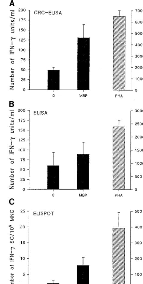
CRC-ELISA compared to conventional ELISA of superna-tants of mononuclear cell suspensions and to ELISPOT assay.
Samples from 10 MS patients were used to compare the num-ber of IFN-gunits recorded spontaneously or after stimulation with MBP or PHA. The levels of production of MBP-reactive IFN-grecorded by the CRC-ELISA were significantly higher than those detected in supernatants of mononuclear cells from the same patients by the conventional ELISA (P,0.05). The total number detected by the CRC-ELISA was about 140 U/ml, compared to 80 U/ml detected by the conventional ELISA. After PHA stimulation, a similar difference between the CRC-ELISA and the conventional ELISA was observed (P, 0.05). Only seven cells secreting IFN-gin response to MBP were recorded by the ELISPOT assay (Fig. 3). However, the difference in the spontaneous IFN-gproduction recorded by the CRC-ELISA and that recorded by the conventional ELISA was not significant. Few spontaneous IFN-g-secreting cells were detected by the ELISPOT assay, while about 400 IFN-g-secreting cells were detected after PHA stimulation (Fig. 3). To study the inter- and intra-assay variations, we repeated the experiments five times. Furthermore, we ran the assay for the same patients several times or incubated the cells
from the same patients in different plates. The variations in the assay were very minor. This was also the case for two other cytokines (interleukin-4 [IL-4] and IL-10) that were used to test the specificity of the assay. Adding secondary antibodies to these cytokines did not give a signal above the background level.
Effects of IFN-b-1b treatment on MBP-specific and
PHA-induced IFN-gproduction. To study the effects of IFN-b-1b
treatment on MS patients, the CRC-ELISA was used to com-FIG. 3. Comparison of the CRC-ELISA with a conventional ELISA of su-pernatants of mononuclear cell suspensions and with the ELISPOT assay. Sus-pensions of PBL from 10 MS patients were plated at 23105cells per well and cultured for 48 h. Triplicate cultures were exposed to the optimal dilution of MBP or PHA. Control cultures received no stimulation. Data are the results of five different experiments. Error bars indicate standard deviations. MNC, mono-nuclear cells; SC, secreting cells.
VOL. 6, 1999 IFN-b SUPPRESSES IFN-g IN MS PATIENTS 417
on August 17, 2020 by guest
http://cvi.asm.org/
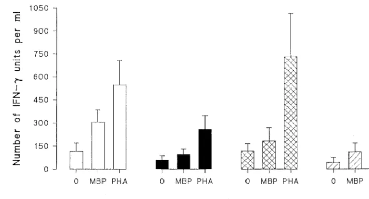
pare MBP-induced IFN-g production in PBL from MS pa-tients treated with IFN-b-1b and PBL from untreated papa-tients. The PHA response reflected by IFN-g production was also studied in both groups. For untreated MS patients, MBP stim-ulation of PBL induced higher IFN-gproduction than no stim-ulation (P, 0.03) did. This MBP-reactive IFN-gproduction was evidently suppressed in MS patients who received IFN-b-1b treatment; there was no significant difference in IFN-g production after MBP stimulation compared to IFN-g produc-tion after culture in the absence of the antigen. Furthermore, the MBP-specific IFN-gproduction in the IFN-b-1b-treated MS patient group was significantly reduced compared to that in untreated MS patients (P , 0.05). PHA stimulation of IFN-ginduction in the IFN-b-1b-treated patients was found to be lower than PHA stimulation of IFN-g induction in the untreated MS patients. However, in both groups, it was still higher than the IFN-ginduction with no stimulation (Fig. 4). OND patients and healthy controls did not show a significant difference in induction of IFN-gin response to MBP compared to induction with no stimulation. Nevertheless, a very high induction of IFN-gwas recorded for both OND patients and healthy controls after stimulation with PHA (Fig. 4).
DISCUSSION
The present work has established a new method to detect cytokine production and has adopted that method to monitor the effects of IFN-b-1b treatment of MS patients. Many studies have emphasized the role of cytokines in modulating immune responses during infections and autoimmunity. However, such studies have focused mainly on the determination of cytokine levels in bodily fluids. Since cytokines act autocrinely or para-crinely, with very short half-lives, and have high affinity for nearby receptors, cellular induction of cytokines has been de-tected, rather than cytokine levels in bodily fluids. To bypass this problem, we used the CRC-ELISA to detect the cytokines immediately after they were released. This was clearly shown in this study, where significant detection of IFN-gwas registered by the CRC-ELISA compared to detection by a conventional ELISA. Other principles used for cellular cytokine detection include enumeration of cytokine mRNA-expressing cells by
the in situ hybridization technique (13) and evaluation of cel-lular production of cytokines by ELISPOT assays, which detect single cells secreting cytokines (15). However, even though both methods can give actual numbers of T cells with a certain functional ability, they are based on subjective analysis. The in situ hybridization technique enables detection of mRNA of a broad range of cytokines. However, mRNA expression does not always correlate to the actual production of cytokines (20). The CRC-ELISA described in this work is an objective assay that rapidly quantifies the amount of produced cytokine. The assay is based on detection of cytokines by the capturing MAb before they are utilized or destroyed by the in vitro conditions of the culture. The CRC-ELISA is also useful in limiting-dilution analysis to measure frequencies of antigen-reactive T cells. In this context, we have initiated studies in an experi-mental allergic encephalomyelitis animal model for MS to compare the number of the MBP-specific IFN-g-secreting cells detected by the classical ELISPOT assay with the frequency of the MBP-specific IFN-g-producing cells detected by the CRC-ELISA. Our preliminary data showed that the CRC-ELISA is more sensitive in the assessment of cell frequencies (1a). The data of the present work also support the above notion, since the levels of IFN-gdetected by CRC-ELISA after MBP stim-ulation (about 140 U/ml) were higher than the number of IFN-g-secreting cells detected by the ELISPOT assay (seven cells). Recording concentrations of a cytokine may be more essential than assessing the number of producing cells in cer-tain situations. In the present study, only seven cells secreted 140 U of IFN-gper ml, suggesting that the number of cells found does not indicate how much of a cytokine is produced. Although CRC-ELISA enables studies of selected cytokines with defined effector or immunoregulatory roles, the inter- and intracellular regulation accomplished by mutual cytokine ef-fects may affect the final cellular production of certain cyto-kines.
Using the CRC-ELISA, we examined the in vivo effects of IFN-b-1b on the production of IFN-g in patients who had received IFN-b-1b treatment and compared the results with those from untreated MS patients, as well as OND patients and healthy controls. The significant suppression of MBP-in-FIG. 4. Effects of IFN-b-1b treatment on MBP-specific and PHA-induced IFN-gproduction. Suspensions of PBL from 15 IFN-b-1b-treated and 29 untreated MS patients, 17 patients with OND, and 15 healthy controls (HC) were plated at a cell number of 23105per well and cultured for 48 h. Triplicate cultures were exposed to the optimal dilution of MBP or PHA or left without stimulation. Note the lower IFN-gproduction in response to MBP and PHA in IFN-b-1b-treated MS patients compared to production in untreated MS patients. Error bars indicate standard deviations.
on August 17, 2020 by guest
http://cvi.asm.org/
duced IFN-gproduction in the IFN-b-1b-treated MS patients reflects the in vivo effects of IFN-b-1b on IFN-gproduction. These effects were previously shown in vitro (9, 14). However, this inhibitory effect is not specific for MBP, since IFN-g pro-duction after PHA stimulation was also reduced in the IFN-b-1b-treated patients, although to a lesser level. The viability of the PBL, as assessed by trypan blue exclusion both at the onset and at the end of the culture, has ruled out the possibility that PBL from IFN-b-1b-treated patients might be less viable and that fewer cells had therefore survived the 48-h incubation period. In support of our results, Brod et al. (3) have recently shown that the capacity of PBL to produce tumor necrosis factor alpha, IFN-g, and IL-4 in response to CD3 MAb was reduced in IFN-b-1b-treated patients, and the capacity of PBL to produce IL-6 was increased. Whether the inhibitory effect of in vivo treatment with IFN-b-1b on IFN-gproduction, in re-sponse to MBP, is related to the reduced number and severity of exacerbations in individual MS patients treated with IFN-b-1b is presently under investigation. Furthermore, we are also investigating the mechanism of action of IFN-b-1b and its effects on other cytokines in MS.
In conclusion, our study has achieved two goals: (i) estab-lishing a new method to detect cytokine production and (ii) using that method to monitor the effects of IFN-b-1b treat-ment of MS patients. These effects were assessed by examina-tion of MBP-specific and PHA-induced IFN-gproduction in treated versus untreated MS patients, compared to patients with OND and healthy subjects. The assay represents a new, objective, sensitive, and rapid approach to detecting cellular production of cytokines and may provide an advantage in cy-tokine detection in the biomedical field.
ACKNOWLEDGMENTS
This study was supported by grants from the Swedish Medical Re-search Council, Karolinska Institute ReRe-search Funds, and NHR.
REFERENCES
1.Adams, D. O., and T. A. Hamilton.1987. Molecular transductional mecha-nisms by which IFN gamma and other signals regulate macrophage devel-opment. Immunol. Rev.97:5–27.
1a.Bakhiet, M., et al.Unpublished data.
2.Banik, N. L. 1992. Pathogenesis of myelin breakdown in demyelinating diseases: role of proteolytic enzymes. Crit. Rev. Neurobiol.6:257–271. 3.Brod, S. A., G. D. Marshall, E. M. Henninger, Jr., S. Sriram, M. Khan, and
J. S. Wolinsky.1996. IFN-b-1b treatment decreases tumor necrosis factor-a and increases interleukin-6 production in multiple sclerosis. Neurology46:
1633–1638.
4.Cruz, M., T. Olsson, J. Ernerudh, B. Ho¨jeberg, and H. Link.1987. Immu-noblot detection of oligoclonal anti-myelin basic protein IgG antibodies in cerebrospinal fluid in multiple sclerosis. Neurology37:1515–1519. 5.Czerkinsky, C., L. Å. Nilsson, W. J. Koopman, J. Metstecky, O¨ . Ouchterlony,
D. M. Kemeny, and S. J. Challacombe (ed.).1988. ELISA and other solid phase immunoassays, p. 217–239. John Wiley & Sons, London, England. 6.Deibler, G. E., R. E. Martenson, and M. W. Kies.1972. Large scale
prepa-ration of myelin basic protein from central nervous tissue of several mam-malian species. Prep. Biochem.2:139–165.
7.Fierz, W., B. Endler, K. Reske, H. Wekerle, and A. Fontana.1985. Astrocytes as antigen presenting cells. I. Induction of Ia antigen expression on astro-cytes by T cells via immune-interferon and its effect on antigen presentation. J. Immunol.134:3785–3793.
8.Goldberg, M., L. S. Belkowski, and B. R. Bloom.1990. Regulation of mac-rophage function by interferon-g. Somatic cell genetic approaches in murine macrophage cell lines to mechanisms of growth inhibition, the oxidative burst, and the expression of the chronic granulomatous disease gene. J. Clin. Investig.85:563–569.
8a.The INFB Multiple Sclerosis Study Group.1993. Interferon-beta-1b is ef-fective in relapsing-remitting multiple sclerosis. I. Clinical results of a mul-ticenter, randomized, placebo-controlled trial. Neurology43:655–661. 9.Kivisa¨kk, P., W. Tian, S. Fredrikson, H. Link, and M. So¨derstro¨m.1997.
Multiple sclerosis: myelin basic protein induced mRNA expression of proin-flammatory cytokines in mononuclear cells is suppressed by interferon-b-1b (IFN-b-1b) in vitro. Eur. J. Neurol.4:460–467.
10. Ling, P. D., M. K. Warren, and S. N. Vogel.1985. Antagonistic effect of interferon-beta on the interferon-gamma-induced expression of Ia antigen in murine macrophages. J. Immunol.135:1857–1863.
11. Link, H.1991. The cerebrospinal fluid in multiple sclerosis, p. 1128–1139.In
M. Swash and J. Oxburry (ed.), Textbook of neurology. Churchill Living-stone, Edinburgh, Scotland.
12. Link, H., O. Olsson, J. Sun, W.-Z. Wang, G. Andersson, H.-P. Ekre, T. Brenner, O. Abramsky, and T. Olsson.1991. Acetylcholine receptor-reactive T and B cells in myasthenia gravis and controls. J. Clin. Investig.87:2191– 2196.
13. Link, J., M. So¨derstro¨m, T. Olsson, B. Ho¨jeberg, J. Ljungdahl, and H. Link.
1994. Increased transforming growth factor-beta, interleukin-4, and interfer-on-gamma in multiple sclerosis. Ann. Neurol.36:379–386.
14. Noronha, A., A. Toscas, and M. A. Jensen.1993. Interferon beta decreases T cell activation and interferon gamma production in multiple sclerosis. J. Neuroimmunol.46:145–154.
15. Olsson, T., J. Sun, J. Hillert, B. Ho¨jeberg, H. P. Ekre, G. Andersson, O. Olerup, and H. Link.1992. Increased numbers of T cells recognizing mul-tiple myelin basic protein epitopes in mulmul-tiple sclerosis. Eur. J. Immunol.
22:1083–1087.
16. Ota, K., M. Matsui, E. L. Milford, G. A. Mackin, H. L. Weiner, and D. A. Hafler.1990. T cell recognition of an immunodominant myelin basic protein epitope in multiple sclerosis. Nature346:183–187.
17. Panitch, H. S., R. L. Hirsch, J. Schindler, and K. P. Johnson.1987. Treat-ment of multiple sclerosis with gamma interferon: exacerbations associated with activation of the immune system. Neurology37:1097–1102.
18. Panitch, H. S.1994. Influence of infection on exacerbations of multiple sclerosis. Ann. Neurol.36:S25–S28.
19. Poser, C. M., D. W. Paty, L. Scheinberg, W. I. McDonald, F. A. Davis, G. C. Ebers, K. P. Johnson, W. A. Sibley, D. H. Silberberg, and W. W. Tourtellotte.
1983. New diagnostic criteria for multiple sclerosis: guidelines for research protocols. Ann. Neurol.13:227–231.
20. Raqib, R., Å. Ljungdahl, A. A. Lindberg, B. Wretlind, U. Andersson, and J. Andersson.1996. Dissociation between cytokine mRNA expression and pro-tein production in shigellosis. Eur. J. Immunol.26:1130–1138.
21. Simmons, R. D., and D. O. Willenborg.1990. Direct injection of cytokines into the spinal cord causes autoimmune encephalomyelitis-like inflamma-tion. J. Neurol. Sci.100:37–42.
22. Skoskiewicz, M. J., R. B. Colvin, E. E. Schneeberger, and P. S. Russel.1985. Widespread and selective induction of major histocompatibility complex determined antigensin vivoby gamma interferon. J. Exp. Med.162:1645– 1664.
23. So¨derstro¨m, M., H. Link, Z. Xu, and S. Fredriksson.1993. Optic neuritis and multiple sclerosis: anti-MBP and anti-MBP peptide antibody-secreting cells are accumulated in CSF. Neurology43:1215–1222.
24. Sun, J. B., T. Olsson, W. X. Wang, B.-G. Xiao, V. Kostalus, S. Fredrikson, H.-P. Ekre, and H. Link.1991. Autoreactive T and B cells responding to myelin proteolipid protein in multiple sclerosis and controls. Eur. J. Immu-nol.21:1461–1468.
25. Tejada-Berges, T., and V. W. Yong.1994. The astrocyte mitogen, tumor necrosis factor-alpha, inhibits the proliferative effect of more potent adult human astrocyte mitogens, gamma-interferon and activated T-lymphocyte supernatants. Brain Res.653:297–304.
26. van Noort, J., L. Nagelkerken, and C. Boog.1994. Immune intervention strategies in EAE. Int. MS J.2:43–50.
VOL. 6, 1999 IFN-b SUPPRESSES IFN-g IN MS PATIENTS 419