Copyright © 1998, American Society for Microbiology
Age-Dependent Levels of Select Immunological Mediators
in Sera of Healthy Children
ULRICH SACK,1* ULLRICH BURKHARDT,2MICHAEL BORTE,3HILTRUD SCHA¨ DLICH,1
KERSTIN BERG,1ANDFRANK EMMRICH1
Institute of Clinical Immunology and Transfusion Medicine,1Department of Anesthesiology and Intensive Care,2
and Hospital for Sick Children,3University of Leipzig, Leipzig, Germany
Received 2 June 1997/Returned for modification 10 July 1997/Accepted 26 September 1997
Serum cytokine levels were measured in 275 healthy children of different ages (3 to 17 years). Interleukin-1 receptor antagonist (IL-1RA), soluble IL-2R (sIL-2R) (sCD25), IL-6, IL-8, tumor necrosis factor alpha
(TNF-a), soluble TNF receptor type II (sTNF-RII) (sCD120b), gamma interferon (IFN-g), soluble intercellular
adhesion molecule 1 (sICAM-1) (sCD54), soluble E selectin (sE-selectin) (ELAM-1; sCD62E), sCD14, and
neopterin were measured with commercial test kits. The mean levels of IL-1RA, sIL-2R, TNF-a, sICAM-1,
sE-selectin, and sCD14 were higher than in healthy adults. In contrast, IFN-gand IL-8 were hardly detectable
in children and thereby significantly lower than in adults. In the case of TNF-a, sICAM-1, sE selectin, and
sCD14, there was a high interindividual variability, apparently unrelated to disease. The profiles of some
cytokines, i.e., IL-1RA, IL-6, and TNF-a, showed age-related increases that overlapped with known patterns of
physical growth. Of note, sIL-2R and sE-selectin instead declined with time. Because of the remarkable age-dependent variability in healthy pediatric subjects, disease-related changes, as well as therapy-dependent alterations, should be considered with caution.
The detection of cytokines and soluble immunological re-ceptors in sera bears diagnostic relevance in many diseases. In specialized diagnostics, the evaluation of cytokine profiles can improve medical care and facilitate therapeutic decisions not only in diseases such as septicemia, tumors, and systemic in-flammation (9) but also in postoperative monitoring of organ transplantation (28). The diagnostic application of these pa-rameters, on the other hand, depends critically on the knowl-edge of normal serum values. In the case of children, system-atic information on normal concentrations is hardly available. The subject of the present study, therefore, was the measure-ment of diagnostically relevant cytokines, soluble receptors, and mediators in serum in healthy children (3 to 17 years of age). Interleukin-1 receptor antagonist (IL-1RA), soluble IL-2R (sIL-2R) (sCD25), IL-6, IL-8, tumor necrosis factor alpha (TNF-a), soluble TNF receptor type II (sTNF-RII) (sCD120b), gamma interferon (IFN-g), soluble intercellular adhesion molecule 1 (sICAM-1) (sCD54), soluble E selectin (sE-selectin) (endothelial leukocyte adhesion molecule 1 [ELAM-1]; sCD62E), sCD14, and neopterin were measured with commercially available enzyme-linked immunosorbent as-say (ELISA) kits. Because the assessment of serum cytokine levels appears to be highly sensitive to the duration and the modality of sample handling, efforts were made to reduce the time and standardize the modalities of the various procedures.
MATERIALS AND METHODS
Sera were obtained from 275 healthy children undergoing corrective surgery or medical checkup (see Table 1 for age distribution). All children had been free of infectious diseases for at least 4 weeks before examination and were not under the influence of any treatment. C-reactive protein was measured to exclude ongoing inflammation. The study was approved by the Research Ethics Com-mittee of the University of Leipzig. Written informed consent was obtained from the parents before entry into the study. All patients with unexpectedly high levels
for any of the cytokines were reexamined to verify that no disease had developed immediately after the test.
Blood samples were collected between 8:00 and 9:00 a.m. by venipuncture and centrifuged within 15 min. Serum aliquots were frozen and stored at270°C until analysis. The specimens were thawed immediately before analysis and used at volumes indicated by the manufacturers.
The analyses were performed with 96-well microtiter plate ELISA kits, except for IL-6 and IL-8, which were analyzed with the Immulite system (DPC Diag-nostic Products Corporation, Los Angeles, Calif.), an automatized random ac-cess immunoassay system developed for the measurement of immune parameters in single samples (3). The ELISAs were obtained from R&D Systems, Minne-apolis, Minn. (IL-1RA, sTNF-RII [p75; sCD120b], and sE-selectin [sCD62E]), Immunotech, Marseille, France (sIL-2R [sCD25] and TNF-a), PerSeptive Di-agnostics, Cambridge, Mass. (IFN-g), T Cell Diagnostics, Cambridge, Mass. (sICAM-1 [sCD54]), and IBL Gesellschaft fu¨r Immunchemie und Immunbiolo-gie, Hamburg, Germany (neopterin and sCD14). Test sensitivity was 6.5 pg/ml for IL-1RA, 5 pM for sIL-2R, 2 pg/ml for IL-6, 6.2 pg/ml for IL-8, 5 pg/ml for TNF-a, 0.5 pg/ml for sTNF-RII, 1 pg/ml for IFN-g, 0.3 ng/ml for sICAM-1, 1 ng/ml for sE-selectin, 1 ng/ml for sCD14, and 0.7 nmol/liter for neopterin.
All ELISA kits provided information on the expected levels in serum in healthy adults, along with technical recommendations for the analyses.
Data were analyzed and plotted with SigmaStat and SigmaPlot scientific soft-ware (Jandel Scientific, Erkrath, Germany), using the Mann-Whitney U rank sum test. Two analyses were performed, the first considering all children as a pooled group (n5275) and the second comparing each age group (Table 1) with adults.
RESULTS
The results of the present analysis in relation to age are summarized in Fig. 1 to 3. Notably, there were no statistically significant differences between females and males (data not shown); accordingly, the data are presented as pooled values.
IL-1RA.The pattern for IL-1RA consisted of an early peak
around 3 years of age (Fig. 3) (P,0.001 in comparison with healthy adults) and a second peak around age 12 (P,0.05). Between 7 and 10 years, and after 14 years of age, the values were comparable to those of adults.
sIL-2R.sIL-2 showed a progressive decline from age 3
on-ward (Fig. 3); however, due to the large variability at very young ages, there was no significant difference relative to adult levels.
IL-6. IL-6 peaked around 3 to 4 years of age (P,0.05 in
comparison with adults), as well as at 15 years (P,0.05) (Fig. 3).
* Corresponding author. Mailing address: Institut fu¨r Klinische Im-munologie und Transfusionsmedizin der Universita¨t Leipzig, Linne´st. 3, D-04103 Leipzig, Germany. Phone: (49.341) 9725506. Fax: (49.341) 9725509. E-mail: [email protected].
28
on August 17, 2020 by guest
http://cvi.asm.org/
IL-8.IL-8 was detectable only rarely and always within the range for normal adults (Fig. 1 and 3).
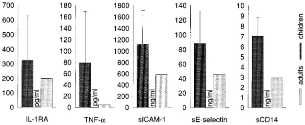
TNF-a.TNF-ashowed a progressive increase with age (Fig. 1), with overall levels significantly higher than those in adults
(Fig. 2) (P,0.01). A clear peak was reached at age 13 to 14 (Fig. 3), with a rather sharp fall to adult levels thereafter.
sTNF-RII.The levels of TNF-RII did not significantly differ
from those of adults, in spite of apparent peaks at 3 to 4 years and at 10 years of age (Fig. 1 and 3).
IFN-g.IFN-gwas hardly detectable in most of the samples; when detected, it remained within the normal adult range (Fig. 1 and 3).
sICAM-1.The concentration of sICAM-1 was significantly
higher in childhood than in adulthood (Fig. 2) (P,0.05), in particular before age 11 (Fig. 1 and 3) (P,0.001 before age 11).
sE-selectin. sE-selectin, on the whole significantly higher
than in adulthood (Fig. 2), showed a progressive fall from age 3 to 5 onward (Fig. 1 and 3) (P,0.05 in comparison with adult levels).
sCD14.sCD14 was significantly higher in childhood than in
adulthood (Fig. 2) (P,0.05), but with no age-related peaks.
Neopterin. Neopterin remained within the normal adult
range throughout childhood (Fig. 1 and 3).
DISCUSSION
The present study provides valuable information on normal ranges for several cytokines, cytokine receptors, or mediators
TABLE 1. Age distribution of children in the study
Age (yr) n
3...10
4...23
5...12
6...21
7...16
8...22
9...17
10...20
11...19
12...21
13...18
14...25
15...18
16...23
17...10
FIG. 1. Age-related expression of immunological mediators in childhood (275 children 3 to 17 years of age) (Table 1). Normal values in adult donors are those provided by the suppliers of the respective kits. Note the remarkable degree of interindividual variability for most of the molecules.
on August 17, 2020 by guest
http://cvi.asm.org/
of potential diagnostic use in pediatrics. The results can be summarized as follows. (i) In general, some molecules (i.e., IL-RA, TNF-a, sICAM-1, sE-selectin, and sCD14) are clearly higher in childhood than in adulthood (Fig. 2), whether or not their profiles present age-related peaks (Fig. 3). (ii) Some cytokines (i.e., IL-RA, IL-6, and TNF-RII) show age-related profiles (Fig. 3), including biphasic patterns that may be re-lated to physical growth (Fig. 3). (iii) Some molecules (i.e., sIL-2R, sICAM-1, and sE-selectin) show a progressive decline after age 3, with no sign of age-related grouping.
The molecules tested in the present study were chosen on the basis of their diagnostic potential. IL-1RA, for example,
antagonizes IL-1 by competitive binding to its receptor, acting thereby as an anti-inflammatory molecule (10, 16). Serum IL-1RA levels are regulated on the basis of serum IL-1 levels, and, albeit in a delayed fashion in comparison to IL-1b, significant levels of IL-1RA can be detected. In adults, the diagnostic use of IL-1RA is mainly in IL-1b-dependent diseases, such as inflammatory and autoimmune diseases, septicemia, and septic shock. In contrast to adults, elevated serum IL-RA levels can be detected in healthy children (Fig. 2), especially around 3 and 10 to 12 years of age (Fig. 1), suggesting that IL-1band/or IL-1RA may play a physiological role in development. Inter-estingly, this age dependency (Fig. 3) seems to closely match the physical growth rates reported by Prader et al. (29). The hypothesis of a link between IL-1 and bone formation is sup-ported by findings showing IL-1 mRNA in developing bone and cartilage (11).
sIL-2R (sCD25), released from activated lymphocytes and resting monocytes by cleavage of the membrane form of IL-2R (38), is detected in the sera of patients with autoimmune or inflammatory diseases, as well as in different forms of leukemia and lymphoma (30, 38). Increased sIL-2R levels are also closely related to rejection episodes after solid-organ engraft-ment (1). In contrast to adults, physiologically high concentra-tions of sIL-R can be found in the sera of healthy children throughout childhood (Fig. 3) (8, 23, 39). At age 15, the levels of sIL-2R decline to those of normal adults (Fig. 1 and 3), suggesting that physiologically high sIL-2R levels are main-tained by the enhanced proliferative activity of the immune system during early childhood, especially in connection to the thymic maturation of T cells. With aging and parallel involu-tion of the thymus, sIL-2R then declines to adult levels.
IL-6 is one of the most promising candidates for use in routine diagnostics. It is produced in inflammatory conditions by monocytes, macrophages, fibroblasts, endothelial cells, ker-atinocytes, lymphocytes, and hepatocytes, as well as in tumors (20). Its secretion is induced by IL-1, TNF, and bacterial en-dotoxins. Serum IL-6 levels are measured in patients with burns, infections, and autoimmune diseases, or else following solid-organ transplantation. In healthy children, serum IL-6 levels are physiologically (and biphasically) elevated around 3 to 4 and 14 years of age, thus confirming previous observations (6). These findings suggest, as in the case of IL-1RA, that IL-6 may play an important physiological role in childhood, in par-ticular in relation to certain stages of physical growth (Fig. 3) (29). In fact, mRNA for IL-6 has been shown to be expressed in developing cartilage and bone (11).
IL-8 is produced by monocytes, macrophages, fibroblasts, endothelial cells, hepatocytes, and chondrocytes. Following FIG. 2. Levels (means with standard deviations) of IL-1RA, TNF-a, sICAM-1, sE-selectin, and sCD14 in serum in healthy children and normal adults (see the legend to Fig. 1). For these select molecules, the pooled childhood values (n5275) were significantly different from the adulthood levels.
FIG. 3. Regression analysis and 95% confidence intervals of measured pa-rameters at different ages. IL-1RA, IL-6, and TNF-ashow not only higher values in children than in adults but also a characteristic pattern which resembles the rates of physical growth in childhood. Of note, sIL-2R, sE-selectin, and sCD14 undergo a progressive decline instead.
on August 17, 2020 by guest
http://cvi.asm.org/
stimulation by other cytokines, IL-8 acts as a chemoattractant especially for polymorphonuclear neutrophils (15). IL-8 is found in high concentrations in the sera of patients with sep-ticemia and also following organ transplantation or during inflammatory and autoimmune diseases. In contrast to the above cytokines, there was hardly any measurable IL-8 in the sera of healthy children (Fig. 1 and 3), indicating that this cytokine is more genuinely involved in inflammation.
TNF-ais produced predominantly by macrophages and lym-phocytes (24, 32). This cytokine acts on fibroblasts and endo-thelial cells, not only inducing inflammatory reactions, fibrosis, and cachexia (5) but also eliciting the production of other inflammatory cytokines and adhesion molecules (5, 22, 37). Because TNF is remarkably unstable, the sera were analyzed very shortly after blood withdrawal, according to a strict stan-dardization protocol. The results of this analysis show that TNF levels are elevated during the entire childhood (Fig. 2), but in a profoundly variable fashion among individuals (Fig. 1). These observations, consistent with similar studies (7, 21), in-dicate that TNF levels in children must be regarded with great-est caution, requiring careful adjustments to take into account the physiological fluctuations.
sTNF-RII (p75) (sCD120b), a member of the TNF receptor family, is expressed on a large variety of cells (33) and is elevated in inflammation, autoimmune diseases, cancer, and sepsis (17, 27). sTNF-RII was slightly but not significantly increased in the sera of healthy children (Fig. 1 and 3), in agreement with other studies (39). Thus, although sTNF-RII is regulated by TNF-a(36), the present study seems to exclude a direct correlation between serum TNF-a and sTNF-RII in childhood.
IFN-gis produced by lymphocytes and macrophages. This cytokine acts on lymphocytes, monocytes/macrophages, and connective-tissue cells as an immunoregulatory, as well as proinflammatory, mediator, with B-cell-differentiating and an-tiviral properties (4). Its levels in serum are mostly low in healthy adults. Indeed, the present study indicates that low levels, or even the absence of immunoreactive IFN-g, also characterize healthy childhood, with little variability among children. In contrast to most of the above cytokines, therefore, there is no elevation corresponding to particular phases of rapid growth (Fig. 1 and 3); IFN-gthus appears to be a gen-uine mediator, and therefore a potential marker, of inflamma-tion and immunity.
sICAM-I (sCD54) is the most important ligand for leukocyte function-associated molecule 1 (LFA-1) and is expressed on a variety of cells including lymphocytes, fibroblasts, keratino-cytes, monokeratino-cytes, and endothelial and dendritic cells (31, 34). The expression of this marker is induced by proinflammatory cytokines, such as IL-1band TNF-a, as well as lipopolysaccha-rides. In immunodiagnostics, sICAM-1 measurements are dicated in several autoimmune and inflammatory diseases, in-cluding multiple sclerosis, as well as in neoplasia and graft rejection following solid-organ transplantation (13). Our study shows enhanced serum sICAM-I concentrations in healthy children (Fig. 2), especially before age 11 (Fig. 3). This obser-vation differs from reports showing low levels in sera of normal children (18).
sE-selectin (sCD62E, or [ELAM-1]) mediates the adhesion of inflammatory cells to the endothelium at sites of inflamma-tion (19). This is followed by lymphocyte extravasainflamma-tion and diapedesis across the vessel wall. High concentrations of sE-selectin are found in a broad range of inflammatory processes (13). Interestingly, there were high levels during normal child-hood (Fig. 2), in particular in early childchild-hood (Fig. 3), whereas other studies found low levels (18). From 12 years of age
onward, sE-selectin declines steadily to normal adult levels (Fig. 1). Possibly, this profile parallels or reflects the increased metabolic turnover of connective tissue and accelerated wound healing in childhood, because sE-selectin appears to play a central role in this process (35).
sCD14, which is released by monocytes and macrophages and interacts with bacterial endotoxins (40), is found in high concentrations in the sera of virally infected patients, as well as in septic, polytraumatic, and severely burned patients (25, 26). In the present study, sCD14 was significantly elevated during childhood (Fig. 2) (P,0.01 except for 6 and 13 years of age), without connection to any disease or trauma.
Neopterin is considered a parameter of increased cellular immunity or inflammatory activity (2). In clinical diagnostics, neopterin is valuable for the monitoring of viral and other infections, malignant diseases, graft survival, and inflammatory diseases (12). In the sera of healthy children, neopterin is always within or slightly below the normal range of adults (Fig. 1 and 3), in agreement with the literature (14), thus represent-ing a quite reliable marker for monitorrepresent-ing the above condi-tions.
Together, the present findings indicate that cytokine con-centrations in the sera of healthy children most often differ from those of adults. In principle, the detection of immuno-logical mediators by ELISA techniques can be influenced by the presence of soluble receptors, inhibitors, antagonists, or binding proteins, as well as by degraded or inactive forms of the cytokines. By investigating a large number of healthy chil-dren, carefully excluding children with ongoing diseases, and following strict protocols for the quick and accurate handling of the specimens, the present study clearly documented eleva-tions of IL-1RA, TNF-a, sICAM-1, sE-selectin, and sCD14. Also, these elevations showed mostly age-dependent longitu-dinal profiles (Fig. 3). Furthermore, the levels of some mole-cules appear to markedly differ between the first 3 years of life and late childhood. However, considering the limited number of small children examined so far, the examination of larger numbers of small children will be helpful in determining reli-able normal cytokine ranges for given age intervals within childhood. These controls, in turn, will be helpful in routine laboratory techniques for the diagnosis and treatment of spe-cific aspects of particular diseases.
ACKNOWLEDGMENTS
We thank Ramona Blaschke, Kati Hofmann, Friedemann Scyman-owski, and Antje Voppmann for technical assistance. We thank Jo¨rg Ermann and Eberhard Keller for assistance in preparing the manu-script. We owe special thanks to Ernesta Palombo-Kinne for critical modification of the reviewed manuscript.
Parts of this work were supported by grants from the Deutsche Forschungsgemeinschaft; the Bundesministerium fu¨r Bildung, Wissen-schaft, Forschung und Technologie; the Interdisziplina¨res Zentrum fu¨r Klinische Forschung; and the Sa¨chsisches Ministerium fu¨r Wissen-schaft und Kunst.
REFERENCES
1. Adams, D. H., L. Wang, S. G. Hubscher, E. Elias, and J. M. Neuberger. 1978. Soluble interleukin-2 receptors in serum and bile of liver transplant recipi-ents. Lancet i:469–471.
2. Anonymous. 1988. Neopterins in clinical medicine. Lancet i:509–511. 3. Babson, A. L. 1991. The Immulite automated immunoassay system. J. Clin.
Immunoassay 14:83–88.
4. Baron, D., S. K. Tyring, W. R. Fleischmann, D. H. Coppenhaver, D. W. Niesel, G. R. Klimpel, G. J. Stanton, and T. K. Hughes.1991. The interfer-ons: mechanisms of action and clinical application. JAMA 266:1375–1383. 5. Beutler, B., and A. Cerami. 1988. Tumor necrosis, cachexia, shock, and
inflammation: a common mediator. Annu. Rev. Biochem. 57:505–518. 6. Brailly, H., F. A. Montero-Julian, C. E. Zuber, S. Flavetta, J. Grassi, F.
Houssiau, and J. van Snick.1994. Total interleukin-6 in plasma measured by
on August 17, 2020 by guest
http://cvi.asm.org/
immunoassay. Clin. Chem. 40:116–123.
7. Cetingul, N., E. Yener, S. Oztop, and G. Nisli. 1994. Serum soluble inter-leukin-2 receptors and tumor necrosis factor-alpha in hematological malig-nancies of childhood. Acta Paediatr. Jpn. 36:49–52.
8. Chan, K. N., A. D. Phillips, J. A. Walker-Smith, and T. T. MacDonald. 1995. Serum interleukin-2 receptors in infants and young children. Acta Paediatr. 84:151–155.
9. Daniel, V. 1995. Diagnostic value of cytokine determination in serum and plasma. Dtsch. Med. Wochenschr. 120:1171–1174.
10. Dinarello, C. D. 1992. Role of interleukin-1 in infectious diseases. Immunol. Rev. 127:119–146.
11. Dodds, R. A., K. Merry, A. Littlewood, and M. Gowen. 1994. Expression of mRNA for IL1 beta, IL6 and TGF beta 1 in developing human bone and cartilage. J. Histochem. Cytochem. 42:733–744.
12. Fuchs, D., G. Weiss, G. Reibnegger, and H. Wachter. 1992. The role of neopterin as a monitor of cellular immune activation in transplantation, inflammatory, infectious, and malignant diseases. Crit. Rev. Clin. Lab. Sci. 29:307–341.
13. Gearing, A. J., and W. Newman. 1993. Circulating adhesion molecules in disease. Immunol. Today 14:506–512.
14. Griffin, D. E., B. J. Ward, E. Jauregui, R. T. Johnson, and A. Vaisberg. 1990. Immune activation during measles: interferon-gamma and neopterin in plasma and cerebrospinal fluid in complicated and uncomplicated disease. J. Infect. Dis. 161:449–453.
15. Grob, P. M., E. David, T. C. Warren, R. P. DeLeon, P. R. Farina, and C. A. Homon.1990. Characterization of a receptor for human monocyte-derived neutrophil chemotactic factor/interleukin-8. J. Biol. Chem. 265:8311–8316. 16. Hannum, C. H., C. J. Wilcox, W. P. Arend, F. G. Joslin, D. J. Dripps, P. L.
Heimdal, L. G. Armes, A. Sommer, S. P. Eisenberg, and R. C. Thompson. 1990. Interleukin-1 receptor antagonist activity of a human interleukin-1 inhibitor. Nature 343:336–340.
17. Haran, N., Y. Bar-Khayim, A. Frensdorff, and G. Barnard. 1991. Tumor necrosis factor (TNF-alpha) binding protein: interference in immunoassays of TNF-alpha. Kidney Int. 40:1166–1170.
18. Hatzistilianou, M., F. Athanassiadou, C. Agguridaki, and D. Catriu. 1997. Circulating soluble adhesion molecules in children with acute lymphoblastic leukaemia. Eur. J. Pediatr. 156:537–540.
19. Hession, C., L. Osborn, D. Goff, G. Chi-Rosso, C. Vassallo, M. Pasek, C. Pittack, R. Tizard, S. Goelz, and K. McCarthy.1990. Endothelial leukocyte adhesion molecule 1: direct expression cloning and functional interactions. Proc. Natl. Acad. Sci. USA 87:1673–1677.
20. Hirano, T., S. Akira, T. Taga, and T. Kishimoto. 1990. Biological and clinical aspects of interleukin 6. Immunol. Today 11:443–449.
21. Ishii, E., S. Ohga, S. Yamada, M. Sako, H. Tasaka, A. Kuwano, M. Sasaki, Y. Tsunematsu, and K. Ueda.1991. Prognosis of children with virus-associ-ated hemophagocytic syndrome and malignant histiocytosis: correlation with levels of serum interleukin-1 and tumor necrosis factor. Acta Haematol. Jpn. 85:93–99.
22. Jacob, C. O. 1992. Tumor necrosis factor alpha in autoimmunity: pretty girl
or old witch? Immunol. Today 13:122–125.
23. Jones, A. C., C. R. Besley, J. A. Warner, and J. O. Warner. 1994. Variations in serum soluble IL-2 receptor concentration. Pediatr. Allergy Immunol. 5:230–234.
24. Jones, E. Y., D. I. Stuart, and N. P. C. Walker. 1989. Structure of the tumour necrosis factor. Nature 338:225–228.
25. Kru¨ger, C., C. Schu¨tt, H. Dietz, J. Lu¨dtke, B. Ruf, A. Langford, H. D. Pohle, and R. O. F. Kunze.1990. Elevated levels of soluble CD14 molecules in serum of HIV-1 infected patients. Immunobiology 181:244.
26. Kru¨ger, C., C. Schu¨tt, U. Obertacke, T. Joka, F. E. Mu¨ller, J. Kno¨ller, W. Kno¨ller, and W. Scho¨nfled.1991. Serum CD14 levels in polytraumatized and severely burned patients. Clin. Exp. Immunol. 85:297–301.
27. Lantz, M., H. Thysell, E. Nilsson, and I. Olsson. 1991. On the binding of tumor necrosis factor (TNF) to heparin and the release in vivo of the TNF-binding protein I by heparin. J. Clin. Invest. 88:2026–2031. 28. Pirenne, J., F. Pirenne-Noizat, D. de Groote, Y. Vrindts, M. Lopez, R. Gathy,
N. Jacquet, M. Meurisse, P. Honore, and P. Franchimont.1994. Cytokines and organ transplantation: a review. Nucl. Med. Biol. 21:545–555. 29. Prader, A., R. H. Largo, L. Molinari, and C. Issler. 1988. Physical growth of
Swiss children from birth to 20 years of age. Helv. Paediatr. Acta 43(Suppl. 52):124–125.
30. Rubin, L. A., and D. L. Nelson. 1990. The soluble interleukin-2 receptor: biology, function, and clinical application. Ann. Intern. Med. 113:619–627. 31. Smith, C. W., R. Rothlein, B. J. Hughes, M. M. Mariscalco, H. E. Rudloff,
F. C. Schmalstieg, and D. C. Anderson.1988. Recognition of an endothelial determinant for CD18-dependent human neutrophil adherence and transen-dothelial migration. J. Clin. Invest. 82:1746–1756.
32. Smith, R. A., and C. Baglioni. 1987. The active form of tumor necrosis factor is a trimer. J. Biol. Chem. 262:6951–6954.
33. Sprang, S. R. 1991. The divergent receptors for TNF. Trends Biochem. Sci. 15:366–368.
34. Springer, T. A. 1990. Adhesion receptor of the immune system. Nature 346:425–434.
35. Subramaniam, M., S. Saffaripour, L. van de Water, P. S. Frenette, T. N. Mayadas, R. O. Hynes, and D. D. Wagner.1997. Role of endothelial selectins in wound repair. Am. J. Pathol. 150:1701–1709.
36. Tartaglia, L. A., and D. V. Goeddel. 1992. Two TNF receptors. Immunol. Today 13:151–153.
37. Tracey, K. J., H. Vlassara, and A. Cerami. 1989. Cachectin/tumour necrosis factor. Lancet i:1122–1126.
38. Waldmann, T. A. 1991. The interleukin-2 receptor. J. Biol. Chem. 266:2681– 2684.
39. Weiss, M., M. Martignoni, T. Petropoulou, B. Solder, and B. H. Belohrad-sky.1996. Increased serum levels of soluble tumor necrosis factor receptor (sTNF-Rs) in children and adolescents with vertically and horizontally trans-mitted HIV infection. Infection 24:301–308.
40. Wright, S. D., R. A. Ramos, P. S. Tobias, R. J. Ulevitch, and J. C. Mathison. 1990. CD14, a receptor for complexes of lipopolysaccharide (LPS) and LPS binding protein. Science 249:1431–1433.