!"###$ %
$& '
' ( )*&'')+, $
- .
-/, - - 0
1 - 0
5! "###
,! "##,6"###7 6
"## 18-,
(, !
#0 5.
! "###### $609$60$9$
' " ! 4:#-234)
Table of Contents
___________________________________________________________________________
Table of Contents... I List of Figures ... III List of Tables ... III List of Abbreviations ... IV Zusammenfassung
………
.
………
.
………
.
……
.
VII Summary ... IX1 Introduction ... 1
1.1 Epidemiology, etiology and clinical picture of spinal cord injury ... 1
1.2 Pathophysiology and clinical management of SCI ... 2
1.3 Preclinical and clinical trials in the field of SCI ... 3
1.4 The failure of successful axonal regeneration in the central nervous system ... 5
1.4.1 The growth inhibitory CNS environment ... 6
1.4.2 The insufficient activation of the intrinsic neuronal growth program upon injury in the CNS ... 8
1.5 STAT3 ... 17
1.5.1 The STAT family ... 17
1.5.2 The JAK/STAT pathway ... 18
1.5.3 The role of STAT3 outside the nervous system ... 20
1.5.4 STAT3 in the context of neuronal survival and regeneration ... 22
1.6 Research question ... 24
2 Material and Methods ... 25
2.1 Material ... 25
2.2 Mice ... 30
2.3 Methods ... 31
2.3.1 Plasmid constructs ... 31
2.3.2 Vector production and purification... 31
2.3.3 Tissue processing and immunohistochemistry ... 32
2.3.4 Quantification of the expression of P-STAT3 and STAT3 ... 33
2.3.5 Gene therapy with recombinant adeno-associated viral vectors ... 33
2.3.6 Confocal microscopy ... 34
2.3.7 In situ analysis of central axonal outgrowth ... 34
2.3.8 In vivo analysis of central axonal outgrowth... 41
2.3.9 Combination therapy with viral gene transfer and chondroitinase ABC ... 41
2.3.10 Statistical analysis ... 42
3 Experiments in the Course of this Study prior to my own Work ... 44
STAT3 depletion impairs the regeneration of PNS axons ... 44
STAT3 deletion impairs initiation but not perpetuation of PNS axon regeneration in vivo 44 4 Results ... 45
4.1 Consequences of a peripheral crush lesion on STAT3 expression ... 45
4.2 STAT3 expression after dorsal column lesion ... 47
4.3 Efficiency of the viral gene therapy ... 48
4.4 Effects of viral gene therapy with STAT3(c) on axon outgrowth after CNS lesion .. 51
Table of Contents II
4.6 Effects of STAT3 deletion on axonal outgrowth after CNS lesion ... 59
4.7 Combination therapy with rAAV-STAT3 and chondroitinase ABC for central axon regeneration ... 62
5 Discussion ... 67
5.1 STAT3 is a key component of the intrinsic growth program ... 67
5.2 STAT3 is an initiator of neurite outgrowth in vivo ... 68
5.3 STAT3 is a phase-specific regulator of axonal regeneration ... 73
5.4 STAT3 is a promising target for therapeutic interventions ... 75
5.4.1 Being in the right place at the right time ... 76
5.4.2 In search of the ideal target in the JAK/STAT pathway ... 77
5.4.3 Bringing STAT3 to neuronal cells: Maximal efficacy and minimal side effects .... 81
5.5 STAT3 in combination with other therapeutic targets ... 82
5.6 Concluding remarks and outlook ... 85
6 References ... 89
7 Acknowledgments ... 105
List of Figures
___________________________________________________________________________
Fig. 1-1 Spinal cord injury ... 2
Fig. 1-2 The inhibitory CNS environment ... 7
Fig. 1-3 The intrinsic neuronal growth program activated upon peripheral injury ... 13
Fig. 1-4 Transcription factors involved in axonal regeneration and their target genes. ... 16
Fig. 1-5 Structure of the STAT3 molecule ... 18
Fig. 1-6 Activation of the JAK/STAT3 pathway ... 20
Fig. 2-1 Equipment for positioning of the animal ... 35
Fig. 2-2 In vivo imaging of fluorescent sensory DRG axons in Thy-GFPs mice... 36
Fig. 2-3 Injection of rAAV into the DRG ... 38
Fig. 2-4 Lesion of the axon emerging from a DRG previously injected with rAAV ... 39
Fig. 2-5 Dissected spinal cord ... 40
Fig. 4-1 Wallerian degeneration and STAT3 expression after peripheral nerve crush lesion . 46 Fig. 4-2 STAT3 expression after dorsal column lesion ... 47
Fig. 4-3 STAT3 expression after dorsal column lesion (Immunohistochemistry) ... 48
Fig. 4-4 Evaluation of the efficiency of the viral gene therapy by immunohistochemistry ... 49
Fig. 4-5 Quantification of the Efficiency of the viral gene therapy) ... 50
Fig. 4-6 Effects of viral gene therapy on CNS outgrowth ... 52
Fig. 4-7 Time course of the experiment ... 53
Fig. 4-8 Effects of viral gene therapy on CNS outgrowth – confocal microscopy ... 54
Fig. 4-9 Gallery of axonal endings ... 55
Fig. 4-10Timecourse of the experiment ... 56
Fig. 4-11Phase specific role of STAT3 for axonal regeneration ... 58
Fig. 4-12Schematic illustration of STAT3 deletion in STAT3 fl/fl mice via cre recombinase .... 59
Fig. 4-13 Sprouting response of STAT3-competent and STAT3-depleted DRG neurons after a CNS lesion and evaluation of P-STAT3 immunoreactivity in the corresponding DRGs. ... 61
Fig. 4-14 Confocal images of axonal outgrowth after combination therapy with rAAV-STAT3 and chondroitinase ABC ... 63
Fig. 4-15 Immunohistochemistry on spinal cords confirming delivery of chondroitinase ABC ... 64
Fig. 4-16Combination therapy with rAAV-STAT3 and chondroitinase ABC ... 65
List of Tables
___________________________________________________________________________ Tab. 1-1 Non-comprehensive Overview of selective non-invasive and invasive therapeutic approaches in SCI therapy ... 5Tab. 1-2 Tissue-specific STAT3-deficient phenotypes outside the nervous system ... 21
List of Abbreviations IV
List of Abbreviations
___________________________________________________________________________
5-HT 5-Hydroxytryptamine = Serotonin
AAV Adeno-associated virus
aCSF Artificial mouse cerebrospinal fluid
ad Adde (fill up to)
ATF-3 Activating transcription factor 3
Bcl-xL B cell lymphoma extra large protein
BDNF Brain-derived neurotrophic factor
BSA Bovine serum albumin
C (+ number) Cervical spinal cord level (+ number) CaCl2 · 2H2O Calcium chloride dihydrate
CaM kinase II, IV Ca2+/calmodulin-dependent protein kinase II, IV
cAMP Cyclic adenosine monophosphate
CAP-23 Cytoskeleton-associated protein 23
CD Cluster of differentiation
C/EBP CCAAT-enhancer-binding protein
ChABC Chondroitinase ABC
CNS Central nervous system
CNTF Ciliary neurotrophic factor
CREB cAMP response element-binding protein
CSPGs Chondroitin sulfate proteoglycans
d/D Day
dH2O Distilled water
DLK Dual leucine zipper-bearing kinase
DNA Deoxyribonucleic acid
DRG Dorsal root ganglion
eCFP Enhanced cyan fluorescent protein
EDTA Ethylenediaminetetraacetic acid
e.g. Exempli gratia = for example
Erk Extracellular signal-regulated kinase
etc. Et cetera
g Gram
GAP-43 Growth-associated protein 43
GDNF Glial cell-derived neurotrophic factor
GFP Green fluorescent protein
gp130 Glycoprotein 130
h Hour(s)
H20 Water/aqua
IGF-I, IGF-II Insulin-like growth factor I, Insulin-like growth factor II
IL-6 Interleukin-6
IRF1 Interferon regulatory factor 1
JAK Janus kinase (or "Just another kinase")
JNK c-Jun N-terminal kinases
KCl Potassium chloride
kDa Kilodalton
KLFs Krüppel-like factor family of transcription factors
l Liter
L (+ number) Lumbar spinal cord level (+ number)
L1CAM L1 cell adhesion molecule
LIF Leukemia inhibitory factor
M Molar mass
MAG Myelin-associated glycoprotein
MAP Microtubule-associated protein
MAPK Mitogen-activated protein kinase
mg Milligram
MgCl2 · 6H2O Magnesium chloride hexahydrate
min Minutes
ml Milliliter
mol Mole
mTOR mammalian Target of Rapamycin
n Number of samples
Na2HPO4 · 2H2O Disodium hydrogen phosphate dihydrate Na2HPO4 · 7H2O Disodium hydrogen phosphate heptahydrate
NaCl Sodium chloride
NaH2PO4 · H2O Sodium dihydrogen phosphate monohydrate
N-cadherin Neural cadherin
N-CAM Neural cell adhesion molecule
NF-κB nuclear factor 'kappa-light-chain-enhancer' of activated B-cells
NGF Nerve growth factor
NT-3; NT-4 Neurotrophin-3; Neurotrophin-4
OES Olfactory ensheathing cells
OMgp Oligodendrocyte myelin glycoprotein
PB Phosphate buffer
PBS Phosphate buffered saline
PFA Paraformaldehyde
PIAS Protein inhibitor of activated STAT
PKA Protein kinase A
PKC Protein kinase C
PNS Peripheral nervous system
P-STAT3 Phosphorylated STAT3
PTEN Phosphatase and tensin homolog
RAGs Regeneration-associated genes
RhoA Ras homolog gene family, member A
RNA Ribonucleic acid
RNA – Ribonucleic acid
ROCK Rho-associated protein kinase
RT Room temperature
s. See
SCI Spinal cord injury
SEM Standard error of the mean
SH2 Src Homology 2
SOCS Suppressor of cytokine signaling
Sox11 Sex determining region Ybox containing gene 11 SPRR1A Small proline-rich repeat protein 1a
STAT3 Signal transducer and activator of transcription 3
List of Abbreviations VI
TBS Tris buffered saline
TF Transcription factor
T (+ number) Thoracic spinal cord level (+ number)
Th1 T-helper cell 1
U Unit
VEGF Vascular endothelial growth factor
wk Week(s)
WT Wildtype
YFP Yellow fluorescent protein
μg Microgram
Zusammenfassung
___________________________________________________________________________
Jedes Jahr erleiden weltweit circa 22 Menschen pro eine Million Einwohner eine Querschnittslähmung, die bei den Betroffenen zu dauerhaften Behinderungen und erheblichen Einschränkungen im Alltag führt. Die schwerwiegenden Defizite nach einer Querschnittslähmung, darunter Lähmungen und chronischer Schmerz, sind darauf zurückzuführen, dass geschädigte Axone im Rückenmark kaum regenerieren und es auch nur in geringem Ausmaß zur funktionellen Reorganisation der noch erhaltenen Nervenverbindungen kommt. Im Unterschied zum peripheren Nervensystem, wo zerstörte Nervenfasern erfolgreich regenerieren, ist die axonale Regenerationskapazität des zentralen Nervensystems (ZNS) spärlich ausgeprägt. Zwar konnte durch intensive Forschung im Verlauf der letzten Jahrzehnte eine Anzahl von „extrinsischen“ wachstumshemmenden
Molekülen von Gliazellen und der extrazellulären Matrix des ZNS identifiziert werden. Es gibt jedoch zunehmend Hinweise darauf, dass zahlreiche dieser „extrinsischen“ Signale letztlich in „intrinsische“ Signalwege der Neuronen selbst einmünden um schließlich die Transkription neuronaler Gene zu verändern. Einer der interessantesten intrinsischen Regulatoren axonaler Regeneration ist der Transkriptionsfaktor ´Signal transducer and activator of transcription 3´ (STAT3). In dieser Arbeit habe ich mithilfe moderner In-vivo-Mikroskopie
Zusammenfassung VIII
einem zunehmend inhibitorischen ZNS-Milieu bedingt ist, wurde die Überexpression von STAT3 zusätzlich mit der Applikation von Chondroitinase ABC, einem Enzym, das die inhibitorischen Moleküle der glialen Narbe neutralisieren kann, kombiniert. Dabei konnte ich zeigen, dass das durchschnittliche Wachstum von Axonen um mehr als das Zweifache gesteigert werden konnte.
Summary
___________________________________________________________________________
Worldwide each year twenty-two in one million people experience a spinal cord injury (SCI) leading to permanent disability and tremendous restrictions in daily life activities. The severe and persistent functional deficits resulting from SCI such as paralysis and chronic pain are due to the limited repair of damaged axonal connections and the poor reorganization of spared nerve connections in the central nervous system (CNS). Whereas damaged axons in the peripheral nervous system (PNS) regenerate successfully, CNS axons fail to regrow. Intensive research over the last decades has identified a number of growth inhibitory molecules in the CNS environment and there is evidence that many of these extrinsic cues converge on intrinsic neuronal signaling pathways that eventually alter gene transcription. One of the most interesting intrinsic regulators of axonal regeneration is the transcription
factor ‘signal transducer and activator of transcription 3’ (STAT3).
Summary X
1
Introduction
___________________________________________________________________________
1.1
Epidemiology, etiology and clinical picture of spinal cord injury
The earliest known description of spinal cord injury was found in an ancient Egyptian text
from the Edwin Smith Surgical Papyrus stating that: “When you examine a man with a
dislocation of a vertebra of his neck, and you find him unable to move his arms and his
legs…..Then you have to say: A disease one cannot treat” (Case et al., 2005).
Introduction 2
Fig. 1-1Spinal cord injury
(A) The spinal cord as part of the central nervous system (green) transmits motor signals from the brain to the periphery via spinal nerves and sends information received from the peripheral nervous system (yellow) to the brain. (B) Motor deficits depending on the level of spinal cord injury. B modified from Thuret et al., 2006.
1.2
Pathophysiology and clinical management of SCI
The mechanical tissue damage upon trauma of the spinal cord is followed by numerous, only incompletely understood mechanisms such as local ischemia, edema formation, disturbance of ion homeostasis, release of free oxygen radicals and excitotoxins as well as recruitment of peripheral inflammatory cells (Schwab et al., 2006; Oyinbo 2011). Astrocytes and microglia become reactive and form a scar around the lesion site, together with extracellular substances such as chondroitin sulfate proteoglycans (CSPGs) and laminin. Within several weeks, macrophages clear the tissue debris and a fluid-filled cyst surrounded by scar tissue cells develops (Schwab et al., 1996; Bareyre et al., 2003; Schwab et al., 2006).
lesion. For example, high cervical lesions can cause pulmonary problems that might require rapid-sequence intubation and lesions above level T6 result in impaired sympathetic innervation of the heart leading to bradycardia. Urinary catheterization is needed to avoid bladder distension and secondary bladder infections. Furthermore, patients should receive thromboprophylaxis due to immobilization and pain killers should be applied early enough to avoid chronification of pain (source: http://www.uptodate.com). Besides, according to a recent meta-analysis, surgical stabilization and decompression of the spinal cord should be considered in all patients from eight to twenty-four hours following acute traumatic SCI (Furlan et al., 2011) to promote neurologic improvement. Moreover, in many therapeutic centers, acute interventions involve application of methylprednisolone as a standard treatment. Methylprednisolone has been found to improve clinical outcome by reducing the secondary damage including cytotoxic edema, inflammation and release of free radicals. Ideally, it is administered at high-dose (30 mg/kg) for twenty-three hours and started within the first eight hours of injury (Bracken 2002; Bracken 2012). After this early pharmacological intervention, further therapy is limited to physiotherapy, which the patient should undergo as early as possible (Edgerton et al., 2006; Mehrholz et al., 2008). In this context, robot-assisted walking training might turn out to significantly ameliorate motor function (Dietz 2010; Wirz et al., 2011).
1.3
Preclinical and clinical trials in the field of SCI
Introduction 4
Non-invasive approaches Substance/
Method
Mechanism Status of the study and results Reference
Erythropoietin (EPO)
Neurons and astrocytes both express and respond to EPO. Amongst others, EPO drives intracellular anti-apoptotic signaling cascades and reduces ischemic damage by autoregulation of blood flow.
Studies investigating behavioral and histological outcome after EPO treatment in rats have yielded inconsistent results. The only human trial found on databases has recently been suspended (Italian multicenter Phase 3 study).
(Goldman et al., 2002; Carelli et al., 2011; Kwon et al., 2011) http://www.clinic altrials.gov
Systemic hypothermia
Hypothermia has neuroprotective effects as it maintains intracellular ATP concentrations by slowing down enzymatic activity.
In rats, reduced apoptosis and tissue damage have been found. Results for hindlimb motor function are contradictory. Clinical trials and case reports show trend towards functional improvement, randomized multicenter trials are still pending.
(Kwon et al., 2008; Dietrich et al., 2011; Kwon et al., 2011)
Minocycline Minocycline exerts inflammatory and anti-apoptotic actions.
The results of a phase II placebo-controlled randomized trial of minocycline in an acute spinal cord injury model could not establish the efficacy of minocycline that has been shown in the majority of animal studies.
(Kim et al., 2009; Kwon et al., 2011; Casha et al., 2012)
Riluzole Riluzole decreases excitotoxicity by blocking voltage-sensitive sodium channels and antagonizing presynaptic calcium-dependent glutamate release.
In rats, there is preclinical evidence for improved tissue protection and ameliorated locomotor scores as well as temporally limited effect on spasticity. Clinical studies are not yet open for patients.
(Cadotte et al., 2011; Kwon et al., 2011) http://www.clinic altrials.gov Invasive approaches Substance/ Method
Mechanism Status of the study and results Reference
Chondroitinase ABC
Enzyme digesting growth inhibitory chondroitin sulfate proteoglycans in the scar after SCI. More information s. section 1.4.1.
Intrathecal application of chrondroitinase ABC improves axonal regeneration and neuroprotection of injured fibers, promotes sprouting/plasticity of uninjured tracts and ameliorates motor function. Clinical trials have not yet been started.
(Bradbury et al., 2011; Kwon et al., 2011)
Anti-Nogo approaches
Mainly antibodies that target Nogo-A which is present in growth- inhibitory myelin of the CNS. More information s. section 1.4.1.
Intrathecal application of anti-Nogo-A antibody in animal models showed upregulation of growth-specific proteins, enhanced sprouting of fibers and the formation of new functional connections. A Phase II trial is currently ongoing.
(Bareyre et al., 2002; Zorner et al., 2010)
Anti-Rho approaches
The Rho signaling pathway regulates the cytoskeleton and exerts an important inhibitory role in neuronal growth
In animal models there is increased axonal sprouting and motor improvement; A phase I/II clinical trial of a recombinant Rho protein antagonist in acute spinal cord injury suggested slightly increased neurological recovery.
(Dergham et al., 2002; Fehlings et al., 2011; Kwon et al., 2011)
Activated macrophages
Activated macrophages eliminate growth
The results of a phase 2 randomized controlled multicenter trial could not
inhibitory myelin debris; They exert neuroprotective and anti-excitatory functions
support the promising results from animal models.
al., 2006; Lammertse et al., 2012)
Olfactory ensheathing cells
These cells have growth permissive properties; Together with regenerating axons they seem to migrate through an unfavorable CNS environment.
No or little effects concerning functional recovery in a phase 1 trial could be found.
(Ramon-Cueto et al., 1998; Dobkin et al., 2006; Mackay-Sim et al., 2008; Chhabra et al., 2009; Lima et al., 2010)
Stem cells (neuronal and mesenchymal stem cells)
Stem cells integrate into the damaged host spinal cord to replace glia and neurons.
Various studies in rodents and primates showed that stem cells reduce apoptosis, promote regeneration by directly inhibiting growth inhibitors in scar and myelin and replace lost cells by differentiating into mature oligodendrocytes. However, the first clinical trial with human embryonic stem cells has been suspended.
(Tetzlaff et al., 2011; Frantz 2012; Sakai et al., 2012; Snyder et al., 2012)
Tab. 1-1 Non-comprehensive overview of selective non-invasive and invasive therapeutic approaches in SCI therapy
1.4
The failure of successful axonal regeneration in the central
nervous system
Although SCI patients can have long life expectancies due to improved intensive medical care and early mobilization, they still suffer from life-long neurological deficits as the spinal cord lacks the capacity to repair injured nerve connections. In contrast to nerve fibers in the spinal cord, peripheral nerves, however, can recover successfully from damage: After peripheral nerve injury, lesioned axons form so called sprouts at the proximal stump and keep growing until they enter the distal stump. They show impressive growth rates from 1-4 mm/d (Hoke 2006).
Thus, the question of why axons regenerate in the peripheral nervous system (PNS), but not in the central nervous system (CNS) has been in the focus of intensive research over the last decades. It has been found that two main obstacles define the poor regeneration in the CNS: a) The growth inhibitory CNS environment and
Introduction 6
1.4.1
The growth inhibitory CNS environment
The environment after a CNS lesion strongly contrasts with the situation seen after axonal damage in the PNS: After lesion of a peripheral nerve, axons distal to the injury degenerate (Wallerian degeneration) and macrophages infiltrate the area of injury to remove myelin and axonal debris. Besides, Schwann cells dedifferentiate within twenty-four to twenty-eight hours from myelinating to non-myelinating cells to help macrophages in clearing up myelin. In addition, they proliferate and form the so called bands of Büngner to guide sprouting axons from the proximal stump into and through the endoneural tubes of the distal nerve stumps. Once, the axon tip has reached the stump and thus successfully re-innervated an end organ, the surrounding Schwann cells start synthesizing myelin sheaths (Fenrich et al., 2004; Johnson et al., 2005). Upon axon injury, Schwann cells switch their gene expression program to promote a pro-regenerative environment. This includes downregulation of myelin genes and upregulation of regeneration-associated genes (RAGs) transcribing for growth-associated protein-43 (GAP-43), neurotrophic factors including neurotrophins (NGF, NT-3, NT-4 and BDNF) and their receptors, neuropoeitic cytokines (e.g IL-6, LIF), IGF-I, IGF-II and GDNF. Also, they enhance expression of cell adhesion molecules such as L1CAM, N-CAM and N-cadherin which guide growth towards the distal nerve stump by enabling adhesion between axons and Schwann cells (Fenrich et al., 2004). A recent study demonstrated that also fibroblasts play a key role when accumulating at the lesion side to sort Schwann cells into cords (Parrinello et al., 2010). Upon this interaction, the Schwann cell cords then guide axonal regrowth across the wound site. Moreover, extracellular matrix proteins including laminin, fibronectin and collagens are secreted to promote the function of the basement membrane (Fu et al., 1997; Fenrich et al., 2004; Johnson et al., 2005). All together, a permissive physical matrix can form around the side injury enabling axonal attachment and growth.
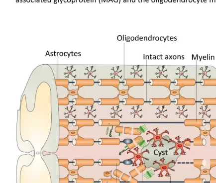
(Filbin 2003; Fenrich et al., 2004). The most prominent myelin inhibitors are Nogo-A, myelin-associated glycoprotein (MAG) and the oligodendrocyte myelin glycoprotein (OMgp).
Fig. 1-2 The inhibitory CNS environment
Schematic representation of the injury site in the spinal cord showing main growth inhibitors such as myelin associated inhibitors (e.g. Nogo) and Chondroitin sulfate proteoglycans (CSPGs) as part of the extracellular matrix of the scar. Illustration modified from Yiu et al., 2006.
In line with this, blockade of Nogo-A by IN-1 antibody has been shown to allow axonal growth on myelin in vitro and in vivo (Caroni et al., 1988; Schnell et al., 1990) moving treatment strategies targeting Nogo-A into promising clinical trials in patients with acute SCI (s. Tab. 1-1).
Introduction 8
physical but also chemical barrier for growing axons. The molecules of the extracellular matrix of the glial scar have been shown to be highly growth inhibitory. Particularly the family of chondroitin sulfate proteoglycans (CSPGs) produced by astrocytes have been shown to be highly growth inhibitory (Silver et al., 2004; Bradbury et al., 2011): In vitro by repelling growing axons and inhibiting growth promoting molecules including laminin, fibronectin and L1 (Dou et al., 1994; Snow et al., 1996) as well as in vivo where development of dystrophic growth cones in regenerating axons was correlated with upregulation of CSPGs (Davies et al., 1997; Davies et al., 1999).
Addressing the growth inhibitory potential of CSPGs, their degradation has been in the focus of investigation (s. Tab. 1-1). The enzyme chondroitinase ABC extracted from the bacterium Proteus vulgaris has turned out to remove the chondroitin sulfate glycosaminoglycan (CS-GAG) chains from the CSPG core protein and thus making the CSPGs less inhibitory (Silver et al., 2004). After promising experiments in vitro, Lemons and colleagues first showed that chondroitinase ABC (ChABC) can degrade CSPGs in scar tissue when applied to spinal cords of contusion injured rats (Lemons et al., 1999). In 2002, Bradbury and colleagues
demonstrated that intrathecal injection of chondroitinase ABC into a lesioned spinal cord promoted axonal sprouting and elongation in dorsal columns and corticospinal tracts. More importantly, a significant improvement in functional locomotor and proprioceptive behavior was observed (Bradbury et al., 2002).
1.4.2
The insufficient activation of the intrinsic neuronal growth program
upon injury in the CNS
Apart from the growth permissive environment, successful axonal regeneration is also dependent on the growth capacity of the injured neuron. Eventually, neurons need to
convert from the “transmitting” into the “growth” mode.
cones where they mediate growth cone elongation (Skene 1989; Benowitz et al., 1997; Frey et al., 2000; Caroni 2001).
Besides, peripherally injured neurons start expressing proteins that are crucial for interactions between growth cones and Schwann cells: They upregulate receptors for neurotrophic factors released by Schwann cells and the neurons themselves release proteins such as neuregulin that – by binding to the erb receptor of Schwann cells – allow close interactions between neurons and glia. Moreover, adhesion molecules and integrins in the growth cone are upregulated to enable its extensions on the surfaces of Schwann cells and basal lamina (Fenrich et al., 2004; Raivich et al., 2007).
The following sections now explain
1) how the intrinsic neuronal growth program is induced in the PNS
2) how the understanding of 1) has already helped to develop approaches that aim to mimic the intracellular growth permissive situation in the CNS and eventually support lesioned CNS axons to regrow.
3) how all the injury signals described in 1) eventually contribute to axonal outgrowth
1.4.2.1 Induction of the intrinsic neuronal growth program after a PNS lesion
As previously mentioned, neurons alter their gene expression pattern when switching to the growth mode. To eventually initiate this transcription program, injury induced signals from the lesion site need to be transmitted to the nucleus (s. Fig. 1-3). In the following the complex mechanisms of injury induced retrograde signaling are briefly outlined (Ambron et al., 1996; Rossi et al., 2007; Rishal et al., 2010):
1.4.2.1.1 Early injury induced signaling
Introduction 10
which in turn regulate the actin cytoskeleton as well as growth related transcription factors such as CREB (Ambron et al., 1996; Swulius et al., 2008). Spira and colleagues could show that intracellular increase of calcium at the lesion site itself induced localized proteolysis of the membrane skeleton (spectrin) and cytoskeleton (actin and microtubules) by calpain proteases. The localized proteolysis is a prerequisite to restructure the cytoskeleton which in turn marks an important step during growth cone formation where post-Golgi vesicles supply membrane material (Spira et al., 2001; Bradke et al., 2012). In line with this, tetrodotoxin, a sodium channel blocker, eventually leads to reduced calcium influx and its related processes (s. above) which in turn goes along with reduced neurite regeneration (Mandolesi et al., 2004). Conversely, electrical stimulation accelerates neurite outgrowth after nerve transection (Brushart et al., 2002). However, the role of calcium and electrical stimulation is complex and remains ambiguous (s. section 1.4.2.2). Apart from calcium, also cAMP – activated by peptide hormones from interneurons and also by calcium – is an integral molecule that determines the regenerative potential already at early phases of regeneration. After peripheral nerve injury, cAMP levels are elevated and by activating the transcription factor CREB via PKA they contribute transcription-dependently to growth initiation (Ambron et al., 1996).
1.4.2.1.2 Interruption of constitutive retrograde signaling (“negative signals”)
regeneration (Cui 2006; Lu et al., 2008). These contradictory findings might be based on different roles of neurotrophins at different time points. Loss of neurotrophic support might help inducing growth, whereas gradual increase in neurotrophins and their receptors stimulate growth in the later course of regeneration (source: Tuszynski, Mark H., Kordower, Jeffrey H.,Ed. CNS Regeneration: Basic Science and Clinical Advances. London: Academic Press, 2nd edition 2008.).
1.4.2.1.3 Induction of growth-promoting signals (“positive signals”)
Following the early phase of injury induced changes (s. above), a large group of “positive retrograde injury signaling” molecules is activated by calcium and other still unknown mechanisms. They are named “positive signals” as they are not active in intact neurons but
produced upon axotomy to trigger the cell body response. The activated molecules such as the transcription factor NF-κB (nuclear factor 'kappa-light-chain-enhancer' of activated B-cells) and mitogen-activated protein kinases (MAPKs) are microtubule-dependently transported towards the cell body where they initiate transcriptional processes. The MAPKs have become interesting candidate regulators of axonal regeneration, as exemplified by the following: Activation of extracellular signal-regulated kinase (Erk) and c-Jun N-terminal kinase (JNK) – two members of the MAPKs – and their interactions with the dynein/dynactin retrograde transport seem to be crucial for axonal regeneration (Abe et al., 2008). In addition, phorphorylated Erk e.g. is detected in axons after peripheral injury (Sheu 2000) and inhibition of Erk prevents adult DRG neurons from spontaneously initiating neurites in culture (Chierzi et al., 2005). Erk - by turning on the transcription factor CREB [cAMP response element-binding protein, (Johannessen et al., 2004)] - and JNK - by activating the transcription factors ATF-3 (Activating transcription factor 3) and c-Jun (Lindwall et al., 2005) - are directly involved in activating growth promoting transcription factors. Recently, dual leucine zipper-bearing kinase 1 (DLK 1), a kinase of a conserved MAPK cascade has been found to positively regulate growth cone formation and axonal regeneration (Hammarlund et al., 2009; Yan et al., 2009; Ghosh-Roy et al., 2010).
Introduction 12
are synthesized locally upon injury (Rossi et al., 2007). The role of this local de novo protein synthesis remains unclear but might contribute to the rapid injury response.
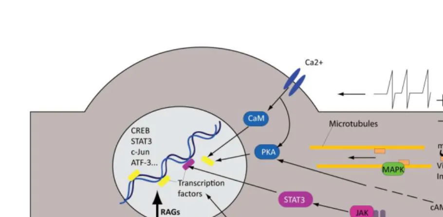
Fig. 1-3The intrinsic neuronal growth program activated upon peripheral injury
The early phase of neuronal response upon nerve damage is characterized by injury induced action potentials and release of neuropeptides (such as 5-HT) from the environment that act via calcium and cAMP. Subsequently, retrograde MAPK and other components activated at the injury site are retrogradely transported to the nucleus. Later on, signals from cytokines and growth factors released by the neuron itself as well as by surrounding cells such as glia and macrophages arrive at the nucleus. Ultimately, all pathways activate transcription factors and alter gene expression resulting in upregulation of growth-associated proteins which in turn orchestrate neurite outgrowth.
1.4.2.2 Failed induction of the intrinsic growth program after a CNS lesion
In contrast to the PNS, where upon injury a cascade of injury signals are relayed to the nucleus and RAGs are upregulated as they are during development (Hoffman 2010), there is no upregulation of RAGs in the CNS (Plunet et al., 2002; Fenrich et al., 2004). It still remains speculative if this lack is due to neuronal inability of activation or transport of signals, failure of overcoming growth inhibition due to maturation (Liu et al., 2011) or reduced response to injury induced cytokines. However, studies on DRG neurons extending one branch to the periphery and the other to the CNS have yielded interesting findings: Lesion of the peripheral branch prior to a central branch lesion promotes regeneration of the central branch (Richardson et al., 1984; Neumann et al., 1999; Ylera et al., 2009). This phenomenon
Introduction 14
injury signals; however, only when the intrinsic program is additionally “primed” by a peripheral lesion via injury related processes such as retrograde travel of injury signals etc. Thus, many efforts have been undertaken to mimic the conditioning lesion effect by specifically modifying one of the above described mechanisms.
Influencing early injury signals has so far led to ambiguous results: Whereas Udina and colleagues could show that electrical stimulation of the sciatic nerve promotes regeneration of injured central axons of the stimulated neurons (Udina et al., 2008), others have not found growth promotion when directly stimulating transected rubrospinal tract axons (Harvey et al., 2005). These differences might be explained by the fact that in contrast to Harvey’s experiment electrical activity in Udina’s experiment mimicked the “conditioning lesion effect”. Addressing cAMP, however, has led to more promising findings: cAMP levels can be therapeutically elevated by conditioning lesion, by administration of cAMP analogues or by treatment with the
phosphodiesterase inhibitor rolipram. Interestingly, it does not only increase the neuron’s
intrinsic growth capacity (s. section 1.4.2.1.1), but does also overcome myelin inhibition both in vitro and in vivo, allowing axons to growth through a spinal cord lesion (Qiu et al., 2002;
Hannila et al., 2008).
1.4.2.3 Molecular anatomy of the intrinsic neuronal growth program
Experiments trying to mimic the growth pattern seen after peripheral lesions by modulating cascades during early injury induced signaling have been performed (s. above). However, more recent attempts aim to directly alter the neuronal growth program on the transcriptional and translational level where different injury induced signals eventually converge.
in gene expression. A selection of TFs and their role for axon regeneration is exemplified in the following(s.Fig. 1-4).
Mice lacking c-Jun in the CNS showed severe defects upon injury and reduced expression of CD44, galanin, and α7β1 integrin, molecules known to be involved in axonal regeneration (Broude et al., 1997; Raivich et al., 2004). Knockout phenotype of the C/EBP transcription factor leads to decreased expression of the growth-associated genes for GAP-43 and
Introduction 16
GAP-43 gene and genes important for synaptogenesis and cytoskeletal dynamics (Moore et al., 2011).
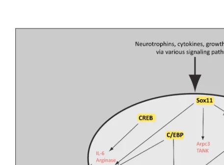
Fig. 1-4Transcription factors involved in axonal regeneration and their target genes.
A variety of intracellular signaling molecules implicated in axon regeneration lead to activation of transcription factors (in yellow boxes). The transcription factors activate target genes (red) in the nucleus and thus promote axonal outgrowth. For example, CREB activates IL-6, Arginase and Tubulin, whereas latter ones are involved in local cytoskeleton assembly. More information as well as references about the transcription factors shown in the illustration can be taken from the text. Image adapted from Tuszynski, Mark H., Kordower, Jeffrey H., Ed. CNS Regeneration: Basic Science and Clinical Advances. London: Academic Press, 2nd edition 2008.
seem to influence axonal regeneration as exemplified by the anaphase-promoting complex (APC) which might degrade pro-regenerative molecules (Lasorella et al., 2006).
1.5
STAT3
Apart from the above mentioned transcription factors, STAT3 has recently emerged as a key candidate regulator of axonal regeneration. Before presenting STAT3 in the context of axonal regeneration, the STAT family, the JAK/STAT pathway as well as the role of STAT3 outside the nervous system is briefly outlined.
1.5.1
The STAT family
Introduction 18
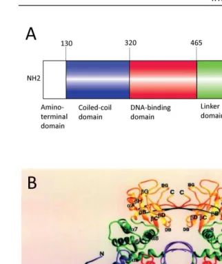
Fig. 1-5Structure of the STAT3 molecule
(A) Linear structure of STAT3 and (B) three-dimensional structure of the STAT3 homodimer bound to DNA showing the different domains of the STAT3 molecule: Amino-terminal domain, coiled-coil domain (blue), DNA-binding region (red), linker domain (green), SH2-domain (yellow) with phosphorylation at a tyrosine residue and the transactivation domain. A adapted from Levy et al., 2002. B adapted from Becker et al., 1998.
1.5.2
The JAK/STAT pathway
STAT3 as well as the other STATs are part of the JAK/STAT signaling pathway (s. Fig. 1-6) transmitting extracellular signals to the nucleus (Schindler et al., 1995; Aaronson 2002; Kisseleva et al., 2002):
catalytic activity: JAKs phosphorylate a distal cytoplasmic tyrosine residue on the receptor thereby creating docking sites for the SH2 domain of STAT3 proteins. Subsequently, STAT3 proteins which are present in the cytoplasm in inactive form are drawn into the receptor complex and are phosphorylated on a single tyrosine residue by the JAKs (Kisseleva et al., 2002). In doing so, also in STAT3 molecules, docking sites for SH2 domains are created. When STAT3 proteins subsequently dissociate from the receptor, the phosphotyrosine residues of two STAT molecules are reciprocally recognized: The SH2 domain of one STAT3 molecule binds to the phosphotyrosine of the other STAT3 molecule, thus forming homodimers. These dimers arerapidly transported from the cytoplasm to the nucleus where they bind to promotor sequences containing the GAS (gamma activated site) element, the pallindromic DNA sequence 5’-TTTCCNGGAAA-3’, and induce gene transcription (Aaronson 2002; Kisseleva et al., 2002).
The JAK/STAT pathway is regulated at several levels (Aaronson 2002; Kisseleva et al., 2002): MAPK e.g. have been shown to amplify STAT3 signaling by additional phosphorylation on serin residues. But there is also negative regulation of the pathway, e.g. phosphatases deactivating STATs and JAKs as well as suppressors of cytokine signaling (SOCS) that inhibit receptors and JAKs. Besides, nuclear regulators control STAT import and export to and from the nucleus and PIAS (Protein inhibitor of activated STATs) prevent STAT3 dimers from binding to the DNA.
A study in human breast cancer cells found that the STAT3 pathway is positively regulated by the above mentioned mTOR signaling pathway, whereas PTEN (s. above) served as a negative regulator of both STAT3 and mTOR signaling (Zhou et al., 2007).
Introduction 20
Fig. 1-6Activation of the JAK/STAT3 pathway
The JAK/STAT3 pathway is activated by cytokines e.g. IL-6, CNTF and LIF. In the course of activation, latent cytoplasmic STAT3 proteins are recruited and phosphorylated by JAKs. Phosphorylated STAT3 molecules dimerize and are translocated into the nucleus where they modulate target genes.
1.5.3
The role of STAT3 outside the nervous system
Promotor for cre
Target tissue/cells
Phenotypes Reference
K5 keratinocytes Altered migration: disrupted wound repair, impaired keratinocyte migration and hair cycle; spontaneous development of ulcers. However, normal proliferation and development of epidermis and hair follicles.
(Sano et al., 1999; Sano et al., 2008)
K5 Thymus epithelium
Altered survival: severe thymic hyoplasia, increased susceptibility to apoptosis-inducing agents such as steroids
(Sano et al., 2001)
Blg Mammary epithelium
Altered apoptosis: delayed mammary involution (Chapman et al., 1999).
Mx Liver Impaired acute-phase response, similar to the phenotype of IL-6 knockout mice
(Alonzi et al., 2001)
Lck T-lymphocytes Altered survival/proliferation: impaired IL-6 dependent T-cell survival and reduced IL-2 induced proliferation
(Akaishi et al., 1998; Takeda et al., 1998). Mlys Monocytes/
Neutrophils
Hypersensitivity to endotoxin shock resulting in augmented production of inflammatory cytokines such as IL-1, IL-6 and IFNγ as a consequence of impaired IL-10 function; chronic colitis; imbalanced T-cell differentiation towards the Th1 type
(Takeda et al., 1999)
Tab. 1-2 Tissue-specific STAT3-deficient phenotypes outside the nervous system
Tissue-specific STAT3 deletion was achieved by taking advantage of the cre/loxP system. Table modified by Levy et al., 2002. In most tissues the functions affected by STAT3 deletion involve cell survival and proliferation
1.5.3.1 STAT3 in cancer
Along with its permissive role for survival and proliferation, STAT3 is also closely associated with oncogenesis (Bromberg 2002): While STAT1 is ascribed a tumor suppressor role, both STAT3 and STAT5 were found to increase tumor cell proliferation, survival and invasion. Constitutive activation of STAT3 has been found in many solid tumors such as breast cancer, prostate cancer and melanoma as well as in leukemias and lymphomas. Moreover, STAT3 is required for the transformation into malignant cells in some malignancies e.g. breast cancer and thyroid cancer.
Introduction 22
1.5.4
STAT3 in the context of neuronal survival and regeneration
The role of STAT3 for the nervous system appears very similar to its effects outside the nervous system where it supports survival and proliferation. STAT3 is an important player during neurodevelopment, where it is e.g. required for axon pathfinding, neurite outgrowth and glial cell differentiation (Dziennis and Alkayed 2008). In rodents, STAT3 expression is detected in neurons and glia from embryonic day fourteen on with increasing levels up to postnatal day twenty-one, when levels start declining (Gautron et al., 2006).
In addition to its role in the developing nervous system, STAT3 has turned out to be an interesting transcription factor in the context of axon regeneration. Several studies demonstrate that increased levels of STAT3 expression and phosphorylation are associated with axonal regeneration in the PNS: After axotomy in rats, STAT3 phosphorylation is induced in facial and hypoglossal neurons already three hours after lesion, whereas this response is not found in non-regenerating Clarke’s nucleus neurons (Schwaiger et al., 2000). A transient activation of STAT3 is also associated with sprouting of septal neurons after entorhinal cortex lesion in rats (Xia et al., 2002). Moreover, a study investigating the different spatial and temporal pattern of STAT3 and Erk phosphorylation in rats could show that nerve transection induced rapid STAT3 phosphorylation particularly in the proximal site adjacent to the injury which decreased to almost baseline when regeneration was complete at six weeks (Sheu 2000). Interestingly, STAT3 is activated at the injury site and then retrogradely transported to the nucleus (Lee et al., 2004).
Apart from regeneration, STAT3 is associated with axonal remodeling in the CNS: Unilateral lesion of the CST and application of IN-1 antibody directed against the growth inhibitor protein Nogo-A triggered reorganization of the remaining CST fibers. Interestingly, IN-1 antibody treatment was among others associated with upregulation of STAT3 expression (Bareyre et al., 2002).
Apart from STAT3 itself, various studies investigating molecules that exert their function via JAK/STAT pathway such as the neuropoietic cytokines IL-6, CNTF, LIF and SOCS3 highlight STAT3 as a good candidate in terms of axonal growth regulation. One study showed that LIF activated STAT3 after nerve injury (Rajan et al., 1995) and that sensory neurons of LIF deficient mice showed a significantly attenuated growth response upon a conditioning injury (Cafferty et al., 2001). Three years later, Cafferty and colleagues demonstrated that in IL-6 deficient mice, a pre-conditioning injury of the sciatic nerve was followed by a total failure of regeneration of dorsal column axons, thus identifying IL-6 as a keyplayer for mediating conditioning lesion-induced increase of regeneration of lesioned axons in the CNS (Cafferty et al., 2004). This goes along with the finding that sensory axons in IL-6 deficient mice show delayed regeneration after crush lesion (Zhong et al., 1999). Interestingly, also hormones such as Erythropoietin (Kretz et al., 2005) and IGF-I (Yadav et al., 2005) as well as neurotrophins such as NGF (Donnerer et al., 2005) and BNDF (Ng et al., 2006) activate STAT3 and have been shown to be involved in axonal regeneration and neurite outgrowth.
Introduction 24
1.6
Research question
In contrast to the PNS, where damaged axons can regenerate successfully, CNS axons fail to regrow. One of the reasons underlying this failure is the insufficient activation of the intrinsic neuronal growth program after a central lesion. To jump-start the intrinsic growth program after CNS injury we first need to identify its molecular key regulators. Especially transcription factors are promising candidate regulators as they can alter the expression of multiple downstream genes. The transcription factor STAT3 is a particularly promising candidate regulator as its expression correlates with the regenerative response (s. section 1.5.4). However, its direct role for axonal outgrowth in vivo has not yet been determined. This study now aimed to directly show a) whether and b) when STAT3 affects central axonal regeneration in vivo by answering the following specific questions (s. Tab. 1-3):
1. Is endogenous STAT3 activation induced after a CNS lesion?
2. Is STAT3 overexpression by viral gene therapy sufficient to induce outgrowth of CNS axons?
3. How does STAT3 affect different phases of axonal outgrowth in the CNS?
4. In case STAT3 overexpression positively influences axonal outgrowth in the CNS, can axon regeneration even be amplified when viral overexpression of STAT3 is combined with the application of chondroitinase ABC?
Tab. 1-3 Research questions to be answered in the course of my study
To address these questions, I used in vivo timelapse fluorescence microscopy and viral gene therapy in transgenic and wildtype mice to investigate the regeneration process of lesioned
2
Material and Methods
___________________________________________________________________________
2.1
Material
1. Materials for surgical experiments
1.1 Reagents
Ketamine hydrochloride 10% (Ketamine)
Bremer Pharma GmbH, Warburg, Germany
Xylariem 20 mg (Xylazine) Riemser Arzneimittel AG, Greifswald-Insel Riems, Germany
Forene (Isoflurane) Abbott AG, Baar, Switzerland
Metacam 1,5 mg/ml Oral Suspension Boehringer Ingelheim, Ingelheim am Rhein, Germany
Sterile artificial mouse cerebrospinal fluid (aCSF)
Solution A:
8,66 g NaCl (Merck) 0,224 g KCl (Merck) 0,206 g CaCl2 · 2H2O (Sigma-Aldrich) 0,163 g MgCl2 · 6H2O (Sigma-Aldrich)
dH20 ad 500 ml
Solution B:
0,214 g Na2HPO4 · 7H2O (Merck)
0,027 g NaH2PO4 · H2O (Merck)
dH20 ad 500 ml
Mixture of solutions A and B in a 1:1 ratio Bepanthen Augen- und Nasensalbe 5
g (eye cream)
Bayer Vital GmbH, Leverkusen, Germany
Ringerlösung Fresenius KabiPac
(Ringer’s solution)
Fresenius KaBI Dtl., Bad Homburg, Deutschland
Cutasept F Lösung 250 ml (disinfectant spray)
Bode Chemie GmbH & Co, Hamburg, Germany
Materials and Methods 26
1.2 Tools and materials
Wella contura W7807 (Hair clipper) Wella, Darmstadt, Germany Micro Pipettes intraMARK Blaubrand
(micropipettes/glass capillaries)
Brand GmbH und Co. KG, Wertheim, Germany
Syringe 3pc 20 ml Omnifix™ luer slip
(syringe for virus injection)
B. Braun Melsungen AG, Melsungen, Germany
Syringe 3pc 5 ml Omnifix™ luer slip (syringe for injection of Ringer’s
solution)
B. Braun Melsungen AG, Melsungen, Germany
BD Plastipak Hypodermic luer slip syringe 1 ml (syringe for
Ketamine/Xylazine injection)
Becton, Dickinson and Company, Franklin Lakes (New Jersey), USA
Safety-Multifly-Set (blood collection system with needle, butterfly, tube and connector to syringe)
SARSTEDT AG & Co., Nümbrecht, Germany
Fine Iris Scissors (for dorsal column lesion)
Fine Science Tools GmbH, Heidelberg, Germany
Feather stainless steel blade (surgical blade)
pfm medical ag, Cologne, Germany
Noyes Spring Scissors (Large spring scissors)
Fine Science Tools GmbH, Heidelberg, Germany
Vannas-Tübingen Spring Scissors (Small angled spring scissors)
Fine Science Tools GmbH, Heidelberg, Germany
Dumont Mini Forceps – Inox Style 3 (Small forceps)
Fine Science Tools GmbH, Heidelberg, Germany
Dumont Mini Forceps – Inox Style 5 (Small forceps, smaller tip than Inox style 3)
Fine Science Tools GmbH, Heidelberg, Germany
Hypodermic Needles BD Microlance 3 30 Gauge (0,3 mm, yellow) for
subcutaneous injection of Ringer’s
solution and anesthesia
Becton, Dickinson and Company, Franklin Lakes (New Jersey), USA
Hypodermic Needles BD Microlance 3 23 Gauge (0,6 mm, blue) for central lesion
Becton, Dickinson and Company, Franklin Lakes (New Jersey), USA
Olsen-Hegar Needle Holder Fine Science Tools GmbH, Heidelberg, Germany Ethicon Ethilon monofil 6-0 size,
667H (skin suture)
Johnson & Johnson Medical GmbH, Norderstedt, Germany
Ethicon Vicryl 4-0 size, MIC101H (intracorporal suture)
Autoclip Wound Clip Applier (stapler) Becton, Dickinson and Company, Franklin Lakes (New Jersey), USA
Autoclip Wound Clips, 9 mm (staples) Becton, Dickinson and Company, Franklin Lakes (New Jersey), USA
Sugi (absorbent triangles) Kettenbach GmbH & Co. KG, Eschenburg, Germany
Metal plate Custom-made
Cast Alnico Button Magnets (magnets)
Eclipse Magnetics Ltd, Sheffield, UK
Rubber bands
Support cushion Custom-made
Osmotic minipump (Model 1007B) Alzet, Cupertino (California), USA Brain Infusion kit 3 Alzet, Cupertino (California), USA 1.3 Technical devices
Olympus KL 1500 LCD (cold light source for stereo microscopy)
Olympus UK Ltd KeyMed House, Southend-on-Sea, Great Britain
Olympus Stereo Microscope SZ51 Olympus UK Ltd KeyMed House, Southend-on-Sea, Great Britain
FST 250 Hot Bead Sterilizer (sterilizer for surgical instruments)
Fine Science Tools GmbH, Heidelberg, Germany
Vertical Micropipette Puller P-30 Sutter Instrument Company, Novato (California), USA
Small Animal Stereotactic Instrument David Kopf Instruments, Tujunga (California), USA T/Pump (Heating pad) Gaymar Industries, Orchard Park (New York), USA
2. Materials for perfusion and immunohistochemistry
2.1 Reagents
PFA (paraformaldehyde) 8% PFA (Sigma-Aldrich) in dH2O, heated up to 55 °C and stirred additional 10 min, filtrated and mixed in a 1:1 ratio with 0,2 M PB (Phosphate buffer), pH adjusted to 7,2-7,8
Agarose Sigma-Aldrich, St.Louis (Missouri), USA
Materials and Methods 28
Sucrose Sigma-Aldrich, St.Louis (Missouri), USA
EDTA (Ethylenediaminetetraacetic acid) Merck KGaA, Darmstadt, Germany Sodium dihydrogen phosphate monohydrate
(NaH2PO4· H2O, M=137,99 g/mol)
Merck KGaA, Darmstadt, Germany
Disodium hydrogen phosphate dihydrate (Na2 HPO4· 2H2O ,M=177,99 g/mol)
Merck KGaA, Darmstadt, Germany
PBS 10x (phosphate buffered saline), pH= 7,2/7,4
2,6 g NaH2PO4 · H2O 14,4g Na2 HPO4· 2H2O 87,5g NaCl (Merck) dH2O ad 1l
TBS 10x (Tris buffered saline), pH=7,6 61 g Tris base (121,14 g/mol), (Sigma-Aldrich)
90 g NaCl dH2O ad 1l
O,2 M PB (phosphate buffer) 27,598 g NaH2PO4· H2O, 35,598 g Na2 HPO4· 2H2O dH2O ad 1l
Anti-STAT3 antibody Cell signaling Technology, Danvers
(Massachusetts), USA
Anti-P-STAT3 antibody Cell signaling Technology, Danvers
(Massachusetts), USA
Goat-anti-rabbit 594 antibody Jackson ImmunoResearch Laboratories, West Grove (Pennsylvania), USA
Goat-anti-mouse 594 antibody Jackson ImmunoResearch Laboratories, West Grove (Pennsylvania), USA
Gibco goat serum Invitrogen GmbH, Darmstadt, Germany
NeuroTrace 435 NeuroTrace 488
Invitrogen GmbH, Darmstadt, Germany Invitrogen GmbH, Darmstadt, Germany Anti-Proteoglycan mouse IgG1, clone 2-B-6
antibody
SEIKAGAKU BIOBUSINESS Company, Tokyo, Japan
2.2 Tools and materials
Microscope slides 76x26 mm Gerhard Menzel Glasbearbeitungswerk
GmbH & Co. KG, Braunschweig, Germany
Microscope cover slips 24x60 mm Gerhard Menzel Glasbearbeitungswerk GmbH & Co. KG, Braunschweig, Germany
Parafilm Brand GmbH & Co. KG, Wertheim
Pipettes, pipette tips and tubes (2ml and 1,5 ml)
Eppendorf AG, Hamburg, Germany
12-well and 96-well cell culture plates Becton, Dickinson and Company, Franklin Lakes (New Jersey), USA
Tissue Tek Cryomold Standard, 25x20x5 mm Sakura Finetek Europe B.V. , Alphen aan den Rijn, The Netherlands
Tissue Tek Cryomold Biopsy, 10x10x5 mm Sakura Finetek Europe B.V. , Alphen aan den Rijn, The Netherlands
Tissue Tek optimal cutting temperature (O.C.T.)
Sakura Finetek Europe B.V. , Alphen aan den Rijn, The Netherlands
Vectashield Mounting Medium Vector Laboratories, Inc., Burlingame (California), USA
Paper filters (185 mm Ø circles) Whatman Schleicher & Schuell GmbH, Dassel, Germany
50 ml centrifuge tubes Greiner Bio-One GmbH, Frickenhausen,
Germany
Erlenmeyer flasks (1l; 0,5 l) Schott, Elmsford (New York), USA 2.3. Technical devices
Leica CM1850 cryostat Leica Microsystems GmbH, Wetzlar,
Germany
Vibratome 1000Plus Intracel LTD, Shepreth, Royston,
Great Britain
Vortex-Genie 2 Scientific Industries, Inc., Bohemia (New
York), USA
KERN EW 150-3M (scales) Kern & Sohn GmbH, Balingen-Frommern, Germany
Laboratory pH meter inoLAB WTW Wissenschaftlich-Technische
Werkstätten, Weilheim, Germany Magnetic stirring hotplate MR 3001K and
stirring bars
Heidolph Instruments GmBH & Co. KG, Schwabach, Germany
Ismatec IP high precision multichannel pump (pump for perfusions)
ISMATEC SA, Labortechnik - Analytik, Glattbrugg, Switzerland
Olympus IX71 inverted fluorescence microscope (evaluation of
immunohistochemistry)
Olympus GmbH, Hamburg, Germany
Olympus SZX16 fluorescence
stereomicroscope (Dissection microscope)
Materials and Methods 30
3. Imaging
Olympus BX51WI upright microscope (Wide-field epifluorescence microscope) for in vivo imaging equipped with x4/0.13 dry, x10/0.3 dry and x20/0.5 dipping cone
water-immersion objectives
Olympus GmbH, Hamburg, Germany
Fast shutter and filer wheel Sutter Instruments, Novato (California), USA Height-adjustable stage with attachment for
metal plate
Custom-made
Cooled SensiCam QE CCD camera The Cooke Corporation, Romulus (Michigan), USA
Standard filter for fluorescent proteins Chroma Technology Corporation, Bellows Falls (Vermont), USA
MetaMorph®software Molecular Devices, Orleans (California), USA
FV1000 confocal system mounted on an upright BX61 microscope, equipped with an x10/0.4 water immersion objective and x20/0.85 and x60/1.42 oil immersion objectives (confocal microscopy)
Olympus GmbH, Hamburg, Germany
Custom-built multiphoton imaging setup based on an Olympus FV 300 scanner equipped with a femto-second pulsed Ti:Sapphire laser (Mai Tai HP,
Newport/Spectra-Physics)
Olympus GmbH, Hamburg, Germany Newport Corporation, Irvine (California), USA
Olympus FV1000 MPE multiphoton microscope
Olympus GmbH, Hamburg, Germany
2.2
Mice
groups. For the evaluation of STAT3 expression in DRG neurons ten, twelve and fourteen days after viral therapy both wildtype (WT) mice on a C57/bl6 background and Thy1-GFPs mice were used. To delete STAT3 I used STAT3 fl/fl mice, maintained on a bl6 background (Takeda et al., 1998). For the nerve crush experiment we used Thy1-YFP16 mice (s. section 4.1). Animals were kept in Eurostandard Type II long cages 365x207x140 mmH (Tecniplast, Hohenpreißenberg, Germany) stored in IVC rack system with a maximum of five mice per cage. Autoclaved food (regular food “Maus” from Ssniff, Soest, Germany) and autoclaved tap
water were supplied ad libitum. Mice were held at a 12 h light/12 h dark cycle. Animals were kept at the breeding room with their parent until day twenty-one postnatal and afterwards weaned and separated by sex. All animal experiments were performed in accordance with regulations of the animal welfare act and protocols approved by the Regierung von Oberbayern.
2.3
Methods
2.3.1
Plasmid constructs
The MCS vector from Stratagene has been used to produce the viral vectors pAAV-STAT3, pAAV-STAT3c and the control viral vectors pAAV-eCFP. For the pAAV-pAAV-STAT3, the STAT3 gene was taken from pcDNA3 STAT3 (Adgene plasmid 8706) by excising it with the restriction enzymes BamHI and XhoI, whereas the STAT3c gene was cut with NotI and SwaI from the pRc/CMV STAT3c Flag (Addgene plasmid 8722). Then, both genes were respectively inserted into the pAAV-MCS at the HincII site. pAAV-eCFP was produced by excising the eCFP gene from the peCFP N1 plasmide at BamHI and NotI and consecutively cloning it in the pAAV- MCS at the HincII site. All cloning was performed by Dr. Florence Bareyre.
2.3.2
Vector production and purification
Materials and Methods 32
copies/ml; rAAV-STAT3c, 5.5x10¹² genome copies/ml; rAAV-eCFP, 9.2x10¹² genome copies/ml; rAAV-cre , 9x10¹² genome copies/ml. Vector production as well as vector purification was performed by Joshua Sanes and In-Jung Kim (Harvard University). rAAV-cre was a kind donation from I.J. Kim (Harvard University).
2.3.3
Tissue processing and immunohistochemistry
Animals were deeply anesthetized with isofluorane and perfused transcardially with 20 ml of saline solution followed by 60 ml of 4% paraformaldehyde (PFA) in 0.1 M phosphate buffer. Spinal cords including the injected DRGs were dissected out and tissues were post-fixed in 4% PFA at 4 °C for 24 h. For consecutive immunofluorescence analysis, DRGs were transferred to 30% sucrose for at least 24 h and embedded/blocked in Tissue-Tek optimal cutting temperature (O.C.T.) compound. Then 20 µm thick coronal sections were cut on a cryostat and mounted on glass slides.
Before immunostaining, sections were dried at room temperature (RT) for at least 2 h and subsequently rinsed 3 times for 10 min in TBS at room temperature. To improve antigen retrieval, slides were heated up to 60 °C in the microwave in Tris/EDTA followed by 15 min at a sub-boiling temperature. Without cooling down the slides, sections were blocked with 5% goat serum in 0,1% Triton X-100 in TBS for 1 h. Afterwards, sections were incubated for 1 h at RT and then overnight at 4 °C with either anti-STAT3 antibody (dilution 1:500) or anti-P-STAT3 antibody (dilution 1:50) diluted in 0.1% Triton X-100-TBS and 2,5% goat serum. Slides were rinsed again 3 times for 10 min with 1xTBS and subsequently incubated with fluorescent secondary antibody (goat-anti-rabbit 594) used at a dilution of 1:500 in TBS for 2 h. Furthermore, nuclei were counterstained using NeuroTrace 435 (dilution 1:500). For the STAT3 deletion experiment in the CNS (s. section 4.6) I used NeuroTrace 488. Before mounting the tissue in Vectashield mounting media, slides were rinsed 3 times for 10 min with TBS.
subsequently cut in 50 µm thick sections by a vibratome. Afterwards, sections were rinsed for 10 min in PBS and blocked with 5% goat serum in 0,1% Triton X-100 in PBS for 1 h. Then, sections were incubated for 1 h at RT and then overnight at 4 °C with Anti-Proteoglycan mouse IgG1 (diluted in 0.1% Triton X-100-PBS and 2,5% goat serum). Sections were rinsed 3 times for 10 minutes with PBS and subsequently incubated overnight with fluorescent secondary antibody (Goat-anti-mouse 594) used at a dilution of 1:500 in 0,1% Triton X-100 in PBS. Eventually, sections were rinsed 3 times for 10 minutes and mounted on glass slides in Vectashield.
2.3.4
Quantification of the expression of P-STAT3 and STAT3
STAT3 and P-STAT3 immunoreactivity was analyzed in DRGs of animals perfused at 10 d, 12 d and 14 d after the injection of rAAV-STAT3 and rAAV-STAT3c into cervical DRGs (C3-C6) of C57/bl6 wildtype or Thy1-GFPs mice. DRG sections were stained for STAT3 and P-STAT3, respectively, and all sections were counterstained with NeuroTrace 435 to reveal the total number of neuronal nuclei in the DRG. Then, images in the red (P-STAT) and blue (NeuroTrace) channel of an Olympus IX71 inverted fluorescence microscope were taken and the proportion of DRG neurons showing STAT3 or P-STAT3 immunoreactivity was determined.
2.3.5
Gene therapy with recombinant adeno-associated viral vectors
Materials and Methods 34
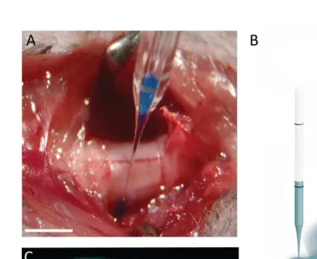
surgical procedure. For those experiments in which the effects of axonal outgrowth after viral STAT3 supplementation in Thy1-GFPs were investigated (s. sections 4.4, 4.5, 4.7) in vivo imaging was performed prior to injection to identify suitable single fluorescently labeled axons with clear visible origin from a DRG. Then, selected DRGs were surgically exposed and injected with rAVV (s. Fig. 2-3).
2.3.6
Confocal microscopy
Confocal images of fixed tissue were obtained on a FV1000 confocal system mounted on an upright BX61 microscope (Olympus) and equipped with an x10/0.4 water immersion objective and x20/0.85 and x60/1.42 oil immersion objectives. Stacks of 12 bit images were recorded and processed using the freeware ImageJ (http://rsbweb.nih.gov/ij).
2.3.7
In situ analysis of central axonal outgrowth
Central branches of fluorescently labeled DRG neurons were identified in vivo and imaged according to the in vivo imaging protocol by the Kerschensteiner and Misgeld labs (Kerschensteiner et al., 2005; Misgeld et al., 2007). The protocols describes how to image individual fluorescently labeled axons in the spinal cord of living transgenic mice expressing the green fluorescent protein (GFP) in a subset of sensory neurons. The decisive steps from the surgical exposure of the spinal cord until the recovery of the animal are briefly outlined in the following.
2.3.7.1 Surgical access to the dorsal surface of the spinal cord
Before starting surgical procedures, mice were given 5 μl Metacam (analgesic) per os by a pipette. Then, mice were intraperitoneally anaesthetized with ketamine-xylazine (ketamine
87 μg per g body weight, xylazine 13 μg per g body weight). Fur was clipped on the upper
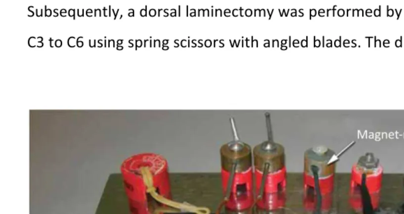
The skin in the surgery area was disinfected by Cutasept. Under the stereomicroscope a midline incision in the skin extending from the occipital bone to the upper thoracic part of the back was performed by a scalpel. Dorsal neck muscles were exposed and cut in the midline using large spring scissors. Customized magnet-mounted retractors were inserted to remove the muscles to the side. Sterile triangles (Sugi) were used to stop bleeding. Subsequently, a dorsal laminectomy was performed by removing the cervical vertebrae from C3 to C6 using spring scissors with angled blades. The dura was kept intact.
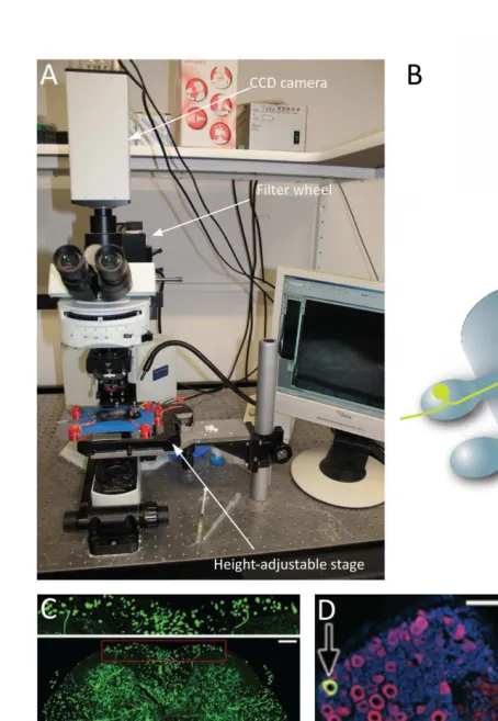
Fig. 2-1Equipment for positioning of the animal
When the surface of the dorsal spinal cord is exposed (not shown), muscles are removed to the side by customized magnet-mounted retractors. A support pillow allows widening of the interlaminar space and rubber bands connected to button magnets attach the animal to the metal plate. Adapted from Misgeld et al., 2007.
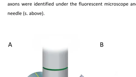
2.3.7.2 Imaging of fluorescently labeled axons and viral injection
Materials and Methods 36