1
In major depression, increased serum dynorphin and kappa opioid receptor levels are positively associated with mu opioid receptor levels and immune activation and are attenuated by nicotine
dependence.
Hussein Kadhem Al-Hakeim (1), Suhaer ZekiAl-Fadhel (2), Arafat Hussein Al-Dujaili (3), Michael Maes (4,5,6).
1 Department of Chemistry, College of Science, University of Kufa, Iraq. E-mail: [email protected].
2 Department of Clinical Laboratory Sciences, College of Pharmacy, University of Kufa, Iraq. E-mail: [email protected].
3 Senior Clinical Psychiatrist at the Faculty of Medicine, University of Kufa, Iraq. E-mail:
4 Department of Psychiatry, Faculty of Medicine, Chulalongkorn University, Bangkok,
Thailand.
5 Department of Psychiatry, Medical University Plovdiv, Plovdiv, Bulgaria.
6 IMPACT Research Center, Deakin University, Geelong, Australia.
Corresponding author
Prof. Dr. Michael Maes, M.D., Ph.D. IMPACT Strategic Research Center
Barwon Health Deakin University
2 Geelong, Vic
Australia.
3 Abstract
Background: There is now evidence that immune and opioid systems show functional reciprocal
relationships and that both systems may participate in the pathophysiology of major depression (MDD).
Objective: The present study was carried out to delineate differences between MDD patients and healthy controls in dynorphin and kappa opioid receptor (KORs) in association with levels of β-endorphins and mu opioid receptors (MORs), interleukin-6 (IL-6) and IL-10.
Method: The present study recruited 60 drug-free male participants with MDD aged 24-70 year and 30 age-matched healthy males as control group and measured serum levels of dynorphin, KOR, β-endorphin, MOR, IL-6 and IL-10.
Results: Serum dynorphin, KOR, β-endorphin and MOR are significantly increased in MDD as compared with controls. The increases in the dynorphin/KOR system and β-endorhin/MOR
system are significantly intercorrelated and are both strongly associated with increased IL-6 and IL-10 levels. Dynorphin, β-endorphin, KOR and both cytokines showed a good diagnostic
performance for MDD versus controls, whereby both opioid peptides and cytokines show a bootstrapped (n=2000) area under the receiver operating curve of 0.972. KOR and the
dynorphin/KOR system are both significantly decreased in depressed subjects with comorbid nicotine dependence.
Conclusion: Our findings suggest that in MDD, immune activation is associated with a simultaneous activation of dynorphin/KOR and β-endorhin/MOR signaling and that these opioid
systems may participate in the pathophysiology of depression by a) exerting immune regulatory
4
Keywords: Depression, cytokines, inflammation, endogenous opioid, opioid receptor.
Significant outcomes
Serum dynorphin and kappa opioid receptor (KOR) levels are significantly increased in
depression (MDD) suggesting that dynorphin/KOR signaling is increased.
In MDD, Dynorphin/KOR and β-endorhin/mu opioid (MOR) signaling are significantly
intercorrelated and associated with immune activation.
Both KOR and MOR systems may participate in the pathophysiology of depression by
exerting immune regulatory as well as emotional and behavioral effects.
Limitations
It would have been more interesting if we had measured more cytokines including those of M1
macrophage, T helper (Th)-1, Th-2, Th-17 and T regulatory phenotypes
5 Introduction
Major depressive disorder (MDD) is a chronic relapsing disorder characterized by the
recurrence of major depressive episodes. A recent meta-analysis reported that, from 1994 to 2014, the lifetime prevalence of depression was 10.8% (1). There is now evidence that MDD is
associated with changes in immune functioning and activation of immune-inflammatory pathways comprising increased production of pro-inflammatory (e.g. interleukin-6 (IL-6)) as well as immune regulatory (e.g.. IL-10) mediators (2, 3, 4, 5, 6, 7, 8). The latter are part of a regulatory system that down-regulates the primary immune-inflammatory response via multiple
negative feedback systems, collectively named the “compensatory immune regulatory system” (CIRS) (8, 9).
Aberrations in endogenous opioid peptides and their receptors are other characteristics of MDD. The opioid system comprises three families of neuropeptides, namely endorphins, enkephalins, and dynorphins, and three cognate receptor subtypes, namely μ (MOR), δ (DOR),
and κ (KOR) receptors (10). β-Endorphin and enkephalins bind to MORs and DORs, while
dynorphin bind predominately to KORs (11). Both opioid receptors and opioid peptides are expressed throughout peripheral and central nervous systems (12). A growing body of research
indicates that those endogenous opioids and their receptors are involved in emotional and behavioral responses to stress, regulation of mood and the pathophysiology of MDD (13, 14). MOR is a key component of reward processing, while acute activation of MOR attenuates
depressive-like behaviors in some but not all studies (15). MDD is accompanied by increased baseline and post-dexamethasone β-endorphin concentrations (2, 16, 17, 18). In animal models, increased β-endorphin secretion is associated with depressive-like behaviors suggesting that the
6
MOR levels were significantly higher in MDD patients than controls (13), while post-mortem studies showed increased MOR binding in suicide victims (20). Positron emission tomography (PET), on the other hand, showed contradictory results with increased binding potential for [11
C]-carfentanil during depression, but decreased biding potential in women with MDD (21). A variety of stressors may increase KOR signaling, which is implicated in stress-induced
changes in brain reward systems, which, in turn, may contribute to despair- and depression-like responses and depressive disorders (15, 22, 23, 24, 25). As such, some authors regard KOR
signaling as an anti-reward dysphoric system increasing risk towards depression (26, 27). The overall conclusion from animal models supports the idea that the KOR system may underpin
aspects of sadness and dysphoria but that the cause of the opioid alterations in MDD remains inconclusive (28). Nevertheless, in a pilot study, no differences in KOR binding (using [11C]GR103545) could be found between MDD patients and controls (29), while prodynorphin
mRNA was significantly lower in MDD and bipolar depression as compared with controls (30). There is now evidence that immune and opioid systems show functional relationships. T
and B-lymphocytes, granulocytes, and monocytes/macrophages express opioid peptides, including β-endorphin and pro-opiomelanocortin (POMC) and all POMC processing enzymes
(31, 32). Opioid peptides are synthesized in circulating leukocytes that, directed by chemokines
and adhesion molecules, may migrate to inflamed tissues where they may exert immune stimulatory as well as immune inhibitory activities (32,33). β-endorphin and dynorphin peptides
modulate functions of lymphocytes and other cells involved in host defense and immunity (34). Associations between both systems are also detected in patients with MDD. For example,
7
reported an increased opioid tone in depression and a concomitant suppression of monocytic
functions (36). Recently, we reported significant associations between MOR levels and IL-10 in
patients with major depression (13). Nevertheless, the associations between immune activation and the KOR/dynorphin system were not studied in patients with MDD.
There is also a strong comorbidity between MDD and tobacco use (NU) or nicotine dependence (ND) with increased inflammatory potential and affiliated nitro-oxidative pathways in comorbid MDD and NU/ND (37, 38, 39). Nicotine has a biphasic effect on the opioid system
with anti-opioid effects at lower doses, while nicotine administration may entrain opioid activity “during the acquisition and re-acquisition of nicotine self-administration” (40). Nevertheless, the
effects of comorbid MDD and ND on the KOR system has not been examined.
Hence, the present study was carried out to delineate a) serum IL-6 and IL-10 levels in association with the KOR/Dynorphin and MOR/endorphin systems in MDD patients as
compared with healthy controls; and b) examine the effects of comorbid MDD and ND on these associations.
Subjects and Methods
Participants
The present study recruited 60 drug-free male participants with MDD aged 24-70 year and 30 age-matched healthy males as a control group. Participants were recruited at “The
8
dynorphin/KOR measurements. The diagnosis was made using criteria of the 4th edition of Diagnostic and Statistical Manual of Mental Disorders (DSM-IV) [American Psychiatric
Association 2000]. Severity of depressive symptoms was assessed using the 24-item Hamilton Depression Rating Scale (HDRS) one or two days before blood was drawn. Only patients with a
total HDRS score >21 were enrolled in the present study. Patients were divided in those with and without ND and all ND patients showed heavy nicotine use of more than 20 cigarettes/day.
Patients were evaluated using a full medical history. We excluded subjects with systemic disease
that may affect immune parameters, including diabetes mellitus, autoimmune disorders, neuro-inflammatory disorders, neuro-inflammatory bowel disorder, and chronic kidney disease. We also
excluded subjects with neurodegenerative/neuroinflammatory disease including stroke, multiple sclerosis, and Alzheimer and Parkinson’s disease. We also excluded MDD patients who were medicated, and subjects with other-axis I diagnosis including schizophrenia, psycho-organic
disorders and substance abuse. To eliminate any effects of overt inflammation from other disorders, serum C-reactive protein (CRP) was evaluated in all samples and we excluded subjects
with CRP values >6 mg/L. Written informed consent was obtained from all participants, according to the guidelines laid down in the current version of the Declaration of Helsinki, after
approval from the ethics committee (IRB) of the College of Science, University of Kufa, Iraq (229-1/2017).
Methods
Five milliliters of venous blood samples were drawn, utilizing disposable needle and
9
then serum was separated and transported into two Eppendorf tubes to be stored at -80 °C until thawed for assay. Serum CRP was measured using a kit supplied by Spinreact®, Spain. This test
is based on the principle of the latex agglutination. Commercial ELISA sandwich kits were used to measure KOR, MOR, β-endorphin, dynorphin, IL-6 and IL-10 (MyBioSource, Inc, CA, USA;
and CUSABIO Co, China). We followed exactly all procedures according to the manufacturer’s instructions. The intra-assay coefficientsofvariation (CV) (precision within-assay) were < 7.0% for all analytes.
Statistical analysis
Differences in scale variables between diagnostic groups were examined using analysis of variance (ANOVA). Associations between nominal variables were assessed using analysis of contingency tables (χ2 test). We used Pearson’s product moment correlation coefficients to check
associations between scale variables. Multivariate general linear model (GLM) analysis was used to assess the effects of diagnosis (MDD with and without ND versus controls) as primary
explanatory variable, while adjusting for extraneous variables (age and BMI). Tests for between-subjects effects were employed to assess the effects of significant explanatory variables on
biomarkers. Multiple post-hoc comparisons among treatment means were assessed using protected LSD values. Differences in the biomarkers among classes are displayed as mean (SE)
values computed on their z-scores. Binary logistic regression analysis was employed to delineate the most important predictors of MDD versus controls, with computation of Odd’s ratios and 95% confidence intervals. We computed the area under the curve (AUC) to determine the
10
specificity (41). Tests were 2-tailed and a p-value of 0.05 was used for statistical significance. All statistical analyses were performed using IBM SPSS windows version 25, 2017. Statistical
analyses were conducted in accordance with the International Conference on Harmonisation E9 statistical principles (November 2005).
Based on the measurements of IL-6, IL-10, β-endorphin, dynorphin, KOR and MOR we computed z unit weighted composite scores as follows:
DYN-KOR: index of KOR signaling computed as z transformation of dynorphin (zDYN) +
zKOR.
DYN-END: integrated index of circulating opioid peptides, computed as zDYN + z β-endorphin.
KOR-MOR: index of opioid receptor status computed as zKOR + zMOR. IL6-IL10: index of immune activation computed as zIL-6 + zIL-10.
Results
Descriptive statistics
Table 1 shows the socio-demographic data as well as the raw values of the biomarkers used in this study. Patients were divided into those with (MDD+ND) and without nicotine
dependence (MDD). There were no significant differences in age, BMI and urban/rural ratio between the three study groups. There were somewhat more married subjects in MDD+ND than in controls, while there were more unemployed people in both depressed subgroups than in
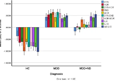
controls. This Table also shows the raw measurements (not adjusted for extraneous variables) of the different biomarkers. Figure 1 shows the differences in biomarker profile between the three
11
Table 2 shows the intercorrelation matrix between different biomarkers in the 90 subjects included in this study. Dynorphin (DYN) was significantly correlated with β-endorphin (END),
MOR, KOR-MOR and IL6-IL10. KOR was significantly associated with END, MOR, DYN-END and IL6-IL10. DYN-KOR was significantly correlated with DYN-END, MOR and IL6-IL10.
END levels were significantly correlated with MOR, KOR-MOR and IL6-IL10. DYN-END was significantly associated with KOR-MOR and IL6-IL10, while KOR-MOR was positively associated with IL6-IL10. Figure 2 shows the association between KOR and IL6-IL10, while
Figure 3 shows the correlation between DYN-END and IL6-IL10. Figure 4 shows the association between KOR-MOR and IL6-IL10.
Biomarker differences between controls and patients groups
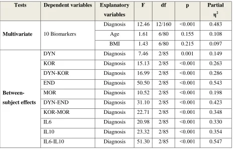
Table 3 shows the outcome of a multivariate GLM analysis with the 10 biomarkers as
dependent variables while adjusting for age and BMI. The dependent variables were DYN, END, KOR, MOR, IL-6, and IL-10 levels as well as z unit weighed composite scores, namely
DYN-KOR, DYN-END, KOR-MOR and IL6-IL10. We found a highly significant effect of diagnosis with an effect size of 0.483, while age and BMI were not significant. There were highly
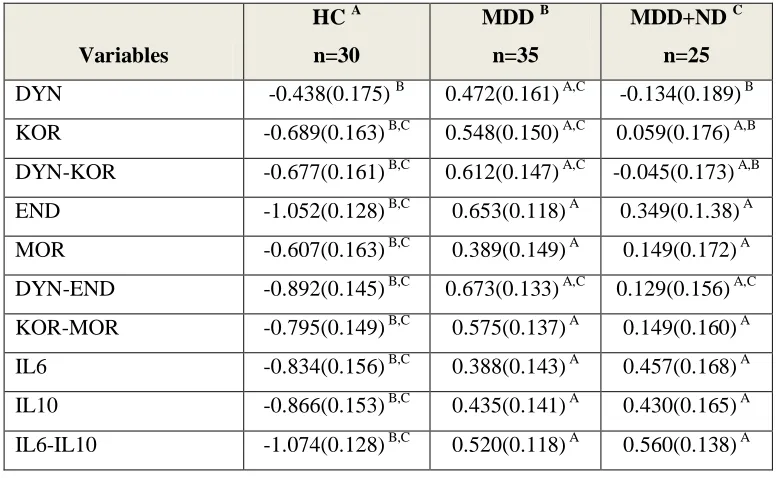
significant associations between all 10 biomarkers and diagnosis with the strongest associations between diagnosis and IL6-IL10, END, DYN-END, IL-10 and KOR-MOR (with effect sizes > 0.300). Table 4 shows the model-generated estimated marginal mean values (in z scores)
obtained by the GLM analyses shown in Table 3. Pair-wise multiple post-hoc analyses showed that DYN was significantly higher in MDD as compared with controls and MDD+ND. KOR,
12
increased from controls MDD+ND MDD. END, MOR, KOR-MOR, IL-6, IL-10 and IL6-IL10 were significantly higher in both MDD groups than in controls.
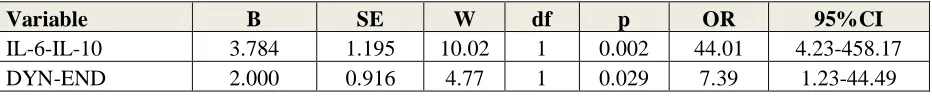
Table 5 shows the outcome of a stepwise binary logistic regression analyses with MDD (versus controls) as dependent variable. IL6-IL10 and DYN-END were the best predictors of MDD (Χ2=77.16, df=2, p<0.001) with an effect size of 0.800 (Nagelkerke value).
Table 6 shows the results of ROC analyses discriminating MDD from controls. The AUC ROC curves for DYN, KOR, DYN-KOR and DYN-END were computed on non-smoking MDD
patients versus controls (because ND has an effect on these biomarkers). The other biomarkers were examined in all MDD patients versus controls. In the same Table we also show the
bootstrapped AUC values after 2000 bootstraps. We found that DYN-END, END, KOR-MOR and IL6-IL10 yielded very high (bootstrapped) AUC (all > 0.849) separating MDD from controls. The best bootstrapped AUC was delineated for the combination of IL6-IL10 and DYN-END (0.972).
Discussion
The first major finding of this study is that serum dynorphin and KOR were significantly
increased in MDD as compared with controls and that increases in those opioid peptides and their receptors are interrelated, suggesting that dynorphin/KOR signaling is significantly increased in MDD. However, two previous clinical studies were unable to observe signs of
increased dynorphin/KOR signaling in depression (see introduction: 29, 30). As described in the Introduction, increased KOR signaling results in anti-reward and dysphoric effects thereby
13
while dynorphin release can be both a cause and consequence of stress hormone release, or may occur as a direct result of stress-induced increases in neuronal activity (42, 43). Evidence
suggests that the acute effects of stress are caused, at least in part, by dynorphin-mediated KOR activation. The predominant effect of KOR stimulation is decreased neuronal activity in cell
populations that express KORs (24). Dendritic dynorphin release in the hippocampus and hypothalamus negatively regulates excitatory inputs via retrograde activation of presynaptic KORs (44). This inhibitory mechanism may generalize to other neuronal populations often
implicated in the regulation of mood and motivation, such as the amygdala and striatum, which express dendritic dynorphin (45). KORs are also involved in the regulation of serotonergic (42)
and noradrenergic (46) systems, which may play a role in MDD. Blockade of KORs decrease immobility time in animal models of the forced swim test, suggesting these receptors may show antidepressant-like effects (47). On the other hand, during acute stress, KOR signaling may
increase physical ability (by producing analgesia) and motivation to escape threat (by producing aversion) thereby facilitating adaptive responses (24).
Our results on dynorphin and KOR are in agreement with our findings that also β-endorphin and MOR are upregulated in MDD and that the increases in β-β-endorphin and MOR
are significantly intercorrelated, indicating increased β-endorphin/MOR signaling in MDD (13). Previously, one paper showed increased expression of MOR in suicide victims, whereas PET scan studies showed controversial results on MOR binding profiles (see Introduction).
Interstingly, MOR (and DOR) activation may elevate mood and therefore may improve depressive states (48).
14
activation. Heightened IRS and CIRS responses are commonly observed in patients with MDD, with increased levels of immune-inflammatory biomarkers and immune-regulatory compounds, (5, 49, 50, 51), including serum IL-6 and IL-10 concentrations (5, 8, 13). Moreover, we observed that these opioid and IRS/CIRS responses yielded a good diagnostic performance for MDD with
a bootstrapped (n=2000) area under the curve of 0.972 (for both opioid peptides combined with both cytokines). In this respect it is intersting to note that recent theories suggest that classical
neurotransmitters convey information between pairs of neurons, whereas neuropeptides and
cytokines convey information and coordinate activities across broader networks of neurons (52, 53). This may explain that combinations of immune and opioid biomarkers have a high
sensitivity, specificity and diagnostic performance for MDD. All in all, our results show that increased dynorphin/KOR and β-endorhin/MOR signaling in MDD are strongly associated with
the peripheral IRS/CIRS response in that illness.
As described in the Introduction, there are many reciprocal relationships between the opioid and immune systems. During inflammation, pro-inflammatory cytokines including IL-1β
and IL-6 may stimulate hypothalamic corticotrophin releasing hormone (CRH) release (2, 54).In response to CRH and cytokines, peripheral blood mononuclear cells may secrete opioids (33). Nevertheless, the major sources of local endogenous opioid ligands including β-endorphin and dynorphin are leukocytes (55). For example, in macrophages, monocytes, granulocytes and
lymphocytes, β-endorphin is present in secretory granules arranged at the cell periphery ready for
exocytosis (11). Quantitative analysis revealed that in early inflammation, granulocytes (especially neutrophils) are the major opioid-containing leukocytes, whereas in later stages of
15
predominate (56, 57). Therefore, leukocytes are able to exert analgesic effects by releasing opioid peptides which bind to opioid receptors of the nociceptors in the periphery (11).
Dynorphins produced during inflammatory conditions display anti-inflammatory effects by attenuating translocation of nuclear factor-ΚB and consequently production of tumor necrosis
factor-α and IL-1β (58). Inhibition of nuclear-factor ΚB is likely a major mechanism explaining the anti-inflammatory effects of opioids (58). Also, activation of KOR by dynorphins negatively regulates many immune cell functions including cytokine production by macrophages (59), T
cell proliferation (60), phagocytoses (61) and antibody production (62). Moreover, dynorphins may exert some of their activities by binding to other receptors, including NMDA receptors,
activating the HPA-axis, inhibiting dopaminergic neurons, and mediating changes in calcium channels leading to increased intracellular calcium levels (63, 64).
Immune cells contain and upregulate signal-sequence encoding mRNA of the β-endorphin precursor POMC and the entire enzymatic machinery necessary for its processing into
the functionally active peptide (55). POMC neuropeptides such as β-endorphin and ACTH are
produced in both the anterior and intermediate lobes of the pituitary, as well as in a cluster of neurons in the hypothalamic arcuate nucleus (65) leading to a concomitant increase in ACTH,
β-endorphin and cortisol in MDD (2, 6, 13, 66, 67). During inflammatory conditions, “CRH stimulates peripheral release of β-endorphin from immune cells” (68) and, therefore, activation
of immune-inflammatory pathways likely plays an important role in contributing to circulating
β-endorphin levels. Animal data show that β-β-endorphin have anti-inflammatory and immunosuppressive properties (69) explaining the positive benefits of CRH-mediated increases in β-endorphin levels under inflammatory conditions (70). Inflammation also induces MOR
16
turn, have anti-infammatory effects and MOR agonists may be used to dampen immune-inflammatory responses during for example immune-inflammatory bowel disease (72). All in all, our findings suggest that in MDD a) immune activation is associated with a simultaneous activation of dynorphin/KOR and β-endorhin/MOR signaling, which both may have CIRS activities
attenuating the primary immune response and b) both increased dynorphin/KOR and β-endorhin/MOR signaling may play a role in depressive symptomatology albeit with divergent effects, namely detrimental (KOR) versus more protective (MOR) effects.
The third major finding of this study is that KOR and the index dynorphin/KOR signaling are both significantly decreased in MDD subjects with comorbid nicotine dependence, while
there are no significant effects on β-endorphin or MOR levels (although there is a trend towards a decrease). Smoking is highly prevalent among people with MDD (73) and is commonly cited as a means to cope with depressed mood and counteract fatigue and inactivity (74). As explained in the introduction, smoking is also a risk factor for depression through activated
immune-inflammatory and oxidative pathways, while comorbid MDD and nicotine dependence are characterized by lowered antioxidant and increased neuro-immune, neuro-oxidative and degenerative biomarkers (38, 39, 73). Isola et al. (2009) proposed that nicotine administration
may influence dynorphin primarily through dopamine release and that glutamate plays a modulatory role (75). Nicotine exposure increases dopamine release in mesolimbic terminal fields (76) and, therefore, one possibility is that the lowered dynorphin/KOR levels in comorbid
MDD and nicotine use may be due to dopamine release in response to nicotine. Moreover, KOR activation may attenuate the reinforcing effects of nicotine (77), while KOR agonists attenuate
17
dynorphin/KOR system plays an inhibitory role in the reinforcing effects of nicotine (79). Therefore, it is aslo possible that MDD patients with releatively lower dynorphin/KOR signaling display increased reinforcement effects of nicotine and thus more nicotine dependence behaviors. Furthermore, due to lowered dynorphin/KOR signaling, heavy smokers may show lowered
anti-inflammatory protection and, therefore, may be more vulnerable to the detrimental effects of smoking-induced IL-6 and TNF-α signaling (73, 80, 81).
Limitations of the study
The results of this study should be discussed with respect to its limitations. One limitation
is that it would have been more interesting if we had measured many more cytokines including those produced by M1 macrophage, T helper (Th)-1, Th-2, Th-17 and T regulatory phenotypes in order to examine the associations of opioids with the full spectrum of IRS and CIRS cytokines
and their ratios (8). Secondly, this study examined male subjects only and therefore our study should be replicated in females.
Conclusion
Serum dynorphin and KOR and β-endorphin and MOR are significantly increased in
MDD as compared with controls. The increases in dynorphin/KOR and β-endorhin/MOR signaling are significantly intercorrelated with signs of IRS/CIRS activation. KOR and the
18
systems may participate in the pathophysiology of depression by exerting CIRS activities attenuating the primary immune response as well as emotional and behavioral effects.
Acknowledgements
None.
Declaration of interest
The authors have no financial conflict of interests.
Funding
There was no specific funding for this specific study.
Authorships.
All authors contributed significantly to the paper and approved the final version.
References
1. LIM GY, TAM WW, LU Y, HO CS, ZHANG MW, HO RC. Prevalence of Depression in the Community from 30 Countries between 1994 and 2014. Sci Rep 2018; 8: 2861.
2. MAES M, JACOBS MP, SUY E, LECLERCQ C, CHRISTIAENS F, RAUS J. An
19
3. CATENA-DELL'OSSO M, ROTELLA F, DELL'OSSO A, FAGIOLINI A, MARAZZITI D. Inflammation, serotonin and major depression. Curr Drug Targets 2013;14(5):571-577.
4. YOUNG JJ, BRUNO D, POMARA N. A review of the relationship between
proinflammatory cytokines and major depressive disorder. J Affect Disord 2014;169:15-20.
5. KÖHLER CA, FREITAS TH, MAES M, et al. Peripheral cytokine and chemokine
alterations in depression: a meta-analysis of 82 studies. Acta Psychiatr Scand 2017;135(5):373-387.
6. AL-HAKEIM HK. Serum cortisol, immunoglobulins and some complements among depressed patients. Indian J Clin Biochem 2008;23(1):76-80.
7. AL-HAKEIM, HK, TWAYEJ AJ, AL-DUJAILI AH. Reduction in serum IL-1β, IL-6,
and IL-18 levels and Beck Depression Inventory-II score by combined sertraline and ketoprofen administration in major depressive disorder: A clinical trial.
Neurol Psychiatry Brain Res 30, 148-153.
8. MAES M, CARVALHO AF. The Compensatory Immune-Regulatory Reflex System (CIRS) in Depression and Bipolar Disorder. Mol Neurobiol 2018;55(12):8885-8903.
9. AL-HAKEIM HK, AL-KUFI SN, AL-DUJAILI AH, MAES M. Serum Interleukin Levels and Insulin Resistance in Major Depressive Disorder. CNS Neurol Disord
Drug Targets 2018;17;626-633.
10.SNYDER SH. Opiate receptors and beyond: 30 years of neural signaling research.
20
11.NINKOVIĆ J. Role of the mu opioid receptor in opioid modulation of immune function. Amino Acids 2013; 45(1): 9-24.
12.LE MERRER J, BECKER JA, BEFORT K, KIEFFER BL. Reward processing by the opioid system in the brain. Physiol Rev 2009;89;1379-1412
13.AL-FADHEL SZ, AL-HAKEIM HK, AL-DUJAILI AH, MAES M. IL-10 is associated with increased mu-opioid receptor levels in major depressive disorder. Eur Psychiatry 2019;57:46-51.
14.PECIÑA M, KARP JF, MATHEW S, TODTENKOPF MS, EHRICH EW, ZUBIETA JK. Endogenous opioid system dysregulation in depression: implications for new
therapeutic approaches. Mol Psychiatry 2019;24:576-587.
15.LUTZ PE, KIEFFER BL. Opioid receptors: distinct roles in mood disorders. Trends Neurosci 2013;36(3):195-206.
16.FACCHINETTI F, PETRAGLIA F, SANCES G, et al. Dissociation between CSF and plasma B-endorphin in major depressive disorders: evidence for a different regulation. J
Endocrinol Invest 1986;9(1):11-14.
17.DARKO DF, IRWIN MR, RISCH SC, GILLIN JC. Plasma beta-endorphin and natural killer cell activity in major depression: a preliminary study. Psychiatry
Res 1992;43(2):111-119.
18.CASTILLA-CORTÁZAR I, CASTILLA A, GURPEGUI M. Opioid peptides and
21
19.DJUROVIC D, MILIC-ASKRABIC J, MAJKIC-SINGH N. Effect of fluvoxamine on the level of beta-endorphin in the sera and nervous tissue of rats. Pharmazie 1998;53: 143-144.
20.ESCRIBA PV, OZAITA A, GARCÍA-SEVILLA JA. Increased mRNA expression of
alpha2A-adrenoceptors, serotonin receptors and mu-opioid receptors in the brains of suicide victims. Neuropsychopharmacology 2004;29(8):1512-1521.
21.PECINA M, ZUBIETA JK. Expectancy Modulation of Opioid Neurotransmission. Int
Rev Neurobiol 2018;138:17-37.
22.CARLEZON WA JR, THOMAS MJ. Biological substrates of reward and aversion: a
nucleus accumbens activity hypothesis. Neuropharmacology 2009; 56(Suppl 1):122-132. 23.BRUCHAS MR, LAND BB, CHAVKIN C. The Dynorphin-Kappa Opioid System as a
Modulator of Stress-induced and Pro-addictive Behaviors. Brain Res 2010;1314C: 44.
24.KNOLL AT, CARLEZON WA. Dynorphin, stress, and depression. Brain Res 2010;1314C: 56.
25.BAILEY S, HUSBANDS S. Targeting opioid receptor signalling in depression: do we need selective kappa opioid receptor antagonists? Neuronal Signaling
2018;2(2):NS20170145.
26.LALANNE L, AYRANCI G, KIEFFER BL, LUTZ PE. The kappa opioid receptor: from addiction to depression, and back. Front Psychiatry 2014;5:170.
27.TAYLOR TT, MANZELLA F. Kappa Opioids, Salvinorin A and Major Depressive Disorder. Current Neuropharmaco 2016; 14:165-176.
22
29.MILLER JM, ZANDERIGO F, PURUSHOTHAMAN PD, et al. Kappa opioid receptor binding in major depression: A pilot study. Synapse 2018;72(9):e22042.
30.HURD YL. Subjects with major depression or bipolar disorder show reduction of prodynorphin mRNA expression in discrete nuclei of the amygdaloid complex. Mol
Psychiatry 2002;7(1):75-81.
31.MOUSA SA, SHAKIBAEI M, SITTE N, SCHÄFER M, STEIN C. Subcellular pathways of beta-endorphin synthesis, processing, and release from immunocytes in inflammatory
pain. Endocrinology 2004;145(3):1331-1341.
32.POMORSKA DK, GACH K, JANECKA A. Immunomodulatory effects
of endogenous and synthetic peptides activating opioid receptors. Mini Rev Med Chem 2014;14(14):1148-1155.
33.RITTNER HL, MACHELSKA H, STEIN C. Leukocytes in the regulation of pain and
analgesia. J Leukoc Biol 2005;78:1215-1222.
34.BIDLACK JM, KHIMICH M, PARKHILL AL, SUMAGIN S, SUN B, TIPTON CM.
Opioid receptors and signaling on cells from the immune system. J Neuroimmune Pharmacol 2006;1(3):260-269.
35.MAES M, VANDERVORST C, SUY E, MINNER B, RAUS J. A multivariate study of simultaneous escape from suppression by dexamethasone of urinary free cortisol, plasma cortisol, adrenocorticotropic hormone and beta-endorphin in melancholic patients. Acta
Psychiatr Scand 1991a;83(6):480-491.
36.CASTILLA A, SUBIRÀ ML, CIVEIRA MP, CUENDE JI, PRIETO J. Monocytic
23
37.NUNES SO, VARGAS HO, PRADO E, et al. The shared role of oxidative stress and inflammation in major depressive disorder and nicotine dependence. Neurosci Biobehav
Rev 2013;37(8):1336-1345.
38.NUNES SO, PICCOLI DE MELO LG, PIZZO DE CASTRO MR, et al. Atherogenic
index of plasma and atherogenic coefficient are increased in major depression and bipolar disorder, especially when comorbid with tobacco use disorder. J Affect Disord 2015a;172:55-62.
39.NUNES SOV, DE CASTRO MRP, MOREIRA EG, GUEMBAROVSKI RL, BARBOSA DS. Association of paraoxonase (PON) 1 activity, glutathione S-transferase GST T1/M1
and STin. 2 polymorphisms with comorbidity of tobacco use disorder and mood disorders. Neuroscience letters 2015b;585:132-137.
40.POMERLEAU OF. Endogenous opioids and smoking: a review of progress and
problems. Psychoneuroendocrinology 1998;23(2):115-130.
41.Bewick V, Cheek L, Ball J. Statistics review 13: receiver operating characteristic curves.
Critical Care 2004;8(6):508-512.
42. BILKEI-GORZO A, RACZ I, MICHEL K, et al. Control of hormonal stress reactivity by
the endogenous opioid system. Psychoneuroendocrinology 2008;33(4):425-436.
43.LAND BB, BRUCHAS MR, LEMOS JC, XU M, MELIEF EJ, CHAVKIN C. The dysphoric component of stress is encoded by activation of the dynorphin kappa-opioid
system. J Neurosci 2008;28(2):407-414.
44.IREMONGER KJ, BAINS JS. Retrograde opioid signaling regulates glutamatergic
24
45.REYES BA, CHAVKIN C, VAN BOCKSTAELE EJ. Subcellular targeting of kappa-opioid receptors in the rat nucleus locus coeruleus. J Comp Neurol 2009; 512(3):419-431.
46.REYES BA, JOHNSON AD, GLASER JD, COMMONS KG, VAN BOCKSTAELE EJ. Dynorphin-containing axons directly innervate noradrenergic neurons in the rat nucleus
locus coeruleus. Neuroscience 2007; 145(3):1077-1086.
47.MAGUE SD, PLIAKAS AM, TODTENKOPF MS, et al. Antidepressant-like effects of kappa-opioid receptor antagonists in the forced swim test in rats. J Pharmacol Exp
Ther 2003;305:323-330.
48.SHIPPENBERG TS, LEFEVOUR A, CHEFER VI. Targeting endogenous mu- and
delta-opioid receptor systems for the treatment of drug addiction. CNS Neurol Disord Drug Targets 2008;7(5):442-453.
49.KRISHNADAS R, CAVANAGH J. Depression: an inflammatory illness? J Neurol
Neuros Psych 2012;83:495-502.
50.AL-HAKEIM HK, AL-RAMMAHI DA, AL-DUJAILI AH. IL-6, IL-18, sIL-2R, and TNFα proinflammatory markers in depression and schizophrenia patients who are free of
overt inflammation. J Affect Dis 2015; 182:106-114.
51.HAAPAKOSKI R, MATHIEU J, EBMEIER KP, ALENIUS H, KIVIMAKI M. Cumulative meta-analysis of interleukins 6 and 1beta, tumour necrosis factor alpha and C-reactive protein in patients with major depressive disorder. Brain Behav Immun
2015;49:206-215.
52.LUDWIG M, LENG G. Dendritic peptide release and peptide-dependent behaviours. Nat
25
53.LEONARD B, MAES M. Mechanistic explanations how cell-mediated immune activation, inflammation and oxidative and nitrosative stress pathways and their sequels
and concomitants play a role in the pathophysiology of unipolar depression. Neurosci Biobehav Rev 2012;36(2):764-785.
54.TURNBULL AV, RIVIER CL. Regulation of the hypothalamic-pituitary-adrenal axis by cytokines: actions and mechanisms of action. Physiol Rev 1999; 79:1-71.
55.MACHELSKA H, SCHOPOHL JK, MOUSA SA, LABUZ D, SCHÄFER M, STEIN C.
Different mechanisms of intrinsic pain inhibition in early and late inflammation. J Neuroimmunol 2003;141:30-39.
56.STEIN C, SCHÄFER M, MACHELSKA H. Attacking pain at its source: new perspectives on opioids. Nat Med 2003;9:1003-1008.
57.RITTNER HL, BRACK A, STEIN C. Pain and the immune system. Br J Anaesth
2008;101:40-44.
58.RAHIMAN SSF, MORGAN M, GRAY P, SHAW PN, CABOT PJ. Inhibitory effects of
dynorphin 3-14 on the lipopolysaccharide-induced toll-like receptor 4 signalling pathway. Peptides. 2017;90:48-54.
59.ALICEA C, BELKOWSKI S, EISENSTEIN TK, ADLER MW, ROGERS TJ. Inhibition of primary murine macrophage cytokine production in vitro following treatment with the kappa-opioid agonist U50, 488H. J Neuroimmunol 1996; 64: 83-90.
60.GUAN L, EISENSTEIN TK, ADLER MW, ROGERS TJ. Inhibition of T cell super antigen responses following treatment with the kappa-opioid agonist U50, 488H. J
26
61.SZABO I, ROJAVIN M, BUSSIERE JL, EISENSTEIN TK, ADLER MW, ROGERS TJ. Suppression of peritoneal mac-rophage phagocytosis of Candida albicans by opioids. J
Pharmacol Exp Ther. 1993; 267: 703-706.
62.MORGAN EL. Regulation of human B lymphocyte activation by opioid peptide
hormones. Inhibition of IgGproduction by opioid receptor class (mu-, kappa-, and delta-) selective agonists. J Neuroimmunol1996; 65: 21-30.
63.JIN W, TERMAN GW, CHAVKIN C. Kappa opioid receptor tolerance in the guinea pig
hippocampus. J Pharmacol Exp Ther 1997;281(1):123-128.
64.SCHWARZER C. 30 years of dynorphins--new insights on their functions in
neuropsychiatric diseases. Pharmacol Ther 2009;123(3):353-370.
65.STENGAARD-PEDERSEN K, LARSSON LI. Comparative immunocytochemical localization of putative opioid ligands in the central nervous system. Histochemistry
1981;73:89-114.
66.MAES M, BOSMANS E, SUY E, MINNER B, RAUS J. A further exploration of the
relationships between immune parameters and the HPA-axis activity in depressed patients. Psychol Med 1991b;21(2):313-320.
67.MAES M, CLAES M, VANDEWOUDE M, et al. Adrenocorticotropin hormone, beta-endorphin and cortisol responses to oCRF in melancholic patients. Psychol Med 1992;22(2):317-329.
68.BERCZI I, QUINTANAR-STEPHANO A. An Update on Neural Regulators of the Hypothalamic-Pituitary-Adrenal Axis. In Insights to Neuroimmune Biology 2nd Edition,
27
69.SILBERSTEIN S, VOGL AM, BONFIGLIO JJ, et al. Immunology, signal transduction, and behavior in hypothalamic-pituitary-adrenal axis-related genetic mouse models. Ann
NY Acad Sci 2009;1153:120-130.
70.KHAIROVA RA, MACHADO-VIEIRA R, DU J, MANJI HK. A potential role for
proinflammatory cytokines in regulating synaptic plasticity in major depressive disorder. Int. J. Neuropsychopharmacol 2009;19:1-18.
71.POL O, ALAMEDA F, PUIG MM. Inflammation enhances mu-opioid receptor
transcription and expression in mice intestine. Mol Pharmacol 2001;60(5):894-899. 72.PHILIPPE D, DUBUQUOY L, GROUX H, et al. Anti-inflammatory properties of the mu
opioid receptor support its use in the treatment of colon inflammation. J Clin Invest 2003;111(9):1329-1338.
73.NUNES SO, VARGAS HO, BRUM J, et al. A comparison of inflammatory markers in
depressed and nondepressed smokers. Nicotine Tob Res 2012;14:540-546.
74.PIPER ME, PIASECKI TM, FEDERMAN EB, et al. A multiple motives approach to
tobacco dependence: the Wisconsin Inventory of Smoking Dependence Motives (WISDM-68). J Consult Clin Psychol 2004;72(2):139-154.
75.ISOLA R, ZHANG H, TEJWANI GA, NEFF NH, HADJICONSTANTINOU M. Acute nicotine changes dynorphin and prodynorphin mRNA in the striatum. Psychopharmacology (Berl) 2009;201(4):507-516
76.DI CHIARA G. Role of dopamine in the behavioural actions of nicotine related to addiction. Eur J Pharmacol 2000;393:295-314.
28
78.HAHN B, STOLERMAN IP, SHOAIB M. Kappa-opioid receptor modulation of nicotine-induced behaviour. Neuropharmacology 2000;39:2848-2855.
79.GALEOTE L, BERRENDERO F, BURA SA, ZIMMER A, MALDONADO R. Prodynorphin gene disruption increases the sensitivity to nicotine selfadministration in
mice. Int J Neuropsychopharmacol 2009;12:615-625.
80.LESPERANCE F, FRASURE-SMITH N, THEROUX P, IRWIN M. The association between major depression and levels of soluble intercellular adhesion molecule 1,
interleukin-6, and C-reactive protein in patients with recent acute coronary syndromes. Am J Psychiat 2004;161:271-277.
81.GASPERSZ R, LAMERS F, WITTENBERG G, et al. The role of anxious distress in immune dysregulation in patients with major depressive disorder. Translational Psychiat 2017;(7):1268.
29
Figure 1 Differences in biomarker profile between controls (HC) and major depressed patients with (MDD+ND) and without (MDD) nicotine dependence.
Shown are the group mean values (±SE) after z transformations were made.
DYN: dynorphin
KOR: kappa opioid receptor
DYN-KOR: index of KOR signaling computed as z transformation of dynorphin (zDYN) + zKOR
END: β-endorphin
MOR: mu opioid receptor
DYN-END: integrated index of circulating opioid peptides, computed as zDYN + zEND. KOR-MOR: index of opioid receptor status computed as zKOR + zMOR.
IL: interleukin
30
Figure 2 Association between kappa opioid receptor (KOR) levels and immune activation as indicated by a z unit weighted composite (IL6-IL10) score based on interleukin (IL)-6 and IL-10
31
Figure 3 Association between opioid peptides as indicated by a z unit weighted composite score based on dynorphin (DYN) and β-endorphin (END) assays (KOR-MOR) and immune activation
32
Figure 4 Association between opioid receptor levels as indicated by a z unit weighted composite score based on kappa (KOR) and mu (MOR) opioid receptor levels (KOR-MOR) and immune
33
Table 1: Socio-demographic, clinical and biomarker data in major depressed patients with nicotine dependence (MDD+ND), patients without ND (MDD), and healthy controls (HC).
Variables HC A
n=30
MDD B
n=35
MDD+ND c
n=25
F/χ2
df p
Age (years) 30.3(8.8) 31.1(11.8) 33.8(9.9) 0.82 2/87 0.443
BMI (kg/m2) 26.2(2.8) 2
4
.2(3.6) 25.3(3.8) 2.60 2/87 0.080Single/Married 16/14 C 14/21 5/20 A 6.41 2 0.041
Urban/Rural 5/25 7/28 8/17 2.02 2 0.364
Employment (N/Y) 4/26 B,C 16/19 A 11/14 A 8.90 2 0.012
Dynorphin (pg/ml) 17.04(2.55) B 18.83(1.86) A,C 17.50(1.79) B 6.35 2/87 0.003
KOR (pg/ml) 8.32(1.71) B,C 11.99(3.99) A,C 10.02(1.81) A,B 13.46 2/87 <0.001
END (pg/ml) 24.90(5.27) B,C 34.95(4.02) A 33.02(2.75) A 49.94 2/87 ˂0.001
MOR (pg/ml) 3.01(0.83) B,C 4.43(1.49) A 4.03(1.27)A 10.94 2/87 ˂0.001
IL-6 (pg/ml) 11.4(2.8) B,C 16.2(3.7) A 16.7(4.3) A 18.78 2/87 ˂0.001
IL-10 (pg/ml) 6.0(1.7) B,C 8.9(2.2) A 8.9(2.0) A 21.78 2/87 <0.001
BDI-II - 48.3(11.0) 50.9(8.5) 0.96 1/58 0.336
BMI: Body mass index
KOR: Kappa opioid receptor
END: β-endorphins
MOR: Mu opioid receptor
IL: interleukin
34
Table 2: Inter-correlation matrix among biomarkers data.
Variables DYN KOR
DYN-KOR
END MOR
DYN-END
KOR-MOR
KOR 0.388**
DYN-KOR 0.833** 0.833**
END 0.396** 0.440** 0.502**
MOR 0.339** 0.328* 0.400** 0.358**
DYN-END 0.835** 0.495** 0.799** 0.835** 0.417**
KOR-MOR 0.209* 0.738** 0.568** 0.477** 0.815** 0.410**
IL6-IL10 0.316** 0.440** 0.454** 0.568** 0.529** 0.529** 0.594**
*: p<0.05 **: p<0.001 (n=90)
KOR: kappa opioid receptor
DYN-KOR: index of KOR signaling computed as z transformation of dynorphin (zDYN) + zKOR
END: β-endorphin
MOR: mu opioid receptor
DYN-END: integrated index of circulating opioid peptides, computed as zDYN + zEND.
KOR-MOR: index of opioid receptor status computed as zKOR + zMOR.
35
Table 3: Results of multivariate GLM analysis with the biomarkers as dependent variables and diagnosis as explanatory variable while adjusting for extraneous variables.
Tests Dependent variables Explanatory
variables
F df p Partial
η2
Multivariate 10 Biomarkers
Diagnosis 12.46 12/160 ˂0.001 0.483
Age 1.61 6/80 0.155 0.108
BMI 1.43 6/80 0.215 0.097
Between-subject effects
DYN Diagnosis 7.46 2/85 0.001 0.149
KOR Diagnosis 15.13 2/85 ˂0.001 0.263
DYN-KOR Diagnosis 16.99 2/85 ˂0.001 0.286
END Diagnosis 50.50 2/85 ˂0.001 0.543
MOR Diagnosis 10.52 2/85 ˂0.001 0.198
DYN-END Diagnosis 31.10 2/85 ˂0.001 0.423
KOR-MOR Diagnosis 22.71 2/85 ˂0.001 0.348
IL6 Diagnosis 20.98 2/85 ˂0.001 0.330
IL10 Diagnosis 23.32 2/85 ˂0.001 0.354
IL6-IL10 Diagnosis 51.30 2/85 ˂0.001 0.547
Diagnosis: Healthy controls versus depression with and without nicotine dependence
DYN: dynorphin
KOR: kappa opioid receptor
DYN-KOR: index of KOR signaling computed as z transformation of dynorphin (zDYN) + zKOR
END: β-endorphin
MOR: mu opioid receptor
DYN-END: integrated index of circulating opioid peptides, computed as zDYN + zEND.
KOR-MOR: index of opioid receptor status computed as zKOR + zMOR.
36
Table 4: Model-generated estimated marginal means of the 10 biomarkers obtained by GLM analysis shown in Table 2 in controls (HC) and major depressed (MDD) patients, divided into those with (MDD+ND) and without (MDD) nicotine dependence.
Variables
HC A
n=30
MDD B
n=35
MDD+ND C
n=25
DYN -0.438(0.175) B 0.472(0.161) A,C -0.134(0.189) B
KOR -0.689(0.163) B,C 0.548(0.150) A,C 0.059(0.176) A,B
DYN-KOR -0.677(0.161) B,C 0.612(0.147) A,C -0.045(0.173) A,B
END -1.052(0.128) B,C 0.653(0.118) A 0.349(0.1.38) A
MOR -0.607(0.163) B,C 0.389(0.149) A 0.149(0.172) A
DYN-END -0.892(0.145) B,C 0.673(0.133) A,C 0.129(0.156) A,C
KOR-MOR -0.795(0.149) B,C 0.575(0.137) A 0.149(0.160) A
IL6 -0.834(0.156) B,C 0.388(0.143) A 0.457(0.168) A
IL10 -0.866(0.153) B,C 0.435(0.141) A 0.430(0.165) A
IL6-IL10 -1.074(0.128) B,C 0.520(0.118) A 0.560(0.138) A
All results are shows as mean (SE) and as z scores
DYN: dynorphin
KOR: kappa opioid receptor
DYN-KOR: index of KOR signaling computed as z transformation of dynorphin (zDYN) + zKOR
END: β-endorphin
MOR: mu opioid receptor
DYN-END: integrated index of circulating opioid peptides, computed as zDYN + zEND.
KOR-MOR: index of opioid receptor status computed as zKOR + zMOR.
37
Table 5: Results of binary logistic regression analysis with major depression (controls as reference group) as dependent variable.
Variable B SE W df p OR 95%CI
IL-6-IL-10 3.784 1.195 10.02 1 0.002 44.01 4.23-458.17 DYN-END 2.000 0.916 4.77 1 0.029 7.39 1.23-44.49
OR: Odd’s ratio with 95% confidence intervals
IL6-IL10: Computed as z(z interleukin-6 (zIL6) + zIL10))
38
Table 6: Results of receiver operating characteristic (ROC) analysis discriminating major depression from normal controls.
* Performed in non-smokers only (all other analyses in all participants)
DYN: dynorphin
KOR: kappa opioid receptor
DYN-KOR: index of KOR signaling computed as z transformation of dynorphin (zDYN) + zKOR
END: β-endorphin
MOR: mu opioid receptor
DYN-END: integrated index of circulating opioid peptides, computed as zDYN + zEND.
KOR-MOR: index of opioid receptor status computed as zKOR + zMOR.
IL6-IL10: index of immune activation computed as z interleukin-6 (zIL6) + zIL10
IL6-IL10 and DYN-END: as obtained using binary logistic regression (shown in Table 5)
Variables Area
ROC SE p 95% CI
Bootstrapped AUC
Boot-AUC 95%CI
DYN * 0.651 0.069 0.036 0.517-0.786 0.664 0.523-0.785
KOR * 0.805 0.053 <0.001 0.702-0.909 0.786 0.670-0.882
DYN-KOR* 0.787 0.056 <0.001 0.677-0.896 0.786 0.677-0.877
DYN-END * 0.936 0.936 <0001 0.883-0.990 0.922 0.852-0.971
END 0.952 0.020 <0.001 0.913-0.990 0.944 0.900-0.978
MOR 0.754 0.053 <0.001 0.651-0.858 0.742 0.633-0.897
KOR-MOR 0.849 0.039 <0.001 0.772-0.925 0.850 0.759-0.911
IL6-IL10 0.947 0.023 <0.001 0.902-0.993 0.931 0.877-0.978