R E S E A R C H
Open Access
Development and evaluation of a one-step
multiplex real-time TaqMan
®
RT-qPCR assay
for the detection and genotyping of equine
G3 and G14 rotaviruses in fecal samples
Mariano Carossino
1,2, Maria E. Barrandeguy
2,3, Erdal Erol
4, Yanqiu Li
5and Udeni B. R. Balasuriya
1*Abstract
Background:Equine rotavirus A (ERVA) is the leading cause of diarrhea in neonatal foals and has a negative impact on equine breeding enterprises worldwide. Among ERVA strains infecting foals, the genotypes G3P[12] and G14P[12] are the most prevalent, while infections by strains with other genomic arrangements are infrequent. The identification of circulating strains of ERVA is critical for diagnostic and surveillance purposes, as well as to understand their molecular epidemiology. Current genotyping methods available for ERVA and rotaviruses affecting other animal species rely on Sanger sequencing and are significantly time-consuming, costly and labor intensive. Here, we developed the first one-step multiplex TaqMan®real-time reverse transcription polymerase chain reaction (RT-qPCR) assay targeting the NSP3 and VP7 genes of ERVA G3 and G14 genotypes for the rapid detection and G-typing directly from fecal specimens. Methods:A one-step multiplex TaqMan®RT-qPCR assay targeting the NSP3 and VP7 genes of ERVA G3 and G14 genotypes was designed. The analytical sensitivity was assessed using serial dilutions of in vitro transcribed RNA containing the target sequences while the analytical specificity was determined using RNA and DNA derived from a panel of group A rotaviruses along with other equine viruses and bacteria. The clinical performance of this multiplex assay was evaluated using a panel of 177 fecal samples and compared to a VP7-specific standard RT-PCR assay and Sanger sequencing. Limits of detection (LOD), sensitivity, specificity, and agreement were determined.
Results:The multiplex G3 and G14 VP7 assays demonstrated high specificity and efficiency, with perfect linearity. A 100-fold difference in their analytical sensitivity was observed when compared to the singleplex assays; however, this difference did not have an impact on the clinical performance. Clinical performance of the multiplex RT-qPCR assay demonstrated that this assay had a high sensitivity/specificity for every target (100% for NSP3, > 90% for G3 VP7 and > 99% for G14 VP7, respectively) and high overall agreement (> 98%) compared to conventional RT-PCR and sequencing. Conclusions:This new multiplex RT-qPCR assay constitutes a useful, very reliable tool that could significantly aid in the rapid detection and G-typing of ERVA strains circulating in the field.
Keywords:Rotavirus A, Equine rotavirus A, ERVA, One-step multiplex RT-qPCR, G-typing, G3, G14, Foal diarrhea
© The Author(s). 2019Open AccessThis article is distributed under the terms of the Creative Commons Attribution 4.0 International License (http://creativecommons.org/licenses/by/4.0/), which permits unrestricted use, distribution, and reproduction in any medium, provided you give appropriate credit to the original author(s) and the source, provide a link to the Creative Commons license, and indicate if changes were made. The Creative Commons Public Domain Dedication waiver (http://creativecommons.org/publicdomain/zero/1.0/) applies to the data made available in this article, unless otherwise stated. * Correspondence:[email protected]
1Louisiana Animal Disease Diagnostic Laboratory and Department of
Pathobiological Sciences, School of Veterinary Medicine, Louisiana State University, Baton Rouge, LA, USA
Background
Equine rotavirus A (ERVA) has been identified as the leading cause of diarrhea in neonatal foals < 3 months of age and is responsible for 20 to 77% of foal diarrhea cases, causing significant economic losses to the equine breeding enterprises [1–7]. Rotaviruses are icosahedral, non-enveloped viruses with a double-stranded, seg-mented RNA genome (dsRNA) that belong to the family Reoviridae (genus Rotavirus) [8, 9]. The ERVA genome consists of 11 double-stranded RNA segments that en-code for six structural proteins (VP1–4, 6 and 7) and six non-structural proteins (NSP1–6). Segment 11 encodes for two non-structural proteins (NSP5 and NSP6) [1,
10]. The rotavirus particle consists of a triple capsid, in-cluding an outer capsid composed of VP7 and VP4, an intermediate layer integrated by VP6 and an inner capsid formed by VP1, VP2 and VP3 [11–13]. The two outer capsid proteins, VP7 and VP4, are the most variable and immunogenic proteins of the virus, which independently elicit neutralizing antibodies following infection [1, 14]. Based on VP6 identity, rotaviruses are classified into eight groups (A-H), from which group A rotaviruses (RVA) are the leading cause of diarrhea in humans and several animal species, including horses [15]. Group A rotaviruses are further classified into G-types and P-types according to the nucleotide sequence of the two outer capsid proteins, VP7 and VP4, encoded by seg-ments 9 and 4 of the genome, respectively [16]. Cur-rently, 27 G-types and 35 P-types of RVA have been recognized in several species including humans [9]. Thus far, seven G-types (G3, G5, G6, G8, G10, G13 and G14) and six P-types (P[1], P[3], P[7], P[11], P[12] and P[18]) have been identified among the RVA affecting horses, with G3P[12] and G14P[12] being the most prevalent and epidemiologically relevant genotypes [1, 2, 17–19]. Other genomic arrangements involving G- and P-types different from G3/G14 and P[12] have been infrequently described as infecting horses [1].
Group A rotaviruses are transmitted through the fecal-oral route and infection in young foals is associated with life-threatening watery diarrhea induced by a com-bination of malabsorptive, osmotic and secretory mecha-nisms [1, 20]. Control of ERVA infection in young foals is achieved by the routine vaccination of pregnant mares with an inactivated vaccine and strict husbandry/hy-gienic practices to reduce the viral burden in the envir-onment [1, 7, 21–23]. ERVA vaccines have been shown to aid in the reduction of the incidence and severity of diarrhea and also in the intensity and duration of viral shedding, however they do not guarantee full protection [1,21,22]. In addition, previous studies have shown that there is significant antigenic variation among ERVA ge-notypes, which leads to emergence of viruses that are not neutralized by antibodies elicited by the current
vaccines [24–29]. Moreover, temporal and spatial varia-tions in the prevalence and distribution of ERVA geno-types has been previously reported [2,29,30]. Therefore, it is important to perform genotypic characterization of ERVA strains in order to understand the molecular epi-demiology of ERVA, identify novel viral reassortants and potential interspecies transmission, and assess vaccine performance in the field. Currently, sequencing of VP7, VP4 and other genome segments are required for geno-typing circulating rotavirus strains. Conventional se-quencing methodologies are generally labor intensive, low throughput and costly. Real-time reverse transcrip-tion quantitative polymerase chain reactranscrip-tion (RT-qPCR) assays, particularly TaqMan® assays, offer a wide spectrum of advantages compared to conventional RT-PCR and sequencing. Some of these advantages in-clude high throughput sample processing, increased sen-sitivity and specificity, faster turnaround time, and ability to multiplex. Even though several singleplex and multiplex RT-qPCR assays have been developed for the genotyping of human RVA genotypes [31–35], none have been developed for the genotyping of animal rotaviruses thus far, including ERVA. Here, we developed and evalu-ated the performance of a one-step multiplex RT-qPCR assay that allows the rapid detection of ERVA and the genotyping of the most frequent G-types affecting horses (G3 and G14) in fecal specimens. Overall, the one-step multiplex RT-qPCR assay developed in this study can sim-ultaneously detect and genotype G3 and G14 ERVA strains with a performance equivalent to that of conven-tional VP7-specific RT-PCR and Sanger sequencing.
Methods
Cell lines and viruses
minimal volume of maintenance media without FBS. After 1 h adsorption at 37 °C, monolayers were overlaid with MA-104 medium containing 0.5μg/ml of trypsin type IX (Sigma-Aldrich, St. Louis, MO) and without FBS, and in-cubated at 37 °C and 5% CO2until 100% cytopathic effect
was observed (48 h post infection). Infected flasks were frozen/thawed, clarified by centrifugation at 1,500X g for 15 min at 4 °C, aliquoted, and stored at−80 °C.
Viral RNA and bacterial DNA
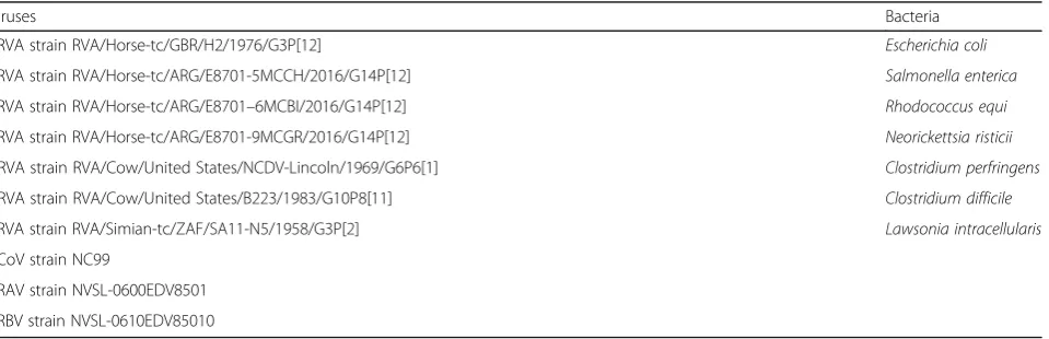
RNA and DNA from the following viruses and bacteria associated with diarrhea in horses were included for specificity evaluation of the ERVA-specific RT-qPCR assay: TCF containing ERVA strains RVA/Horse-tc/ GBR/H2/1976/G3P[12], RVA/Horse-tc/ARG/E8701-5M CCH/2016/G14P[12], RVA/Horse-tc/ARG/E8701–6M CBI/2016/G14P[12] and RVA/Horse-tc/ARG/E8701-9MCGR/2016/G14P[12] [29]; TCF containing bovine RVA (BRVA) strains RVA/Cow/United States/NCDV-Lincoln/1969/G6P6[1] and RVA/Cow/United States/ B223/1983/G10P8[11], TCF containing simian RVA strain RVA/Simian-tc/ZAF/SA11-N5/1958/G3P[2], TCF containing equine coronavirus strain NC99 [36], and TCF containing equine rhinitis A (NVSL-060 0EDV8501) and B (NVSL-0610EDV85010) viruses. ERVA strain H2, BRVA strains NCDV-Lincoln and B223, and simian RVA strain SA11 were kindly pro-vided by Dr. Viviana Parreño (INTA, Buenos Aires, Argentina). Equine rhinitis viruses were obtained from the National Veterinary Services Laboratories, United States Department of Agriculture, Ames IA. DNA samples from Escherichia coli, Salmonella enterica, Rhodococcus equi, Neorickettsia risticii, Clostridium perfringens, Clostridium difficileandLawsonia intracel-lularis were obtained from the University of Kentucky Veterinary Diagnostic Laboratory (Table1).
Fecal samples
A total of 177 fecal samples from diarrheic foals were used in this study. Among these, 112 fecal samples were collected from farms in central Kentucky [29] while 65 were from outbreaks of diarrhea that occurred in Argentina between 2009 and 2014 [29,30]. Ten percent fecal suspensions in serum-free EMEM were prepared, centrifuged at 2500 X g for 15 min at 4 °C, then filtered through a 0.45μm syringe filter. Aliquots of fecal suspensions were stored at−80 °C.
Nucleic acid isolation
Nucleic acid isolation was performed using the taco™ mini nucleic acid extraction system (GeneReach USA, Lexington, MA, USA) as previously described [37]. Two hundred microliters of 10% fecal suspension or tissue culture supernatant was used as sample input and elu-tion was performed with 200μl of elution buffer and stored at−80 °C for future use.
RT-PCR amplification of ERVA VP7 gene (segment 9) We established a VP7-specific (gene segment 9) standard RT-PCR assay using the Qiagen One-Step RT-PCR kit (Qiagen, Valencia, CA, USA) as previ-ously described [38]. This assay was used as the gold-standard method for ERVA detection in fecal specimens [2, 39]. Briefly, a 25μl reaction mixture was composed of 5μl 5X One-Step RT-PCR Buffer, 1μl dNTP Mix, 1μl of VP7-specific forward and re-verse primers (Table 2, 20μM, final concentration 0.8μM), 1μl of One-Step RT-PCR Enzyme Mix, 11μl of RNase-free water and 5μl of template previously subjected to a denaturing step at 95 °C for 5 min. The cycling conditions included a reverse transcription step (50 °C for 30 min) followed by a PCR activation step at 95 °C for 15 min; 35 cycles of denaturation (94
Table 1A panel of viruses and bacteria associated with diarrhea in horses, cattle and simians was used to assess the specificity of the singleplex and multiplex RT-qPCR assays for detection and genotyping of ERVA
Viruses Bacteria
ERVA strain RVA/Horse-tc/GBR/H2/1976/G3P[12] Escherichia coli
ERVA strain RVA/Horse-tc/ARG/E8701-5MCCH/2016/G14P[12] Salmonella enterica
ERVA strain RVA/Horse-tc/ARG/E8701–6MCBI/2016/G14P[12] Rhodococcus equi
ERVA strain RVA/Horse-tc/ARG/E8701-9MCGR/2016/G14P[12] Neorickettsia risticii
BRVA strain RVA/Cow/United States/NCDV-Lincoln/1969/G6P6[1] Clostridium perfringens
BRVA strain RVA/Cow/United States/B223/1983/G10P8[11] Clostridium difficile
SRVA strain RVA/Simian-tc/ZAF/SA11-N5/1958/G3P[2] Lawsonia intracellularis
ECoV strain NC99
ERAV strain NVSL-0600EDV8501
ERBV strain NVSL-0610EDV85010
°C for 1 min), annealing (47 °C for 1 min) and exten-sion (72 °C for 2 min); and a final extenexten-sion at 72 °C for 2 min. PCR amplification products yielded a 1062 bp band following electrophoretic separation in a 1% agarose gel.
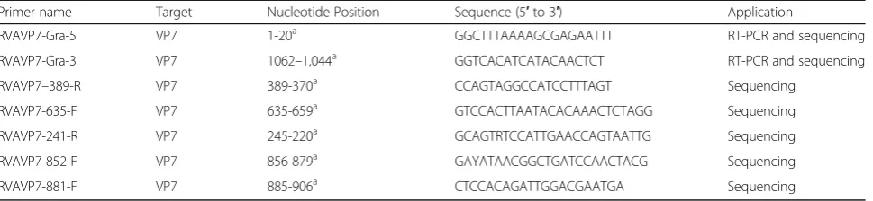
Sequencing of ERVA VP7 gene for G-typing
Sequencing of the full-length VP7 gene (genome seg-ment 9) was performed using a high fidelity One-Step RT-PCR kit (Qiagen One-Step Ahead RT-PCR kit) and the forward and reverse primers RVAVP7-Gra-5 and RVAVP7-Gra-3 (Table 2) as previously described [29]. Briefly, a 25μl reaction mixture was composed of 10μl 2.5X One-Step Ahead RT-PCR Master Mix, 1μl of VP7-specific forward and reverse primers (20μM, final concentration 0.8μM), 1μl of 25X One-Step Ahead RT-Mix, 7μl of RNase-free water and 5μl of template previously subjected to a de-naturing step at 95 °C for 5 min. The cycling condi-tions included a reverse transcription step (45 °C for 15 min) followed by a PCR activation step at 95 °C for 5 min; 40 cycles of denaturation (95 °C for 15 s), an-nealing (47 °C for 15 s) and extension (68 °C for 2 min); and a final extension at 68 °C for 5 min. PCR products (1062 bp) were gel-purified using the QIA-quick® Gel Extraction kit (Qiagen) according to the manufacturer’s recommendations. DNA was submitted for Sanger sequencing to a commercial company (Eurofins Genomics LLC, Louisville, KY, USA). Both DNA strands of VP7 amplicons were sequenced using a panel of primers specified in Table 2. Sequence ana-lysis was performed using Geneious R7 (Biomatters Inc., Newark, NJ, USA). G-types were identified using an automated genotyping tool for RVA (RotaC 2.0,
http://rotac.regatools.be/) [40].
Accession numbers
The nucleotide sequences derived from the fecal samples and tissue culture fluid corresponding to ERVA strains
RVA/Horse-tc/ARG/E8701-5MCCH/2016/G14P[12], RVA/ Horse-tc/ARG/E8701–6MCBI/2016/G14P[12] and RVA/ Horse-tc/ARG/E8701-9MCGR/2016/G14P[12] utilized in this study were deposited in GenBank under accession numbers MG970165-MG970197, MH458234-MH458237, KP116019-KP116049 and MF074190-MF074212.
Primer and probe design
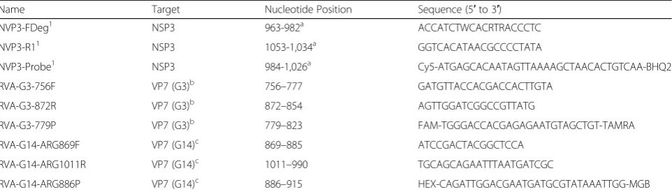
Multiple alignments of full-length ERVA G3 (n= 17) and G14 (n= 39) VP7 nucleotide sequences derived from Gen-Bank were performed and consensus sequences obtained using Geneious R7 software. G-type specific forward and reverse primers and probes were designed towards conserved regions specific to G3 VP7 and G14 VP7 gene sequences using the PrimerQuest tool (https://www. idtdna.com/Primerquest/home/Index) (Table3). The pri-mer and probe sequences were checked for specificity using the NCBI Basic Local Alignment Search Tool (BLAST; https://blast.ncbi.nlm.nih.gov/Blast.cgi?PROGR AM=blastn&PAGE_TYPE=BlastSearch&LINK_LOC=blas thome) while self-annealing sites, hairpin loop formation and 3′complementarity were verified using the IDT Oli-goAnalyzer tool (https://www.idtdna.com/calc/analyzer).
Synthesis of target NSP3 and VP7 genes and preparation of in vitro transcribed RNA
ERVA-specific in vitro transcribed (IVT) RNA was synthe-sized in order to determine the analytical sensitivity of the ERVA-specific multiplex RT-qPCR assay. For this purpose, a 493 nt insert containing the targeted regions (NSP3 [nt position 963–1053], G3 VP7 [nt position 756–872] and G14 VP7 [nt position 869–1011] derived from ERVA strain H2 (NSP3 and G3 VP7) and ERVA strain FI23 (G14 VP7) (GenBank Accession numbers KM454500.1, KM454497.1 and KM454508.1, respectively) were chem-ically synthesized (GeneArt™ Gene Synthesis, Thermo-Fisher Scientific, Regensburg, Germany) and cloned into the pGEM®-3Z vector (Promega, Madison, WI) down-stream of the T7 promoter (pRVA_NSP3G3G14) by a commercial company. Subsequently,E. coli K12 DH10B™
Table 2Primers used for RT-PCR amplification and sequencing of VP7 (genome segment 9) of ERVA
Primer name Target Nucleotide Position Sequence (5′to 3′) Application
RVAVP7-Gra-5 VP7 1-20a GGCTTTAAAAGCGAGAATTT RT-PCR and sequencing
RVAVP7-Gra-3 VP7 1062–1,044a GGTCACATCATACAACTCT RT-PCR and sequencing
RVAVP7–389-R VP7 389-370a CCAGTAGGCCATCCTTTAGT Sequencing
RVAVP7-635-F VP7 635-659a GTCCACTTAATACACAAACTCTAGG Sequencing
RVAVP7-241-R VP7 245-220a GCAGTRTCCATTGAACCAGTAATTG Sequencing
RVAVP7-852-F VP7 856-879a GAYATAACGGCTGATCCAACTACG Sequencing
RVAVP7-881-F VP7 885-906a CTCCACAGATTGGACGAATGA Sequencing
a
T1R were transformed with the construct. Trans-formed bacteria were cultured overnight at 37 °C with shaking (270 rpm). Plasmid DNA was purified using QIAprep Spin Miniprep kit (Qiagen, Valencia, CA) following the manufacturer’s instructions and screened by restriction digestion using the unique EcoRI, BamHI, and HindIII restriction sites within and flanking the insert. Sequence authenticity was con-firmed by Sanger sequencing using T7 and SP6 promoter-specific primers. Plasmid DNA (1μg) was linearized using HindIII, purified using the High Pure PCR Product Purification kit (Roche, Indianapolis, IN) as instructed, and 0.5μg of plasmid DNA was used for in vitro transcription of the pRVA_NSP3G3G14 insert using the Megascript®T7 Transcription kit (ThermoFisher Scientific, Waltham, MA) following the manufacturer’s rec-ommendations. Residual plasmid DNA was removed by di-gestion with TURBO™DNase (ThermoFisher Scientific) for 15 min at 37 °C. The IVT RNA product was analyzed by agarose gel electrophoresis, subjected to a clean-up proced-ure using the MEGAclear™ Transcription Clean-Up kit (ThermoFisher Scientific), and quantified using a Nano-Drop 2000 spectrophotometer (ThermoFisher Scientific). The pRVA_NSP3G3G14 IVT RNA was stored at −80 °C until used. The number of ERVA IVT RNA molecules per microliter (copies/μl) was calculated according to the fol-lowing formula:
The concentration of ERVA IVT RNA was adjusted to 107copies/μl using nuclease-free water containing 40 ng/μl of Ambion® Yeast tRNA (ThermoFisher Scientific), and serially ten-fold diluted (107- 0.1 IVT RNA copies/μl) using nuclease-free water containing Ambion®Yeast tRNA.
ERVA-specific singleplex TaqMan®real-time RT-PCR assays targeting G3 VP7, G14 VP7 and NSP3 genes
Primers and probes specific for ERVA G3 VP7 and G14 VP7 were designed as described above (Table3). The reac-tion was set up using the QuantiTect™Probe RT-PCR kit (Qiagen) following the manufacturer’s recommendations. Briefly, the 25μl reaction contained 12.5μl of 2X Quanti-Tect™ Probe RT-PCR Master Mix with ROX, 0.25μl QuantiTect™RT Mix, 200 nM TaqMan®fluorogenic probe, 500 nM of each primer, and 5μl of template RNA (previ-ously subjected to a denaturing step at 95 °C for 5 min). Reverse transcription and amplification were carried out in an ABI 7500 Fast Real-time PCR System (Applied Bio-systems®, Life Technologies, Grand Island, NY). The pro-gram included 30 min at 50 °C (reverse transcription step), 15 min at 95 °C (PCR initial activation step), followed by 45 cycles at 94 °C for 15 s (denaturation) and 60 °C for 1 min (combined annealing/extension). The NSP3-specific (gene segment 7; pan-rotavirus A) assay was established in the laboratory as previously described (Table3) [41].
Number of IVT RNA molecules=μL¼Avogadro
0s number 6ð :0221023Þ IVT RNA concentration gð =μLÞ
IVT RNA molecular weight gð Þ
Table 3Primers and probe combinations for the detection of rotavirus A (pan-rotavirus A, targeting the NSP3 gene) and specific amplification of the VP7 gene of equine rotavirus A G3 and G14 genotypes
Name Target Nucleotide Position Sequence (5′to 3′)
NVP3-FDeg1 NSP3 963-982a ACCATCTWCACRTRACCCTC
NVP3-R11 NSP3 1053-1,034a GGTCACATAACGCCCCTATA
NVP3-Probe1 NSP3 984-1,026a Cy5-ATGAGCACAATAGTTAAAAGCTAACACTGTCAA-BHQ2
RVA-G3-756F VP7 (G3)b 756–777 GATGTTACCACGACCACTTGTA
RVA-G3-872R VP7 (G3)b 872–854 AGTTGGATCGGCCGTTATG
RVA-G3-779P VP7 (G3)b 779–823 FAM-TGGGACCACGAGAGAATGTAGCTGT-TAMRA
RVA-G14-ARG869F VP7 (G14)c 869–885 ATCCGACTACGGCTCCA
RVA-G14-ARG1011R VP7 (G14)c 1011–990 TGCAGCAGAATTTAATGATCGC
RVA-G14-ARG886P VP7 (G14)c 886–915 HEX-CAGATTGGACGAATGATGCGTATAAATTGG-MGB
1
Primers and probe name and sequences derived from Freeman et al., 2008
a
nucleotide position based on GenBank Accession number X81436
b
nucleotide position based on GenBank Accession number KM454497.1
c
nucleotide position based on GenBank Accession number KM454508.1
ERVA-specific multiplex TaqMan®real-time RT-PCR assays targeting G3 VP7, G14 VP7 and NSP3 genes
The G3 VP7, G14 VP7 and NSP3-specific assays were multiplexed for the simultaneous identification of all genotypes (pan-rotavirus A) and G-typing of ERVA. The reaction was set up using the QuantiTect™ Multiplex RT-PCR kit (Qiagen) following the manufacturer’s recom-mendations. Briefly, the 25μl reaction contained 12.5μl of 2X QuantiTect™ Multiplex RT-PCR Master Mix with ROX, 0.25μl QuantiTect™ RT Mix, 200 nM of each Taq-Man®fluorogenic probe, 200 nM of each primer, and 5μl of template RNA (denatured at 95 °C for 5 min before be-ing added into the reaction well). Reverse transcription and amplification were carried out in an ABI 7500 Fast Real-time PCR System (Applied Biosystems®). The pro-gram included 20 min at 50 °C (reverse transcription step), 15 min at 95 °C (PCR initial activation step), followed by 40 cycles at 94 °C for 45 s (denaturation) and 60 °C for 75 s (combined annealing/extension).
Statistical analysis
Standard curves were performed using IVT RNA (107 to 0.1 IVT RNA copies/μl). Coefficients of determination (R2) were used to assess curve fitness. PCR amplification effi-ciencies (%) were calculated after regression analysis using the following formula: E¼ ½10−slope1 −1 100. Limit of detec-tion with 95% confidence (LOD95%) was determined by
stat-istical probit analysis (a non-linear regression model) using the commercial software SPSS 14.0 (SPSS Inc., Chicago, IL, USA) for all assays with 9 replicates per dilution ranging from 105 to 1 IVT RNA copies/μl. Cycle threshold (Ct) cut-off values were determined as the average Ct + 3 stand-ard deviations of nine replicates of the endpoint dilution [42]. Precision (within-run and between-run imprecision) of the ERVA multiplex RT-qPCR assay was determined by per-forming 9 replicates of IVT RNA containing 105, 104and 103RNA copies/μl on the same run (within-run impreci-sion) or three replicates of IVT RNA containing 105, 104 and 103RNA copies/μl tested on two different operational days. The coefficient of variation (%) was determined for each target concentration (G3 VP7, G14 VP7, and NSP3). The performance of the ERVA multiplex RT-qPCR assay was evaluated in fecal specimens and compared to the VP7-specific RT-PCR and G-typing by Sanger sequencing. Contingency tables (2 × 2) were generated to determine the sensitivity, specificity and agreement (kappa statistic) of each target within the multiplex RT-qPCR assay.
Results
Analysis of fecal samples by VP7-specific RT-PCR and sequencing for determination of G-types
A total of 177 fecal samples were included in the study, from which 92 samples were confirmed negative for
ERVA, while 85 were positive as determined by VP7-specific standard RT-PCR [29, 30]. From the 85 ERVA-positive samples, 58 were collected in Argentina and 27 were collected from the USA (Kentucky). Among these, 41 were confirmed as G3 genotype while 44 were confirmed as G14 genotype by sequencing of the VP7 gene. Extensive genetic and phylogenetic analysis of these samples was recently published in a separate article [29].
Analytical sensitivity and specificity of ERVA-specific singleplex and multiplex RT-qPCR assays targeting G3 VP7, G14 VP7 and NSP3 genes
Analytical sensitivity of ERVA-specific singleplex RT-qPCR assays
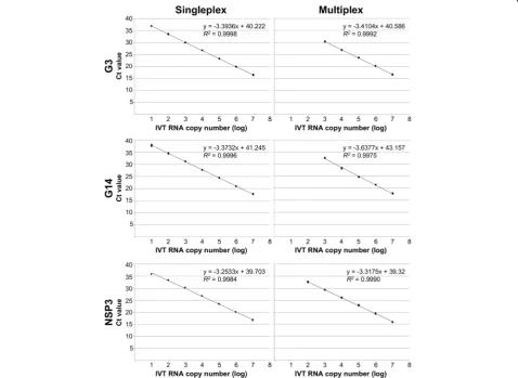
The analytical sensitivity of the ERVA-specific singleplex and multiplex RT-qPCR assays was determined using a ten-fold dilution series (3 replicates per dilution) of IVT RNA (107to 0.1 IVT RNA copies/μl) containing the target sequences. Standard curves generated for the three targets (G3 VP7, G14 VP7 and NSP3) under singleplex conditions demonstrated perfect linearity (R2 > 0.99, Table 4 and Fig. 1). Amplification efficiencies for the G3 VP7, G14 VP7 and NSP3 targets under singleplex conditions were 97, 98% and 103%, respectively. Detection rates (100%) for the singleplex RT-qPCR assays are shown in Table4. Pro-bit analysis determined that the limits of detection 95% (LOD95%) of the G3 VP7, G14 VP7 and NSP3 RT-qPCR
assays under singleplex conditions were 2.6, 5.7 and 27 copies/μl of IVT RNA and cycle threshold (Ct) cut-off points were determined at 38, 39 and 34, respectively.
Analytical sensitivity of ERVA-specific multiplex RT-qPCR assay
Standard curves generated for the three targets (G3 VP7, G14 VP7 and NSP3) under multiplex conditions also demonstrated perfect linearity (R2> 0.99, Table4and Fig.
1). However, while the amplification efficiencies for the G3 VP7 and NSP3 targets were ± 10% of that determined under singleplex conditions (96 and 100%, respectively), a lower amplification efficiency was determined for the G14 VP7 target when multiplexing (88%). Detection rates (100%) for the multiplex RT-qPCR assay are shown in Table4. While the 100% detection rate limit for the NSP3 assay was equal between the singleplex and multiplex for-mats, a 100-fold difference was observed for the G3 VP7 and G14 VP7 assays when these were multiplexed (Table
4). In comparison to the singleplex format, the LOD95%
Analytical specificity of ERVA-specific singleplex and multiplex RT-qPCR assays
To evaluate the analytical specificity of the singleplex and multiplex RT-qPCR assays, a panel of rotavirus strains along with other viruses and bacteria associated with diarrhea in horses was used (Table 1). The ERVA-specific G3 and G14 VP7 primer-probe combina-tions were exclusively specific for the respective ERVA
genotype, did not cross-react between each other, did not amplify other rotavirus genotypes from other species and, interestingly, did not amplify the simian SA11 strain (G3P2). The NSP3-specific primer-probe combination in both singleplex and multiplex format was specific for RVA and amplified the reference G3 and G14 strains of ERVA as well as bovine and simian rotavirus strains as previously reported [34]. None of the assays (G3 VP7, G14
Table 4Analytical sensitivity analysis of singleplex and multiplex RT-qPCR assays for the detection and genotyping of equine rotavirus A
Parameter Singleplex Multiplex
G3 G14 NSP3 G3 G14 NSP3
Slope −3.3936 −3.3732 −3.2533 −3.4104 −3.6377 −3.3175
Linearity (R2) > 0.99 > 0.99 > 0.99 > 0.99 > 0.99 > 0.99
Efficiency (%) 97 98 103 96.4 88 100
LOD95%(copies/μl) 2.6 5.7 27 716 215 47
Detection rate limit (100%, copies/μl) 10 10 100 1000 1000 100
Ct cut-off 38 39 34 32 34 34
LOD95%, limit of detection 95%; Ct, cycle threshold
VP7 and NSP3) amplified other viruses or bacteria associ-ated with diarrhea in horses.
Precision assessment of the ERVA-specific multiplex RT-qPCR assay
To evaluate the precision of the multiplex RT-qPCR assay, within-run and between-run imprecision was de-termined as recommended [42]. In all cases, the coeffi-cient of variation was less than 3%, indicating that the multiplex assay has a high repeatability (within-run) and reproducibility (between-run) within the range of detec-tion (Table5).
Clinical performance of the ERVA-specific multiplex RT-qPCR assay targeting G3 VP7, G14 VP7 and NSP3
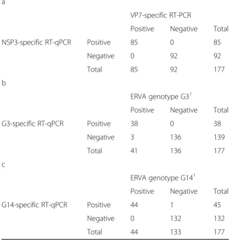
The clinical performance of the ERVA-specific multiplex RT-qPCR assay was evaluated in a total of 177 fecal samples. The NSP3 (pan-RVA) assay was able to suc-cessfully detect ERVA in all positive samples (85/85) while no non-specific amplifications were observed in negative samples (n= 92; Table6a). Therefore, the assay presented 100% sensitivity and specificity when com-pared to the VP7-specific standard RT-PCR assay, along with perfect agreement (kappa = 1). In the case of the G3 VP7 assay, the assay was able to correctly genotype 38/41 ERVA G3 samples while non-specific amplifica-tions were not observed in G3-negative samples (n = 136, Table 6b). Only three ERVA G3 positive samples were unable to be genotyped by the multiplex assay, however these were correctly genotyped by the G3-specific singleplex RT-qPCR assay. Overall, the G3 VP7 assay presented a 92.7% sensitivity and 100% speci-ficity when compared to the VP7-specific standard RT-PCR assay, and a high agreement (98.31% [kappa = 0.951]). Finally, the G14 VP7 assay was able to correctly identify 44/44 ERVA G14-positive samples and did not amplify 132/133 ERVA G14 negative samples (Table6c). Consequently, the G14 VP7 assay presented a 100% sen-sitivity and 99.2% specificity when compared to the VP7-specific standard RT-PCR assay. The agreement be-tween assays was high (99.44% [kappa = 0.985]). Regard-ing the presumed false positive sample, although this sample was determined to be an ERVA G3P[12] by Sanger sequencing, it yielded a concurrent positive
amplification by the G3 and G14-specific RT-qPCR as-says in both their singleplex and multiplex formats, sug-gesting a possible co-infection with both genotypes of ERVA.
Discussion
Group A rotaviruses are a primary cause of diarrhea in children and animal species, including horses [1–6, 43,
44]. Even though seven G-types and six P-types of ERVA have been identified in horses, the G3P[12] and G14P[12] constitute the most epidemiologically relevant genotypes [1,2,17–19]. Spatial as well as temporal fluc-tuations between these predominant G-types (G3 and G14) of ERVA circulating in equine populations have been reported around the world [2, 30]. Interestingly, the emergent pattern of G14 ERVA and the temporal shift in the prevalent genotype has been observed in as-sociation with the implementation of widespread vaccin-ation programs in Argentina, Japan and Ireland [2, 30,
45, 46], which rely on the use of inactivated vaccines containing only the H2 or HO-5 (G3P [12]) strains of ERVA. The difficulties faced to date in establishing cell-culture adapted G14P[12] or other strains of ERVA has precluded their inclusion into vaccine formulations. However, we have recently isolated and cell-culture adapted, three G14P[12] ERVA strains with the potential
Table 5Replication experiment to evaluate precision (within-run and between-(within-run imprecision) of the multiplex RT-qPCR assays for the detection and genotyping of equine rotavirus A
Concentration of target (IVT RNA copies/μl)
Within-run imprecision Between-run imprecision
G3 G14 NSP3 G3 G14 NSP3
100,000 2.63% 1.29% 0.89% 2.92% 1.56% 0.97%
10,000 1.75% 0.74% 0.42% 2.19% 1.24% 0.83%
1,000 2.01% 0.51% 0.56% 2.52% 0.63% 0.68%
Table 6Evaluation of the clinical performance of the multiplex RT-qPCR assay for the detection and genotyping of equine rotavirus A in fecal samples compared to VP7-specific RT-PCR and sequencing (gold standard). (a) NSP3 (b) G3 VP7 and (c) G14 VP7
a
VP7-specific RT-PCR
Positive Negative Total
NSP3-specific RT-qPCR Positive 85 0 85
Negative 0 92 92
Total 85 92 177
b
ERVA genotype G31
Positive Negative Total
G3-specific RT-qPCR Positive 38 0 38
Negative 3 136 139
Total 41 136 177
c
ERVA genotype G141
Positive Negative Total
G14-specific RT-qPCR Positive 44 1 45
Negative 0 132 132
Total 44 133 177
1
to be used as reference G14P[12] strains to study the molecular biology of this genotype and perform vaccine efficacy studies following heterologous challenge in the future [29].
In light of the antigenic differences between ERVA ge-notypes, their spatial and temporal distribution and their impact on vaccine efficacy, molecular surveillance and genotypification of circulating strains is critical. Since genomic arrangements of ERVA other than G3P[12] and G14P[12] are rare and the outer capsid protein VP7 con-tains the major neutralizing epitopes, we developed a one-step multiplex TaqMan® real-time RT-PCR for the rapid detection and G-typing of the most prevalent ge-notypes of ERVA (G3 and G14) in fecal specimens. Compared to the conventional methods for ERVA geno-typing (RT-PCR and Sanger sequencing), the multiplex RT-qPCR assay has a significantly faster turnaround time, is high-throughput, less labor-intensive and exhibits a high sensitivity, specificity and agreement as demonstrated in this study. While multiplexing did not have an impact on the detection limit of the NSP3-tar-get, the G3 and G14 targets demonstrated a 100-fold dif-ference in their analytical sensitivity under multiplex conditions. However, this difference in analytical sensi-tivity did not have a significant impact on their clinical performance on fecal specimens and only three G3 ERVA-positive samples were unable to be typed by the multiplex RT-qPCR assay (false negatives). Interestingly, these samples were correctly G-typed when the G3 VP7-specific assay was performed under singleplex con-ditions. Such differences are probably due to a combin-ation of low target nucleic acid in these fecal specimens along with the 100-fold higher analytical sensitivity of the singleplex compared to the multiplex assay. Despite the low number of false negative samples (n = 3), all three targets (G3 VP7, G14 VP7 and NSP3) showed a high sensitivity and specificity (> 90%) along with a high level of agreement (> 98%) in the clinical specimens tested under multiplex conditions.
Noteworthy, a single sample, G-typed as G3 by means of conventional methods (RT-PCR and Sanger sequen-cing), exhibited specific amplification of both the G3 VP7 and G14 VP7 targets simultaneously under single-plex and multisingle-plex conditions. Although confirmation would require RT-PCR using genotype-specific primers or next generation sequencing, due to the fact that both G3 and G14 ERVA strains were identified to be co-circulating in the same farm during the same time period, these results suggest that this dual-positive fecal specimen most likely derived from a foal that was co-infected with both G3 and G14 ERVA strains. Conse-quently, this may indicate that the multiplex RT-qPCR assay developed can be advantageous for the diagnosis of co-infections with G3 and G14 strains of ERVA that
are currently challenging to identify. Further assessment using spiked specimens is required in order to analyze this multiplex RT-qPCR assay’s capability to identify co-infected animals. Due to the lack of reference strains and uncommon occurrence of other ERVA G-types, these were not included in this study. Therefore, it is im-perative to perform Sanger sequencing on those samples that test positive for ERVA by amplification of NSP3 but are not genotyped as G3 or G14 by the current assay. In this regard, the genotyping assay developed here will fa-cilitate rapid genotyping of circulating strains and iden-tify rare G-types that can then be incorporated into this assay depending on their epidemiological relevance.
Conclusions
In conclusion, the study presented herein describes the development and evaluation of a one-step multiplex TaqMan® RT-qPCR assay for the detection and genotyp-ing of the most frequent G-types of ERVA infectgenotyp-ing horses. This assay demonstrated to have a high sensitiv-ity, specificity and agreement compared to conventional RT-PCR and sequencing, providing rapid and reliable G-typing of ERVA strains. Therefore, this assay is highly suitable for routine diagnostics as well as to aid current surveillance programs of ERVA by rapidly characterizing circulating strains. Finally, the number of specific targets included in this assay can be updated and expanded as other genomic arrangements of ERVA emerge and be-come prevalent in equine populations.
Abbreviations
BRVA:bovine rotavirus A; DNA: deoxyribonucleic acid; dNTP: deoxynucleotide
triphosphate; EMEM: Eagle’s minimum essential medium; ERVA: equine
rotavirus A; IVT: in vitro transcribed; LOD: limit of detection; RNA: ribonucleic acid; RT-qPCR: reverse transcription real-time polymerase chain reaction; RVA: rotavirus A; SRVA: simian rotavirus A; TCF: tissue culture fluid
Acknowledgements
The authors would like to kindly acknowledge the veterinarians who participated from this study by submitting fecal specimens from diarrheic foals. The authors would also like to thank Dr. Kenton Morgan, Equine Technical Services, 16009 Knorpp Road, Pleasant Hill, MO 64080, USA for initiating and coordinating the field sample collection and Dr. Viviana Parreño, INCUINTA, Instituto Nacional de Tecnología Agropecuaria (INTA), Buenos Aires, Argentina, for providing reference strains for specificity testing.
Funding
This study was supported by Zoetis Animal Health (Kalamazoo, MI, USA), the Gluck Equine Research Foundation (GERF) competitive grant No. 1215351520 and the INTA-HARAS agreement (CVT 123, INTA, Hurlingham, Buenos Aires, Argentina).
Availability of data and materials
The nucleotide sequences derived from the fecal samples and tissue culture fluid corresponding to ERVA strains RVA/Horse-tc/ARG/E8701-5MCCH/2016/
G14P [12], RVA/Horse-tc/ARG/E8701–6MCBI/2016/G14P [12] and
Authors’contributions
MC designed the study, performed the experiments, analyzed the data and wrote the manuscript; MEB provided a large number of sequenced fecal specimens to be used in this study and revised the manuscript; EE provided reference bacterial DNA for specificity testing and revised the manuscript; YL contributed to sample archiving, isolation of nucleic acids and revised the manuscript; UBRB conceived and designed the study, obtained funding, analyzed the data and wrote the manuscript.
Ethics approval and consent to participate
Not applicable.
Consent for publication
Not applicable.
Competing interests
The authors declare that they have no competing interests.
Publisher’s Note
Springer Nature remains neutral with regard to jurisdictional claims in published maps and institutional affiliations.
Author details
1Louisiana Animal Disease Diagnostic Laboratory and Department of
Pathobiological Sciences, School of Veterinary Medicine, Louisiana State University, Baton Rouge, LA, USA.2Escuela de Veterinaria, Universidad del
Salvador, Champagnat 1599, Ruta Panamericana km54.5 (B1630AHU), Pilar, Buenos Aires, Argentina.3Instituto de Virología, CICVyA, INTA. Las Cabañas y
Los Reseros s/n, (1712) Castelar, Buenos Aires, Argentina.4Department of
Veterinary Science, University of Kentucky Veterinary Diagnostic Laboratory, University of Kentucky, Lexington, KY, USA.5Maxwell H. Gluck Equine Research Center, Department of Veterinary Science, University of Kentucky, Lexington, KY, USA.
Received: 27 December 2018 Accepted: 20 March 2019
References
1. Bailey KE, Gilkerson JR, Browning GF. Equine rotaviruses--current understanding and continuing challenges. Vet Microbiol. 2013;167(1–2):135–44.
2. Garaicoechea L, Mino S, Ciarlet M, Fernandez F, Barrandeguy M, Parreno V. Molecular characterization of equine rotaviruses circulating in Argentinean foals during a 17-year surveillance period (1992-2008). Vet Microbiol. 2011; 148(2–4):150–60.
3. Dickson J, Smith VW, Coackley W, McKean P, Adams PS. Rotavirus infection of foals. Aust Vet J. 1979;55(4):207–8.
4. Conner ME, Darlington RW. Rotavirus infection in foals. Am J Vet Res. 1980; 41(10):1699–703.
5. Slovis NM, Elam J, Estrada M, Leutenegger CM. Infectious agents associated with diarrhoea in neonatal foals in Central Kentucky: a comprehensive molecular study. Equine Vet J. 2014;46(3):311–6.
6. Imagawa H, Sekiguchi K, Anzai T, Fukunaga Y, Kanemaru T, Ohishi H, Higuchi T, Kamada M. Epidemiology of equine rotavirus infection among foals in the breeding region. J Vet Med Sci. 1991;53(6):1079–80.
7. Magdesian KG, Dwyer RM, Gonzalez Arguedas M: Viral Diarrhea. In Equine
Infectious Diseases. Edited by Sellon DC, Long MT. St. Louis, MO: Saunders; 2014: 198–203.
8. Carstens EB. Ratification vote on taxonomic proposals to the international committee on taxonomy of viruses (2009). Arch Virol. 2010;155(1):133–46. 9. Matthijnssens J, Ciarlet M, McDonald SM, Attoui H, Banyai K, Brister JR, Buesa
J, Esona MD, Estes MK, Gentsch JR, et al. Uniformity of rotavirus strain nomenclature proposed by the rotavirus classification working group (RCWG). Arch Virol. 2011;156(8):1397–413.
10. Newman JF, Brown F, Bridger JC, Woode GN. Characterisation of a rotavirus. 20b. Nature. 1975, 258(5536):631–633.
11. Eren E, Zamuda K, Patton JT. Modeling of the rotavirus group C capsid predicts a surface topology distinct from other rotavirus species. Virology. 2016;487:150–62.
12. Chen JZ, Settembre EC, Aoki ST, Zhang X, Bellamy AR, Dormitzer PR, Harrison SC, Grigorieff N. Molecular interactions in rotavirus assembly and
uncoating seen by high-resolution cryo-EM. Proc Natl Acad Sci U S A. 2009; 106(26):10644–8.
13. Settembre EC, Chen JZ, Dormitzer PR, Grigorieff N, Harrison SC. Atomic model of an infectious rotavirus particle. EMBO J. 2011;30(2):408–16. 14. Dyall-Smith ML, Lazdins I, Tregear GW, Holmes IH. Location of the major
antigenic sites involved in rotavirus serotype-specific neutralization. Proc Natl Acad Sci U S A. 1986;83(10):3465–8.
15. Matthijnssens J, Otto PH, Ciarlet M, Desselberger U, Van Ranst M, Johne R. VP6-sequence-based cutoff values as a criterion for rotavirus species demarcation. Arch Virol. 2012;157(6):1177–82.
16. Matthijnssens J, Ciarlet M, Heiman E, Arijs I, Delbeke T, McDonald SM, Palombo EA, Iturriza-Gomara M, Maes P, Patton JT, et al. Full genome-based classification of rotaviruses reveals a common origin between human Wa-like and porcine rotavirus strains and human DS-1-Wa-like and bovine rotavirus strains. J Virol. 2008;82(7):3204–19.
17. Matthijnssens J, Mino S, Papp H, Potgieter C, Novo L, Heylen E, Zeller M, Garaicoechea L, Badaracco A, Lengyel G, et al. Complete molecular genome analyses of equine rotavirus a strains from different continents reveal several novel genotypes and a largely conserved genotype constellation. J Gen Virol. 2012;93(Pt 4:866–75.
18. Nemoto M, Tsunemitsu H, Imagawa H, Hata H, Higuchi T, Sato S, Orita Y, Sugita S, Bannai H, Tsujimura K, et al. Molecular characterization and analysis of equine rotavirus circulating in Japan from 2003 to 2008. Vet Microbiol. 2011;152(1–2):67–73.
19. Matthijnssens J, Ons E, De Coster S, Conceicao-Neto N, Gryspeerdt A, Van Ranst M, Raue R. Molecular characterization of equine rotaviruses isolated in Europe in 2013: implications for vaccination. Vet Microbiol. 2015;176(1–2): 179–85.
20. Ramig RF. Pathogenesis of intestinal and systemic rotavirus infection. J Virol. 2004;78(19):10213–20.
21. Barrandeguy M, Parreno V, Lagos Marmol M, Pont Lezica F, Rivas C, Valle C, Fernandez F. Prevention of rotavirus diarrhoea in foals by parenteral vaccination of the mares: field trial. Dev Biol Stand. 1998;92:253–7. 22. Powell DG, Dwyer RM, Traub-Dargatz JL, Fulker RH, Whalen JW, Jr.,
Srinivasappa J, Acree WM, Chu HJ. Field study of the safety,
immunogenicity, and efficacy of an inactivated equine rotavirus vaccine. J Am Vet Med Assoc 1997, 211(2):193–198.
23. Sheoran AS, Karzenski SS, Whalen JW, Crisman MV, Powell DG, Timoney JF. Prepartum equine rotavirus vaccination inducing strong specific IgG in mammary secretions. Vet Rec. 2000;146(23):672–3.
24. Papp H, Matthijnssens J, Martella V, Ciarlet M, Banyai K. Global distribution of group a rotavirus strains in horses: a systematic review. Vaccine. 2013;31(48): 5627–33.
25. Browning GF, Chalmers RM, Fitzgerald TA, Snodgrass DR. Serological and genomic characterization of L338, a novel equine group a rotavirus G serotype. J Gen Virol. 1991;72 ( Pt 5:1059–64.
26. Browning GF, Fitzgerald TA, Chalmers RM, Snodgrass DR. A novel group a rotavirus G serotype: serological and genomic characterization of equine isolate FI23. J Clin Microbiol. 1991;29(9):2043–6.
27. Hardy ME, Woode GN, Xu ZC, Williams JD, Conner ME, Dwyer RM,
Powell DG. Analysis of serotypes and electropherotypes of equine rotaviruses isolated in the United States. J Clin Microbiol. 1991;29(5): 889–93.
28. Nemoto M, Tsunemitsu H, Murase H, Nambo Y, Sato S, Orita Y, Imagawa H,
Bannai H, Tsujimura K, Yamanaka T, et al. Antibody response in vaccinated pregnant mares to recent G3BP[12] and G14P[12] equine rotaviruses. Acta Vet Scand. 2012;54:63.
29. Carossino M, Barrandeguy ME, Li Y, Parreno V, Janes J, Loynachan AT, Balasuriya UBR. Detection, molecular characterization and phylogenetic analysis of G3P[12] and G14P[12] equine rotavirus strains co-circulating in Central Kentucky. Virus Res. 2018;255:39–54.
30. Miño S, Adúriz M, Barrandeguy M, Parreño V. Molecular characterization of equine rotavirus group a detected in Argentinean foals during 2009–2014. J Equine Vet Sci. 2017;59:64–70.
31. Anaya-Molina Y, De La Cruz Hernandez SI, Andres-Dionicio AE, Teran-Vega HL, Mendez-Perez H, Castro-Escarpulli G, Garcia-Lozano HA. One-step real-time RT-PCR helps to identify mixed rotavirus infections in Mexico. Diagn Microbiol infect dis. 2018.
32. Andersson M, Lindh M. Rotavirus genotype shifts among Swedish
33. Esona MD, Gautam R, Tam KI, Williams A, Mijatovic-Rustempasic S, Bowen MD. Multiplexed one-step RT-PCR VP7 and VP4 genotyping assays for rotaviruses using updated primers. J Virol Methods. 2015;223:96–104.
34. Gautam R, Mijatovic-Rustempasic S, Esona MD, Tam KI, Quaye O, Bowen
MD. One-step multiplex real-time RT-PCR assay for detecting and genotyping wild-type group a rotavirus strains and vaccine strains (Rotarix(R) and RotaTeq(R)) in stool samples. PeerJ. 2016;4:e1560. 35. Kottaridi C, Spathis AT, Ntova CK, Papaevangelou V, Karakitsos P. Evaluation
of a multiplex real time reverse transcription PCR assay for the detection and quantitation of the most common human rotavirus genotypes. J Virol Methods. 2012;180(1–2):49–53.
36. Zhang J, Guy JS, Snijder EJ, Denniston DA, Timoney PJ, Balasuriya UB. Genomic characterization of equine coronavirus. Virology. 2007;369(1):92–104. 37. Carossino M, Lee PA, Nam B, Skillman A, Shuck KM, Timoney PJ, Tsai Y, Ma
L, Chang HG, Wang HT, Balasuriya UBR. Development and evaluation of a reverse transcription-insulated isothermal polymerase chain reaction (RT-iiPCR) assay for detection of equine arteritis virus in equine semen and tissue samples using the POCKIT system. J Virol Methods. 2016;234:7–15. 38. Mino S, Barrandeguy M, Parreno V, Parra GI. Genetic linkage of capsid
protein-encoding RNA segments in group a equine rotaviruses. J Gen Virol. 2016;97(4):912–21.
39. Chang KO, Parwani AV, Saif LJ. The characterization of VP7 (G type) and VP4 (P type) genes of bovine group a rotaviruses from field samples using RT-PCR and RFLP analysis. Arch Virol. 1996;141(9):1727–39.
40. Maes P, Matthijnssens J, Rahman M, Van Ranst M. RotaC: a web-based tool for the complete genome classification of group a rotaviruses. BMC Microbiol. 2009;9:238.
41. Freeman MM, Kerin T, Hull J, McCaustland K, Gentsch J. Enhancement of detection and quantification of rotavirus in stool using a modified real-time RT-PCR assay. J Med Virol. 2008;80(8):1489–96.
42. Burd EM. Validation of laboratory-developed molecular assays for infectious diseases. Clin Microbiol Rev. 2010;23(3):550–76.
43. Parashar UD, Gibson CJ, Bresee JS, Glass RI. Rotavirus and severe childhood diarrhea. Emerg Infect Dis. 2006;12(2):304–6.
44. Vlasova AN, Amimo JO, Saif LJ. Porcine rotaviruses: epidemiology, immune responses and control strategies. Viruses. 2017;9(3).
45. Tsunemitsu H, Imagawa H, Togo M, Shouji T, Kawashima K, Horino R, Imai K, Nishimori T, Takagi M, Higuchi T. Predominance of G3B and G14 equine group a rotaviruses of a single VP4 serotype in Japan. Arch Virol. 2001; 146(10):1949–62.