ABSTRACT
NELSON, STEEVENSON. Characterization of Furin Protease Sensitive Site Processing and Its Effects on Sindbis Virus Assembly and Budding (under the direction of Dr. Dennis Brown.)
Sindbis virus particles are composed of three structural proteins (C/E2/E1). The E1 glycoprotein is organized into a highly constrained, energy-rich conformation. It’s hypothesized that this energy is utilized to drive events that deliver the viral genome to the cytoplasm of a host cell. The extraction of the E1 glycoprotein from virus membranes results in disulfide-bridge rearrangement and the collapse of the protein to a low-energy, non-native configuration. In a new approach to the production of membrane glycoproteins, furin protease recognition motifs were installed at various positions in the E1 glycoprotein ectodomain. Proteins containing the furin sensitive sites undergo normal folding and assembly in the endoplasmic reticulum and only experience the consequence of the mutation after transport to the cell surface. Processing by furin in the Golgi results in the release of the protein from the membrane from which they are assembled. This processing also impacts the envelopment of the nucleocapsid in the modified plasma membrane.
CHARACTERIZATION OF FURIN PROTEASE SENSITIVE SITE PROCESSING AND ITS EFFECTS ON SINDBIS VIRUS ASSEMBLY AND BUDDING
by
STEEVENSON NELSON
A dissertation submitted to the Graduate Faculty of North Carolina State University
in partial fulfillment of the requirements for the Degree of
Doctor of Philosophy
BIOCHEMISTRY
Raleigh, NC
2005
APPROVED BY:
_________________________ _________________________
__________________________ _________________________
DEDICATIONS
Everything I do and will ever do in life is done for four simple reasons. First, I’ve been the recipient of countless blessings. Second, I have a great wife and four wonderful kids that have allowed me to pursue countless dreams and achieve them. This doctoral degree is no exception. Third, I have had a wonderful mother who has sacrificed more than anyone will ever know or understand. And lastly, an unwavering desire to leave behind a good legacy. When I’m dead and my kids are asked the question, who was your dad? They should be able to answer with pride.
BIOGRAPHY
I was born on November 6th 1976 as Steevenson Augustin in Port-au-Prince, Haiti but lived most of my life in the nearby suburb of Petion-Ville. Due to immigration purposes, my last name was changed to Nelson with Nelson being my mother’s maiden name. I have an older brother, (Jean Richard Nelson) and a younger brother (Pierre Jr. St. Louis) who is also the youngest. Sabrina Nelson is my only female sibling and is approximately 6 years younger than I. My earliest childhood memories of Haiti involve us living at the bottom of a church (St. Therese) in Petion-Ville. We were homeless and living at the bottom of this church as a charitable case. However, to put it in perspective, we were considered extremely fortunate by several other families in the surrounding area because we had shelter, very little food no and running water. My mom left Haiti for the United States when I was months old and returned in February 1986.
Aspiring for a better life for her and for her children, my mom moved to the United States soon after my birth and subsequently I was raised by my various family members from my mother’s side for the first eleven years of my life. I left Haiti and moved to Boston with my mother on February 7th 1986 and thus began the Americanization chapter of my life. Skipping all the ‘nonessentials’, I married Nikole and we had 4 four wonderful kids over the years: (DeVaughn-January 3rd 1995, Dominique-September 24th 1998, Elizabeth-December 27th 1999, and Steevenson Jr.-July 11th 2003). I lived in Boston with my entire immediate family from 1986 to 2001 after which I moved to North Carolina for graduate school with my wife and kids. During my years as a graduate student, I succeeded academically and professionally but endured tremendous hardships emotionally and personally. Still, the family managed to survive all the health scares and other issues that rattle us to our deepest cores. With this chapter is now over…we move on.
Now that you’re all caught up, there are few questions that have to be answered in order for this to be considered a ‘good biography’. So, please read below and take care.
1. What makes me special or interesting?--Nothing. I was born, I will die and in
between I paid taxes.
iii
2. What kind of effect did I have on the world? Other people?-- My effect on the world
is yet to be determined but I hope I’ve affected most of the people in my life in a positive way. But only God knows what they say behind my back.
3. What are the adjectives I would use to describe me as a person?-- Short, annoying,
responsible and driven.
4. What examples from my life illustrate those qualities? --Wife, four kids, and a Ph.D.
by 29.
5. What events shaped or changed my life? --Moving to the U.S and having kids.
6. Did I overcome obstacles? Take risks? Get lucky? --There are too many obstacles to
list, too many risks to list and blessed not lucky.
7. Would the world be better or worse if I hadn't lived? How and why? It all depends if
you liked me or not.
iv
ACKNOWLEDGMENTS
I would like to thank everyone who has helped me throughout the years. Starting with my elementary school principal Sister Paula, to my mentors at UMass Dr. Steven Ackerman, Dr. Hagar and the Ronald E. McNair program and especially Millicent Riggins and the other fellows. A very special thank you is also due to Drs. Dennis Brown and Raquel Hernandez for their immense patience and support during my years in the lab. They had no reason to let me into their lab but they did. For that, I’m eternally grateful and they are eternally mournful.
Above all, the greatest thanks go out to my wife, kids, mom and sister. For everything that’s obvious but most importantly for what’s not so obvious.
v
Table of Contents
TABLE OF CONTENTS Page
LIST OF TABLES ……….. .viii
LIST OF FIGURES………... iv
1. CHAPER ONE………1
INTRODUCTION- GENERAL INFORMATION ON SINDBIS VIRUS.…... 1.1 Togaviridae family……….….………2
1.2 Host receptor recognition and genome introduction …….………...2
1.3 Nonstructural proteins-nsP1 to nsP4 .………...8
1.4 Structural proteins-Polyprotein precursor synthesis and processing…...……8
1.5 Nucleocapsid assembly and budding...………...10
1.6 E1 and E2 transmembrane glycoproteins………...10
1.7 E1 and E2 on the surface of a mature virion...……….……...13
1.8 Summary of this thesis………...……….…...…...14
References………...…..……….…...……….…...…...16
2. CHAPTER TWO ………. ……….19
Rapid preparative purification of West Nile and Sindbis virus PCR products utilizing a microbore anion-exchange column. Journal of Virological Methods (August 2004). Hernandez, Raquel. Nelson, Steevenson. Salm, Jeffery. Brown, Dennis and Andrew J. Alpert. References..………...……….…...……….…...…...27
3. CHAPTER THREE ………. ………..29
In vivo processing and isolation of furin protease sensitive alphavirus glycoproteins:
A new technique for producing mutations in virus assembly. Nelson, Steevenson.
Hernandez, Raquel. Ferreira, Davis. Brown, Dennis. (Virology. 2005 Feb 20;332(2):629-39).
References…..………...……….…...……….…...…...40
4. CHAPTER FOUR ………. ………..……. 41
Structural characterization of E2 glycoprotein of Sindbis by lysine biotinylation and
LC-MS/MS. Joshua S. Sharp, Steevenson Nelson, Dennis Brown and Kenneth B.
Tomer (Submitted to Virology-September 2005).
References…..………...……….…...……….…...…...61
5. CHAPTER FIVE ………..….………… 72
Insertion of Furin sensitive sites into the Sindbis Virus E2 glycoprotein. Nelson,
Steevenson. Hernandez, Raquel. Ferreira, Davis. Brown, Dennis. (To be submitted to Virology-December 2005).
References………...………...……….…...……….…...…...90
6. CHAPTER SIX …………..……….…….105
Summary of conclusion and future directions.
List of Tables
Page
Table 3.1 Infectious virus production from BHK-21 and CHO RPE.40 cell lines. ……….………33 Table 5.1 Infectious virus production from BHK-21 and CHO RPE.40 cell
lines. ……….………94
viii
List of Figures
Page
Figure 1.1 Cryo-EM reconstruction of a Sinbis virus particle ……….. 3 Figure 1.3 RNA replication of the SIN genome. ……….……….. 7 Figure 1.2 Translation of the structural proteins of SIN, and the assembly
of new virus particles..……….……….... 5 Figure 2.1 Electrophoretic analysis of ethidium bromide-stained agarose gels
of template DNA (A) and PCR product mixtures (B and C)…………...22 Figure 2.2 Chromatograms of the test PCR mixtures and the complete PCR
reactions. Shown in (A) is the chromatogram produced from the PEI column of a PCR reaction for Sindbis virus without DNA or primers…...22 Figure 2.3 Peak fraction analysis of the [-32P] labeled PCR reaction………...23 Figure 2.4 Chromatographic separation of PCR products generated by a mixture
of mutant template DNAs.……….………...23 Figure 2.5 Chromatographic separation and agarose gel analysis of WNV PCR
products……….….26 Figure 3.1 Schematic representation of Sindbis virus E1 and E2 membrane
glycoproteins depicted in tubular form………...32 Figure 3.2 Particle/pfu ratios of BHK-21 grown wild type virus and furinsensitive
mutants………....33 Figure 3.3 Tricine and SDS-PAGE analysis of proteins produced by furin-sensitive
mutants……….………...34 Figure 3.4 Electron micrographs of BHK-21 monolayers infected with furin-sensitive
mutants and wild type virus.……….………..…35 Figure 3.5 SDS-PAGE of fractions from linear sucrose gradients of wild type
virus and the furin-sensitive mutant 393……….36 Figure 3.6 SDS-PAGE of supernatants from furin-sensitive and wild type virus
infected BHK-21 cells……….38 Figure 4.1 Cryo-EM reconstruction of a Sinbis virus particle………..64 Figure 4.2 Log-log plot of the number of plaque-forming units of Sindbis virus
ix
per mL. ……….…...22 Figure 4.3 Transmission electron micrograph of purified Sindbis virus particles……..67 Figure 4.4 Representative MS/MS spectrum of a biotinylated peptide from the E2
glycoprotein………69 Figure 4.5 Sites of biotinylation mapped onto the sequences of the E1 and E2
glycoproteins….……….…71 Figure 5.1 Schematic representation of Sindbis virus E1 and E2 membrane
glycoproteins depicted in tubular form with the location of the installed furin protease sensitive sites numbered within the open circles.…………...92 Figure 5.2 Graph indicating the Particle/pfu ratios of BHK-21 grown wild type virus and furin sensitive E2 mutants. ……….……….……...95 Figure 5.3 SDS-PAGE analysis of viral structural proteins produced by furin-sensitive
mutants……….………...97 Figure 5.4 Electron micrographs of BHK-21 monolayers infected with furin-sensitive
E2 mutants and wild type virus.……….……….…99 Figure 5.5 Model of budding process and the hypothesized stages of aberrant virus
assembly process of the furin sensitive E2 mutants ………...101 Figure 5.6 Electron micrographs of purified virus and virus like particles produced from
infected BHK-21 monolayers infected with wild type and furin-sensitive E2 mutants……….……….……….…103
x
CHAPTER 1
INTRODUCTION-GENERAL INFORMATION ON SINDBIS VIRUS
INTRODUCTION
1.1 Togaviridae family
The Togaviridae family is comprised of two genera, the Alphavirus genus and the
Rubivirus genus. The Alphavirus genus consists of approximately 25 different viruses
including Eastern/Western Equine Encephalitis viruses (EEE/WEE), Semliki Forest virus (SFV), Ross River virus (RRV), and the prototype Sindbis virus (SV). Primarily vectored by mosquitoes, these arthropod borne viruses are relatively small with an average diameter of 70 nm. Alphaviruses are enveloped and contain a 49S (+) single stranded RNA genome as well
as possess a T = 4 icosahedral symmetry (1). First isolated in Egypt, Sindbis has subsequently been shown to thrive in both vertebrate and invertebrates as host organisms. Sindbis infection in humans is rather innocuous and results in general malaise (fever, rash and diarrhea) with these symptoms often are misdiagnosed and attributed to other more prominent enteric and dermal virus infections. The same is not true however for all members of the Alphavirus genus. For example, Venezuelan Equine Encephalitis virus (VEE), Western
Equine Encephalitis virus (WEE), and Eastern Equine Encephalitis virus (EEE) are known to cause fatal encephalitis thus posing a great veterinary risk in certain regions of the world (1,4). Although infections in humans are rare, these viruses are neurotropic and pose a global threat to humans. The ability to generate and manipulate infectious Sindbis virus RNA, and the ability of the virus to grow to very high titers, as well as the low morbidity associated with Sindbis infections are all important reasons for the use of Sindbis as a prototype to study various aspects of Alphavirus lifecycle.
1.2 Host receptor recognition and genome introduction
Binding of the 423 amino acid E2 glycoprotein to a receptor(s) at the plasma membrane of the host is the primary step in Sindbis virus infection (3.4). Neither the cellular receptor(s) nor the precise E2 amino acids involved in the binding step are concretely known. Several methods of analyses have been employed in deciphering the architecture of the 80 heterotrimeric spike complexes seen adorning the surface of the
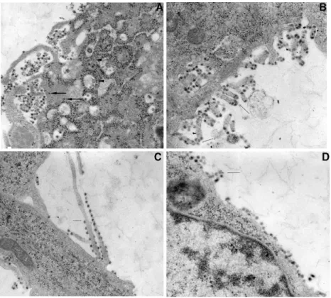
Figure 1. Cryo-EM reconstruction of a Sindbis virus particle. The inner most capsid shell of the virus
particle is shown in blue and the outer most E1/E2 glycoprotein shell is shown in yellow. The host derived lipid bilayer is sandwiched between the layers and is shown in red. The 5 fold axes of rotation are shown in the middle figure located in the bottom row of images. (Image provided by Dr. Angel Parades, Baylor College of Medicine).
virus. The spikes were reveled to be formed predominantly by E2 (27,31) with little E1 exposed. E2 has also been demonstrated to possess the major neutralizing antigenic sites for Sindbis virus by the ability of anti-E2 antibodies to inhibit virus binding to cells (3,4). Using anti-idiotypic antibodies directed against E2-specific antibodies (5,8), serial passage (6,8), antibody treatments combined with Electron Microscopy (EM) and image reconstruction analysis, several attempts have been made to identify the cell receptor which is recognized by E2 on target host cells (7,8). Despite these intense efforts, identification of the specific
Alphavirus receptor(s) has remained enigmatic. This process is complicated by the ability of
Sindbis and related viruses to evolve into lab adapted strains as well as interact transiently with host factors at the plasma membrane (9). Due to these difficulties, candidate receptors such as Heparin Sulfate, DC-SIGNS, and Laminin proteins have all been suggested as receptors utilized by Sindbis. However, none have proven to be the definitive partner for E2. The ability of this virus to infect a broad range of dissimilar host organisms suggests that the interaction between E2 and the receptor(s) is malleable or non-specific. In addition, it’s also been hypothesized that E2 recognizes and attaches to a receptor family. This hypothesis is extremely intriguing but also very difficult to prove nonetheless (1).
Once receptor recognition has occurred, genome delivery is the second major step in Sindbis virus infection. The mechanism of delivery of the Alphavirus genome to a host cell
remains the most debated aspect of the Togaviridae lifecycle (7,9,32). To date, infection by
enveloped viruses is proposed to require fusion of the viral envelope with a cell membrane after virus-receptor interaction (33). Infection by the membrane containing Influenza virus is the most well described virus entry mechanism requiring low pH mediated fusion (34). This (-) sense RNA containing virus is comprised of a host derived lipid-bilayer and three integral membrane proteins: hemagglutinin (HA), neuraminidase (NA) and M2. Underlying the host derived lipid membrane is a matrix layer composed of M1 proteins. In the Influenza fusion model, the virus infects cells via a multi-step process with the primary step being the internalization of the virus via receptor-mediated endocytosis. The internalized viruses are trafficked along the endocytic pathway to late endosomes where exposure to low pH triggers protein rearrangements resulting in the HA-catalyzed fusion between the viral and endosomal membranes and subsequent release of the ribonucleoprotein complexes (vRNPs) into the cytoplasm. The released complexes are later imported into the nucleus for viral gene expression and replication (34). Several lines of analyses have described a fusion function for Sindbis virus and produced data consistent with several models for the delivery of the
Alphavirus genome. Thus, the role of low pH and disulfide bond shuffling in the entry
process of Sindbis virus have been investigated and deemed necessary for infection (32,33,35).
Figure 2. Translation of the structural proteins of SIN, and the assembly of new virus particles.
A direct comparison between SV, SFV, TBE and Influenza is often drawn with regard to the mechanisms involved in genome delivery (1,37). Like Influenza, Alphaviruses are proposed
to undergo fusion of the viral membrane to cellular membranes within acidic endosomes after receptor mediated cellular uptake of virus particles. Once inside endosomes, the acidic pH is proposed to trigger drastic conformational rearrangements of the structural proteins exposing the putative type-2 fusion peptide located in E1 (aa 79-98) (1,33,37). The ability to fuse Alphaviruses to liposomes and host cell membranes in tissue culture are the primary
pieces of evidence put forth to argue that membrane fusion is involved in the infection process. In addition, the use of drugs that block endosome acidification and thus block viral RNA delivery and synthesis have also been utilized as evidence to support the receptor mediated-membrane fusion model for genome delivery.
Although accurate in many ways, there are some experimental inconsistencies in this infection model. Unlike Influenza, Alphaviruses are not pleomorphic membrane containing
structures with associated fusion proteins. They are comprised of very rigid T = 4 icosahedral shells that are interconnected and sandwich the lipid bilayer needed for fusion (32). For SV and other members of the Alphavirus genus to utilize the Influenza model, the
inner and outer T = 4 icosahedral shells formed by the viral structural proteins would need to disassemble and reassemble. This would result in a novel set of lateral associations between these structural proteins in order to fulfill all the requirements demanded by the Influenza model. An alternate model to Sindbis virus infection by low pH mediated fusion has been put forth and this model proposes that the delivery of the viral genome involves a mechanism independent of receptor mediated endocytosis.
Sindbis virus has been proposed to attach to the plasma membrane of the host cell and form a proteinaceous pore through which the viral genome is delivered (32). The data presented by the authors to support this model suggest a process of direct penetration at the plasma membrane of the host independent of acid pH, disassembly of the icosahedral protein shells, or virus-host membrane fusion. In this model, receptor binding induces conformational changes and results in the rearrangement of the viral glycoproteins and the exposure of critical domains needed for the formation of a pore structure. Cryo-EM reconstruction analysis of pH 5.3 treated virus revealed the presence of a novel protruding structure at the 5-fold rotational axis of the particle induced by the low pH treatment. This proteinaceous structure is suggested to be comprised primarily of E1 proteins. In conjunction with the host membrane bilayer, this protruding structure is proposed to form a pore structure. It was further suggested that conformational changes induced by E2 binding to host cell receptors might result in a similar rearrangement of the virus glycoproteins and ultimately the formation of the pore structure through which the RNA genome of the virus is delivered directly to the cytoplasm of the host. Additionally, quantitative virus infectivity assays conducted at 40C and 150C (conditions
know to prevent the formation of endosomes) resulted in the production of normal virus titers (32).
In the low pH membrane fusion model of Sindbis virus infection, the question of
Figure 3. A schematic diagram of the Sindbis virus RNA replication and translation processes.
nucleocapsid breakdown remains unresolved. As a part any proposed infection model for
Alphaviruses, nucleocapsid uncoating and host cytoplasmic genome exposure is required in
order to translate the viral genome. It has been proposed that Sindbis nucleocapsids interact with the cytoplasmic ribosomes of the host and this interaction serves at the trigger for the uncoating process with capsid protein residues 94 to 106 suggested to be the site of interaction (10). The proposed penetration model would alleviate several issues related to genome uncoating and answer numerous questions which the low pH fusion model cannot. Nonetheless, the penetration model dismisses a tremendous body of work that supports the low pH fusion model and that in itself leads to several questions that the penetration model cannot answer. Taken together, these data would suggest that Sindbis virus genome introduction into a host does not require membrane fusion and is a complicated process with several intriguing questions that need to be resolved.
1.3 Nonstructural proteins-nsP1 to nsP4
The (+) polarity single stranded RNA genome of Sindbis (11,703 bp) is methyl capped at the 5’end and contains a poly-A tail at the 3’ end (11,13). The genome encodes four nonstructural proteins (nsP1-ns4) in addition to three structural proteins. Upon delivery, the 49S genomic viral RNA serves as the template for direct translation of the nonstructural proteins. These four nonstructural proteins are located at the 5’ two-thirds of the genome and are numbered according to their order in the genome (Fig 2). These proteins are translated as polyprotein precursors nsP123 or nsP1234 with the latter being generated in lower abundance by read-through of an opal codon between P3 and P4 (11). The polyproteins undergo a stepwise series of cleavages producing individualized functional nsPs. The nucleotide sequence encoding nsP1 is 1620 base pairs in length and is translated to a protein of 540 residues. This 60kDa protein contains a methyltransferase domain, guanylyltransferase activity (12,13,14) and has been implicated in the association of the replicase complex to membranes (15). The 90kDa, 807 residues nsP2 protein has been demonstrated to play in
integral role in the early lifecycle of the virus. This protein has been shown to contain NTP-binding ability, helicase motifs (16,17), as well as the protease activity that cleaves the nsP123 and nsP1234 polyproteins (18). nsP2 has also been shown to be involved in the regulation of (-)strand RNA synthesis (19) and in the initiation of subgenomic 26S RNA synthesis (20). The phosphoprotein nsP3 is 549 residues long and has no know function in the Alphavirus lifecycle in mammalian cells. Identified as the viral polymerase (21), nsP4 is a
616 residue protein with a molecular weight of 70kDa. Taken together, these nonstructural proteins along with unknown cellular factors are directly responsible for the replication of the (+)strand genomic RNA, the (-)strand RNA and the 26S subgenomic mRNA.
1.4 Structural proteins-Polyprotein precursor synthesis and processing
The 26S subgenomic RNA promoter is a conserved sequence located between the nonstructural and structural genes (22) and overlaps nsP4. This promoter serves as the binding site for the polymerase complex that transcribes the 26S subgenomic mRNA which in turn serves at the template for the translation of the structural proteins (Fig.2). The structural proteins are synthesized as a polyprotein precursor (NH2-capsid-PE2 (E3-E2)-6K-E1-COOH) with the primary processing step resulting in the release of capsid by an
autoproteolitic activity (18). This results in the generation of a polyprotein intermediate composed of NH2-PE2 (E3-E2)-6K-E1-COOH. The capsid protein is 264 residues long and encapsidates the RNA genome of the virus to form nucleocapsids. Once translated, a serine protease activity associated with the carboxyl terminus of the capsid protein is utilized to release it from the remaining polyprotein (23, 24). This cleavage results in the inactivation of this serine protease because of the presence of the C-terminal tryptophan residue in the active site (25). Once the capsid protein is cleaved, the signal recognition particle (SRP) recognizes and binds to the amino-terminus of PE2 and comprises the early translation complex. The binding of the SRP results in a temporary halt of translation and initiates translocation of the complex to the membranes of the Endoplasmic Reticulum (ER). In the ER, the SRP is removed and insertion of the polyprotein into the ER membrane proceeds as translation of the polyprotein is completed, and processing of the polyprotein begins. While still residing in the ER, subsequent processing by signalase releases 6K from PE2 and E1, allowing the dimerization of E1 and PE2 and subsequent trimerization of these dimers. Also, within the ER lumen, mannose groups are added en bloc to the PE2 and E1.E1 is glycosylated at amino acids 139 and 245 while PE2 is glycosylated at positions 196 and 318. Various functions have been attributed to the carbohydrates on E1 and E2 (1). These modifications have been shown to be critical to several functional aspects including increasing the solubility of the proteins as well as possible involvement in receptor recognition.
During transport of the homotrimers from the ER, PE2 is processed by the Trans Golgi Network (TGN) endoprotease furin which releases E3 from E2. In addition, further modifications of the mannose groups also occur in the Golgi apparatus. At some unknown location within the exocytic pathway the endodomain of E2 is withdrawn from the membrane, exposing this structure to the cytoplasm and permitting association of the nucleocapsid to the virus protein modified membrane. After recognition of the E2 endodomain by the nucleocapsid, envelopment of the virus particle is initiated and
protein-protein associations drive the budding process. Once envelopment is completed, a mature virus particle is released from the plasma membrane of the host.
1.5 Nucleocapsid assembly and budding
As discussed above, E1 associates with PE2 in the ER to form heterodimers that oligomerize into homotrimeric complexes prior to being exported through the secretory pathway to the plasma membrane (35) with this resulting in the formation of a viral protein modified host plasma membrane. After the autoproteolitic release of capsid into the cell cytoplasm, genomic RNA and capsid self-assemble into nucleocapsid structures. Nucleocapsids are formed from newly synthesized capsid proteins in the cytoplasm of the host binding to one or more encapsidation sequences located within the nsP1 sequence (from nucleotide 945 to 1076) (25). Sequential association of capsid proteins with viral RNA sequences ultimately results in the formation of a nucleocapsid core. The nucleocapsid associates with the 33 amino acid endodomain tail of E2 in the homotrimeric complexes at the plasma membrane and this interaction is believed to initiate the envelopment and budding processes. The budding particles will exit the plasma membrane of the host containing exactly 240 copies of each of the transmembrane glycoproteins and capsid protein in a 1:1:1 stocheometric ratio (1). Analysis of the lipid composition associated with budding particles have revealed a membrane consisting of approximately 25% sphingomyelin, 27% phosphatidylcholine, 19% phosphatidylserine, and 26% phosphatidylethanolamine (28) with a cholesterol:phospholipid ratio of approximately 1:1 (29, 30).
The exact mechanism responsible for the initiation of envelopment and budding remains furtive. However, in this thesis evidence will be provided that shows that insertions of truncated E1 and E2 proteins into developing virions arrest these processes at different steps along the pathway.
1.6 E1 and E2 transmembrane glycoproteins
In a mature virion there are 240 copies of the glycoproteins E2 and E1 arranged as trimers of heterodimers. Upon translation, E1 is found in a relaxed, extended conformation referred to as E1α (38). A series of disulfide-bridge folding intermediates are formed as the
E1 protein matures to a final, metastable, energy-rich conformation referred to as E1ε. E1ε is found in the homotrimers which are exported from the ER to the TGN (35). In the TGN, the PE2 precursor is processed to E2 by the endoprotease furin and E1 is converted to a metastable high energy protein. The endoprotease furin is a member of the PACE enzyme
superfamily (Paired basic Amino acid Cleaving Enzyme) (39). Furin is a calcium dependent subtilisin-like Kex-2 analog endoprotease that carries out post-translational modifications within vesicular membranes of the secretory pathways of mammalians cells and is also found in insect cells. Furin cleaves PE2 at the conserved amino acid sequence Arg-X-Arg/Lys-Arg to E2 and E3. The E3 protein is not found in mature SV particles.
E1 is a glycoprotein that spans the membrane with its carboxy-terminal region exposing two basic amino acids on the internal side of the plasma membrane. The ectodomain of the E1 protein has been proposed to be folded into an elongated structure consisting almost exclusively of antiparallel beta-sheets and composed of two disulfide-bridge constrained domains; a functional domain (amino acids 1-129) and structural domain (amino acids 130-436) separated at amino acid 129 (40) based on structural analysis and on sensitivity to reducing agent (37,41). These two domains contain intramolecular disulfide-bridges involving 12 cysteine residues which stabilize the protein into an energy-rich conformation. This energy may be used to breach the cell membrane of the host to initiate genome delivery. The mechanism by which cell penetration takes place is unclear. There is data supporting the model that penetration takes place by membrane fusion in acidic endosomes (33). Other data however suggests penetration at the cell surface by formation of a proteinaceous pore in the absence of membrane fusion (42.
The disulfide-bridges formed by the cysteine residues in the E1 ectodomain are important for proper structure and function of the virus. In previous studies it was shown that the disulfide-bridges that contribute to Sindbis infection are reduced upon exposing purified SV to the mild reducing agent dithiothreitol (DTT) for a short period of time (41). This brief exposure did not affect the structural integrity of the virus but drastically reduced infectivity. Prolonged exposure of the virus to the reducing agent did result in complete loss of infectivity and near complete loss of structural integrity. These results were verified by electron microscopy (EM) and plaque assays and the data suggests that the disulfide-bridges essential for infection are readily accessible to the reducing agent while disulfide-bridges responsible for maintaining the three-dimensional structure of the virus are less accessible (41). Detergent extraction from mature virions followed by proteolytic removal of the transmembrane domain has allowed crystallization of the E1 glycoprotein ectodomain and
has yielded a structure at atomic resolution (37). The extraction protocol utilized exposed the E1 protein to conditions that have been previously demonstrated to cause rearrangement of the disulfide–bridges resulting in the formation of numerous low energy non native conformations (38). It is therefore possible that the crystal structure produced is one of a non-native low energy form of the protein. In other experiments the ectodomain of the Dengue virus E protein (the Flavivirus equivalent of the Alphavirus E1 protein) has been crystallized after expression in a baculovirus system as a truncated protein without the membrane-anchoring domain (43). This produced a structure similar to the detergent extracted E1 protein. This protocol makes the assumption that a membrane spanning protein will fold correctly when not associated with a membrane and also assumes that the protein will fold into a native conformation in the absence of other virus proteins. There is current evidence that suggests that these assumptions may not be entirely correct and that membranes play an important role in determining how proteins fold (44).
Structural protein-protein interactions are required to generate the three-dimensional surface of the virus and this necessitates E1 and E2 to interact specifically. E1-E1 protein interactions serve to stabilize the protruding spikes seen on the surface of the virus and compose the skirt region surrounding the base of the spike (27,31). These E1 interactions are crucial for the formation of the three-dimensional icosahedral surface of a mature metastable virus particle (31). One function of the E2 protein within the infectious particle includes docking of the virus to the host receptor. Previous analyses of the spike complexes seen adorning the surface of the virus using various approaches reveled these to be formed predominantly by E2 (27,31). One approach, using mutants which disrupted cleavage of the naturally occurring furin site between E3 and E2 in the precursor PE2 were shown to have additional electron densities lying at the periphery of the spike complex which were absent in wild type virus (27). Additional data from our lab using a site specific biotinylation labeling strategy also indicate that E2 it is more exposed than E1 in a mature particle (36). These data support previous studies demonstrating the spike to be predominantly composed of E2 with little E1 exposed. E2 has also been shown to posses the major neutralizing antigenic sites for Sindbis virus which underscores the utmost importance of full characterization of all
structural and functional aspects of this protein. These data are required for a complete understanding of Sindbis virus assembly and infection.
1.7 E1 and E2 on the surface of a mature virion
In order to generate an accurate protein map of an Alphavirus surface, a trace of the
E1 protein (from SFV) derived from an electron density map (3.5Å resolution) was carried out and the authors described E1 interactions at the surface of the virus (37). It was surmised that the Flavivirus E and Alphavirus E1 proteins evolved from a common ancestor. This was
rationalized in part by the observed similarities between the placements of the fusion peptides within crystallized proteins from each genus. The data generated showed both E and E1 to contain three domains (I-III), each with a secondary structure consisting almost
exclusively of antiparallel β-sheets. Domain II, the dimerization domain, was shown to
contain the putative fusion peptide, while domain I serves as the centralized eight-stranded β barrel domain. The carboxy-terminal domain III, is an IgG-like domain that contains the major neutralizing sites for the virus and as well as a connecting loop leading directly to the transmembrane segment of E and E1. On the surface of the virion, the placement of E1 subunits displayed overall similarities to the placement of the E protein. E proteins are suggested to dimerize at domain II with the globular domain III at opposite ends of the dimer and the putative fusion peptides buried within the dimer interface. The E1 placements are dissimilar from the E placements in that the E1 fusion peptides are exposed and make contacts with E2. In this model, E1 makes up a majority of the three-dimensional flat surface of the virus particle with domains I and III forming the skirt region around the base of the protruding spike structures. This implies that the 80 protruding spike structures seen on the surface of the virions are composed primarily of a globularly shaped E2 with some contribution from domain III of E1. This model also suggests that the main difference between E1 and E is founded in the dual function of E (receptor recognition and fusion) versus the singular function of E2 (receptor recognition).
The structural data generated from these studies are in agreement with much of the Cryo-EM data available for Alpha and Flaviviruses. However, other credible data (see above)
suggest that the extraction methods utilized to obtain the proteins crystallized can result in the rearrangement of the disulfide bridges present in E1. This could account for the
remarkable similarities found between the SFV E1 and SV E1 proteins. Overall, a critical understanding of the precise location of the glycoproteins on the surface of Alphaviruses is
needed for a more accurate model of the protein-protein associations required for assembly of infectious particles and elucidation of the mechanism of infection. From a pharmacological perspective, a precise structure is necessary for the development of anti-viral therapeutic agents for this group of viruses.
1.8 Objectives and Summary of this thesis
Mutational analysis of the viral glycoproteins has greatly increased our understanding of many of the mechanisms involved in the processes of assembly and budding of the
Alphavirus Sindbis. In the studies described in this thesis, we attempt to shed some light on
the debatable native conformations of the Sindbis virus glycoproteins, the effects of truncated proteins on virus assembly and budding as well as the identification of specific amino acids on the surface of intact virions. The First Chapter of this thesis deals with the general
characteristics of the virus. Information on the Alphavirus genus, virus entry, genome
replication and budding is provided.
In Chapter 2, a detailed description of the preparative purification of PCR products
by microbore anion-exchange by HPLC using polyethyleneimine is described. This method is invaluable for the purification and separation of nanoscale amounts of DNA for mutagenic purposes. Furthermore, the role and relevance of this method in the overall cloning process is discussed and evidence is provided for its usefulness. Overall, this method provides a novel and alternate way of separating and purifying nanoscale amounts of DNA for further genetic manipulation and other purposes.
In Chapter 3, in depth details of the in nivo processing and characterization of furin
protease sensitive Alphavirus mutants are presented. The effects of installing the R-X-R/K-R
conserved furin recognition motif in E1 on virus assembly and budding is discussed. Additionally, the production and effects of truncated E1 proteins is discussed.
In Chapter 4, site specific labeling and structural characterization of the E2
glycoprotein from Sindbis by lysine biotinylation and LC-MS/MS is discussed. These studies expanded our understanding of Sindbis virus amino acids exposed on the surface of intact
infectious virus particles. The precise orientation of specific lysine residues is demonstrated and their importance on virus infectivity discussed.
In Chapter 5, the preliminary studies in Chapter 3 are expanded and the effects of
installed furin protease sensitive sites on Sindbis virus E2 glycoprotein processing and virus assembly are characterized. These studies proved valuable in understating the complexities associated with processing of these sites contained in the protein E2 as compared to those expressed in E1. Taken together, these studies point to the advantages and difficulties associated with using this methodology for the production of truncated proteins.
In Chapter 6, the results and conclusions of this thesis are summarized and
discussed. Furthermore, future directions are also discussed and future experiments proposed.
References
1. Strauss, J. H., and E. G. Strauss. 1994. The Alphaviruses: gene expression, replication, and evolution. Microbiol. Rev. 58:491-562.
3. Byrnes, A. P., and D. E. Griffin. 1998. Binding of Sindbis virus to cell surface heparin sulfate. J. Virol. 72:7349-7356.
4. Ludwig, G. V., J. P. Kondig, and J. F. Smith. 1996. A putative receptor for Venuzuelan equine encephalitis virus from mosquito cells. J. Virol. 70:5592-5599.
5. Strauss JH, Wang KS, Schmaljohn AL, Kuhn RJ, Strauss EG. Host-cell receptors for Sindbis virus. Arch Virol Suppl. 1994;9:473-84.
6. Jolanda M. Smit, Barry-Lee Waarts, Koji Kimata, William B. Klimstra, Robert Bittman, and Jan Wilschut Adaptation of Alphaviruses to Heparan Sulfate: Interaction of Sindbis and Semliki Forest Viruses with Liposomes Containing Lipid-Conjugated Heparin.
7 . Ubol, S., and D. E. Griffin. 1991. Identification of a putative Alphavirus receptor on mouse neural cells. J. Virol. 65:6913-6921.
8. Wang, K., A. L. Schmaljohn, R. J. Kuhn, and J. H. Strauss. 1991. Antiidiotypic antibodies as probes for the Sindbis virus receptor. Virology 181:694-702.
9. Klimstra, W. B., K. D. Ryman, and R. E. Johnston. 1998. Adaptation of Sindbis virus to BHK-21 cells selects for use of heparan sulfate as an attachment receptor. J. Virol. 72:7357-7366.
10. Wengler, G., D. Würkner, and G. Wengler. 1992. Identification of a sequence element in the Alphavirus core protein which mediates interaction of cores with ribosomes and the disassembly of cores. Virology 191:880-888.
11. Lopez S, Bell JR, Strauss EG, Strauss JH. The nonstructural proteins of Sindbis virus as studied with an antibody specific for the C terminus of the nonstructural read through polyprotein. Virology. 1985 Mar;141(2):235-47.
12. Rozanov MN, Koonin EV, Gorbalenya AE. Conservation of the putative methyltransferase domain: a hallmark of the 'Sindbis-like' supergroup of positive-strand RNA viruses. J Gen Virol. 1992 Aug;73 ( Pt 8):2129-34.
13. Wang HL, O'Rear J, Stollar V. Mutagenesis of the Sindbis virus nsP1 protein: effects on methyltransferase activity and viral infectivity. Virology. 1996 Mar 15;217(2):527-31.
14. Scheidel LM, Durbin RK, Stollar V. Sindbis virus mutants resistant to mycophenolic acid and ribavirin. Virology. 1987 May;158(1):1-7.
15. Ahola, T., A. Lampio, P. Auvinen, and L. Kaarianen. 1999. Semliki Forest virus mRNA capping enzyme requires association with anionic membrane phospholipids for activity. EMBO J. 18:3164-3172.
16. Lain S, Riechmann JL, Martin MT, Garcia JA. Homologous potyvirus and flavivirus proteins belonging to a superfamily of helicase-like proteins. Gene. 1989 Oct 30;82(2):357-62.
17. Company, et al., 1991
18. Shirako Y, Strauss JH. Regulation of Sindbis virus RNA replication: uncleaved P123 and nsP4 function in minus-strand RNA synthesis, whereas cleaved
products from P123 are required for efficient plus-strand RNA synthesis. J Virol. 1994 Mar;68(3):1874-85.
19. Sawicki, D. L., and S. G. Sawicki. 1993. A second nonstructural protein functions in the regulation of alphavirus negative-strand RNA synthesis. J. Virol. 67:3605-3610.
20. Suopanki, J., D. L. Sawicki, S. G. Sawicki, and L. Kaarianen. 1998. Regulation of Alphavirus 26S mRNA transcription by replicase component nsP2. J. Gen. Virol. 79:309-319.
21. Sawicki, D. L., D. B. Barkhimer, S. G. Sawicki, C. M. Rice, and S. Schlesinger. 1990. Temperature sensitive shut-off of Alphavirus minus strand RNA synthesis maps to a nonstructural protein nsP4. Virology 174:43-52.
22. Ou JH, Strauss EG, Strauss JH. Comparative studies of the 3'-terminal sequences of several alpha virus RNAs. Virology. 1981 Mar;109(2):281-9.
23. Barth, B. U., M. Suomalianen, P. Liljeström, and H. Garoff. 1992. Alphavirus assembly and entry: role of the cytoplasmic tail of the E1 spike subunit. J. Virol. 71:7857-7865.
24. Liljeström, P., and H. Garoff. 1991. Internally located cleavable signal sequences direct the formation of Semliki Forest virus membrane proteins from a polyprotein precursor. J. Virol.65:147-154. 24 Chapter 1
25. Choi, H. K., L. Tong, W. Minor, P. Dumas, U. Boege, M. G. Rossmann, and G. Wengler. 1991. Structure of Sindbis virus core protein reveals a chymotrypsin-like serine proteinase and the organization of the virion. Nature 354:37-43.
26. Cheng, R. H., R. J. Kuhn, N. H. Olson, M. G. Rossmann, H. K. Choi, T. J. Smith, and T. S. Baker. 1995. Nucleocapsid and glycoprotein organization in an enveloped virus. Cell 80:621-630.
27. Paredes, A. M., H. W. Heidner, P. Thuman-commike, B. V. Venkararam Prasad, R. E. Johnston and W. Chiu. 1998. Structural localization of the E3 glycoprotein in attenuated Sindbis virus mutants. J. Virol. 72:1534-1541.
28. Allan, D., and P. Quinn. 1989. Membrane phospholipid asymmetry in Semliki Forest virus grown in BHK cells. Biochim. et Biophys. Acta 987:199-204.
29. Laine, R., H. Soderlund, and O. Renkonen. 1973. Chemical composition of Semliki Forest
30. Renkonen, O., L. Kaarainen, K. Simons, and C. G. Gahmberg. 1971. The lipid class composition of Semliki Forest virus and plasma membranes of the host cells. Virology 46:318-326.
31. Paredes, A. M., Brown, D. T., Rothnagel, R., Chiu, W., Schoepp, R. J., Johnston, R. E., and Prasad, B. V. (1993). Three-dimensional structure of a membrane-containing virus. Proc Natl Acad Sci U S A90(19), 9095-9.
32. Paredes, A. M., Ferreira, D., Horton, M., Saad, A., Tsuruta, H., Johnston, R., Klimstra, W., Ryman, K., Hernandez, R., Chiu, W., and Brown, D. T. (2004). Conformational changes in Sindbis virions resulting from exposure to low pH and interactions with cells suggest that cell penetration may occur at the cell surface in the absence of membrane fusion. Virology324(2), 373-86.
33. Kielian, M. (1995). Membrane fusion and the alphavirus life cycle. Adv Virus Res
45, 113-51.
34. Lakadamyali M, Rust MJ, Zhuang X. Endocytosis of influenza viruses. Microbes Infect. 2004 Aug;6(10):929-36. Review.
35. Carleton, M., Lee, H., Mulvey, M., and Brown, D. T. (1997). Role of glycoprotein PE2 in formation and maturation of the Sindbis virus spike. J Virol 71(2),
1558-66.
36. Sharp et al October 2005 Submitted to Virology.
37. Lescar J, Roussel A, Wien MW, Navaza J, Fuller SD, Wengler G, Wengler G, Rey FA. The Fusion glycoprotein shell of Semliki Forest virus: an icosahedral assembly primed for fusogenic activation at endosomal pH. Cell. 2001 Apr 6;105(1):137-48.
38. Mulvey, M., and Brown, D. T. (1994). Formation and rearrangement of disulfide bonds during maturation of the Sindbis virus E1 glycoprotein. J Virol68(2),
805-12.
39. Moehring, J. M., Inocencio, N. M., Robertson, B. J., and Moehring, T. J. (1993). Expression of mouse furin in a Chinese hamster cell resistant to Pseudomonas exotoxin A and viruses complements the genetic lesion. J Biol Chem 268(4),
2590-4.
40. Phinney, B. S., and Brown, D. T. (2000). Sindbis virus glycoprotein E1 is divided into two discrete domains at amino acid 129 by disulfide bridge connections. J Virol74(19), 9313-6.
41. Anthony, R. P., Paredes, A. M., and Brown, D. T. (1992). Disulfide bonds are essential for the stability of the Sindbis virus envelope. Virology190(1), 330-6.
42. Paredes, A. M., Ferreira, D., Horton, M., Saad, A., Tsuruta, H., Johnston, R., Klimstra, W., Ryman, K., Hernandez, R., Chiu, W., and Brown, D. T. (2004). Conformational changes in Sindbis virions resulting from exposure to low pH and interactions with cells suggest that cell penetration may occur at the cell surface in the absence of membrane fusion. Virology324(2), 373-86.
43. Modis, Y., Ogata, S., Clements, D., and Harrison, S. C. (2004). Structure of the dengue virus envelope protein after membrane fusion. Nature427(6972), 313-9.
44. Tamm LK, Hong H, Liang B. Folding and assembly of beta-barrel membrane proteins.Biochim Biophys Acta. 2004 Nov 3;1666(1-2):250-63. Review.
CHAPTER 2
Rapid preparative purification of West Nile and Sindbis virus
PCR products utilizing a microbore anion-exchange column
Raquel Hernandez a,∗, Steevenson Nelson a, Jeffery R. Salma,
Dennis T. Browna, Andrew J. Alpert b
a Department of Molecular and Structural Biochemistry, North Carolina State University, Campus Box 7622, Raleigh, NC 27695 7622, USA
b PolyLC Inc, 9151 Rumsey Road, Ste. 180, Columbia, MD 21045, USA
Received 22 December 2003; received in revised form 19 April 2004; accepted 20 April 2004 Available online 19 June 2004
19
Journal of Virological Methods 120 (2004) 141–149
Rapid preparative purification of West Nile and Sindbis virus PCR
products utilizing a microbore anion-exchange column
Raquel Hernandeza,∗, Steevenson Nelsona, Jeffery R. Salma, Dennis T. Browna, Andrew J. Alpertb
aDepartment of Molecular and Structural Biochemistry, North Carolina State University, Campus Box 7622, Raleigh, NC 27695 7622, USA bPolyLC Inc, 9151 Rumsey Road, Ste. 180, Columbia, MD 21045, USA
Received 22 December 2003; received in revised form 19 April 2004; accepted 20 April 2004
Available online 19 June 2004
Abstract
Analysis and purification of specific PCR products from PCR reactions can be problematic due to several issues relating to amplification and low product yield. The use of HPLC as a preparative tool in PCR product analysis is common but has not replaced traditional electrophoretic techniques for purifying DNA to be used in subsequent experiments. Gel purification of PCR products can result in a net loss greater than 50% of the starting DNA amount. Thus, this method of recovery can become the limiting factor in the overall cloning protocol. This paper describes a simple and relatively inexpensive micro-preparative HPLC method to purify and analyze nM quantities of DNA. A microbore polyethyleneimine-based anion-exchange column fractionates PCR mixtures in less than 40 min with a recovery of the purified specific product as high as 80%, thus eliminating the need for gel purification. Using this method, nested PCR products from Sindbis virus differing by 18 bp in some cases and a 277 bp fragment from West Nile virus were resolved and quantified. This method differs from existing methodologies because separation is based on size and charge as well as the overall G+C content of the PCR product.
© 2004 Elsevier B.V. All rights reserved.
Keywords: Sindbis virus; West Nile virus; PCR products; Preparative anion-exchange HPLC purification
1. Introduction
As a prototype of the Alphavirus, Sindbis virus (SV) is used as a model system to study assembly of icosahedral membrane containing viruses. Sindbis virus is encoded by a positive sense single stranded RNA genome of 11 703 nt
enclosed in highly symmetrical nested T = 4 icosahedral
protein shells of both virus and host composition (Hernandez et al., 2003). The geometric organization of Sindbis virus proteins has underscored the value of this model in the study of virus architecture (Ferreira et al., 2003; Pletnev et al., 2001; Strauss and Strauss, 2001).
West Nile virus (WNV) belongs to the distantly re-lated Flaviviruses, which contain many arthropod-borne human pathogens. West Nile virus strains are encoded by a positive sense single stranded RNA genome of
approxi-∗Corresponding author. Tel.:+1-919-515-5765;
fax:+1-919-515-2047.
E-mail address: raquel [email protected] (R. Hernandez).
mately 10 000 nt. This group of viruses causes significant encephalitic, hemorrhagic and fibrile diseases in humans and economically important domestic animals (Strauss and
Strauss, 2002; Strauss and Strauss, 1994). While other
members of the Flavivirus genus have traditionally been the focus of worldwide epidemiological surveillance, West Nile virus has recently been established in the USA as a significant emerging disease for which no licensed human vaccine is available (Campbell et al., 2002). For these rea-sons, WNV has come into higher focus in the research community (Pletnev et al., 2002).
Genetic manipulation of viruses, and other organisms has been revolutionized by the increasingly sophisticated ap-plications and variations of the polymerase chain reaction (PCR) (Coen, 1987; Lanciotti et al., 2000; Mackay et al., 2002). Amplification of G+C rich genomes such as those which occur in the Alpha and Flaviviruses (Jenkins et al., 2001) has been facilitated as well but continues to represent a significant challenge for specific genes in these related RNA virus families.
0166-0934/$ – see front matter © 2004 Elsevier B.V. All rights reserved. doi:10.1016/j.jviromet.2004.04.013
20
R. Hernandez et al. / Journal of Virological Methods 120 (2004) 141–149
The use of PCR has evolved from the original concept of using E. coli DNA polymerase I in multiple cycles
of DNA amplification (Kleppe et al., 1971; Templeton,
1992), to modern applications using a wide variety of ther-mophilic polymerases (Cline et al., 1996). The simplicity and versatility of this technique makes it the method of choice to manipulate DNA, and it has been successfully adapted for automated purposes such as cycle sequencing (Daniels, 1996). Many variations of the standard PCR re-action are in use routinely with new applications described with increasing frequency (Lanciotti et al., 2000). Sufficient PCR product recovery however can be limited by inefficient
amplification of stable G+C rich sequences compounded
by poor product recovery from agarose gels.
The majority of PCR reactions produce large amounts of the specific DNA of interest with relatively minor side reaction products. However, some DNA templates are not amplified efficiently and produce a large mixture of prod-ucts or a low specific product yield. These templates tend to display significant secondary structure and thermal stability (Mathews et al., 2000). Theoretical product yields from these difficult templates are often not achieved and mixtures of specific and nonspecific products are obtained. This prob-lem is a function of the specific template sequence and the specific primer pair sequences that are required. These fac-tors complicate efficient product production and require op-timization of each of the PCR segments. In addition, adjust-ment of parameters such as segadjust-ment time, ramp rate, segadjust-ment temperature, ionic strength, and component concentrations can become necessary for optimal productivity (Saiki, 1989). Often, after extensive optimization of all these required pa-rameters, specific product yield still remains quite low. For applications which require the specific amplified product to be applied for uses other than in end-point analysis, the purification method used can become the limiting factor (Mackay et al., 2002). PCR products are routinely purified
by agarose gel electrophoresis (Shaw-Bruha and Lamb,
2000). Agarose gel electrophoresis simultaneously allows sizing and separation of the desired product(s) from the tem-plate DNA, nonspecific products, primers, dNTPs and other contaminants. The DNA band of interest is excised from the gel and can be extracted by a variety of methods (Vogelstein and Gillespie, 1979). Under the best of circumstances, a recovery of about 50% for many DNA fragments can be ex-pected. This recovery can require up to 2 days in preparatory time and often proves insufficient for many further cloning applications.
HPLC is a reliable and powerful alternative method to agarose gels for nucleic acid purification. Both reversed-phase (Huber, 1998; Oefner et al., 1992; Wong et al., 2000) and anion-exchange (Huber, 1998; Kato et al., 1988, 1989; Katz and Dong, 1990; Katz et al., 1990; Maa et al., 1990) HPLC have been used to resolve mixtures of dsDNA frag-ments. HPLC in tandem with other sophisticated instrumen-tation (LC/MS) (Lin et al., 2002) has been applied to the iso-lation and analysis of complex mixtures of DNA products.
Micropreparative DNA isolation methods using com-mercially available columns for less than g quantities of DNA are lacking. In this study, we assess the use of an anion-exchange column on a miniaturized scale suitable for analysis and purification of PCR products. Separation uti-lizing this method is simple and relies on the size, charge,
and overall G +C content of the product. The goal of
the present work is to develop a reproducible alternative method for the purification and concentration of specific DNA products without the need for preparative agarose gel electrophoresis. Our methodology is proposed as a simple alternative method that allows ng quantities of DNA to be analyzed, purified and recovered for further experiments. The advantages of this technique are illustrated by our abil-ity to separate PCR products of nested sequences 247 and 229 bp in size into concentrated fractions ready for further molecular cloning.
2. Materials and method
2.1. PCR amplifications, PCR reaction parameters and instrumentation
PCR reactions were run using five different template plasmid DNAs. The first set of reactions shown inFigs. 1–3
were done using Sindbis virus TM 25 (a deletion mutant) containing a 3 bp deletion (nt9761 to nt9764) in the E2 gly-coprotein gene of the cDNA (Hernandez et al., 2003). The wild-type plasmid is 13 703 bp in length and encodes a full length viral cDNA used in the in vitro production of infec-tious viral RNA (Rice et al., 1987). The mutant DNAs shown inFig. 4also contain additional deletions in the E2 glyco-protein gene of the cDNA. TM 10 contains a 48 bp deletion (nt9734 to nt9782), TM 16 contains a 30 bp deletion (nt9743 to nt9773) while the parental plasmid cDNA which contains no deletion produces a product 277 bp in length (Hernandez et al., 2003).
The second type of mutant template was derived from a plasmid which encodes a 2219 bp insert containing the two structural membrane proteins of West Nile virus strain 385-99 [nt934 (BamHI)–nt3142 (NotI)] in the cDNA 3.1 expression vector (Stratagene, La Jolla, CA), a generous gift of Alan Barrett, UT Medical Branch, Galveston, TX. All plasmid DNAs are isolated by standard purification protocols (Maniatis et al., 1982) and purified through ce-sium chloride gradients. The purified DNA concentration is estimated from 1% agarose gels containing DNA mass standards. Each PCR reaction contains 40 ng of the spe-cific template DNA in PCR buffer (10 mM Tris–HCl [pH
8.3], 50 mM KCl, 5 mM MgCl2), 200M each
deoxynu-cleotide triphosphate (dNTP) (NEB, Beverly, MA), and 600 ng of each of the sense and antisense primers. For each of the Sindbis wild type and deletion TM mutants
the sense primer was 5 GCA TTA CTA CCA TCG CCA
TCC 3, the antisense primer was 5 CAA AGG TAT
R. Hernandez et al. / Journal of Virological Methods 120 (2004) 141–149
Fig. 1. Electrophoretic analysis of ethidium bromide-stained agarose gels of template DNA (A) and PCR product mixtures (B and C). (A) Sindbis virus plasmid template DNA (cDNA), TM 25 analyzed on 1% agarose gels. Lane 1 shows the total mass in ng above the closed arrow for each DNA fragment. Lane 2 contains a 1 Kb ladder with an open headed arrow designating the largest fragment of 10 Kb. Lane 3 (1l∼50 ng total) and lane 4 (5l∼250 ng total) reveal a small amount of RNA contamination in the larger aliquot. In (B) is shown the 4% agarose gel analysis of the test PCR reaction mixes. Lane 1 is the same as in (A). Lane 2 contains a 123 bp (bottom arrow) ladder used to size DNA products in higher concentration gels. Lane 3 displays the products of a PCR reaction without template DNA. No specific product is seen although a small primer generated product appears at the bottom of the gel. No products are generated in the absence of primers (lane 4) and specific products are only generated from complete reaction mixtures (lanes 5 and 6). Lane 6 is essentially the same PCR reaction as lane 5, labeled with [␣-32P] dATP. An autoradiogram of (B) lane 6 is displayed in (C) lane 1 showing the 274 bp product and minor products (mp).
0 50 100 150 200 250
0 10 20 30 40 50 60
Time (min) Abs260 0 50 100 150 200 250 300 350
0 10 20 30 40 50 60 Time (min) As260b 0 50 100 150 200 250
0 10 20 30 40 50 60 Time (min)
Abs260
(A) (B) (C)
11 10 9 8 7 6 5 4 3 2 1 7 6 5 4 3 2 1 9 8 7 5 4 3 2 1 9 8
Fig. 2. Chromatograms of the test PCR mixtures and the complete PCR reactions. Shown in (A) is the chromatogram produced from the PEI column of a PCR reaction for Sindbis virus without DNA or primers. As detailed in Method 1, 20l of the PCR reaction was added to 20l of mobile phase A and loaded onto the column. PCR reactions fromFig. 1Blanes 3 and 4 containing only template or primers produced the profile in (B). The chromatogram displayed is representative for both of these PCR mixtures. A representative chromatogram is shown in (C) for the PCR reaction composed of the complete reagent mix or the reaction with the [␣-32P] dATP tracer (Fig. 1Blanes 5 and 6). Comparison of the control reactions in (A) and (B) with the complete PCR reaction in (C) identified peak 11 as the reaction product. Fraction 10 is a minor product (compare toFig. 3Bfraction 10).
R. Hernandez et al. / Journal of Virological Methods 120 (2004) 141–149
Fig. 3. Peak fraction analysis of the [␣-32P] labeled PCR reaction. In (A), agarose gel electrophoresis through 4% agarose of the fractions collected from the separation described inFig. 2Cshows that no DNA products are detected by ethidium bromide stain prior to peaks 10 and 11. An autoradiogram,
Fig. 3Bof the gel shown in (A) further demonstrates that no absorbance peaks observed inFig. 2Care the result of DNA not detected by staining.
Fig. 4. Chromatographic separation of PCR products generated by a mixture of mutant template DNAs. In (B) is shown a 4% agarose gel of the PCR mixture before and after LC. Lane 2 contains 5l of the reaction mixture. The bottom band in this lane is a product of 229 bp, the middle band is 247 bp and the top band is 277 bp (arrows). As seen on the gel fractions 9 and 10 contain minor products. Fraction 11 displays a large absorbance but only a smear of nonspecific products is detected. Fractions 12 (33 min) and 13 (35–37 min) show the specific products. Fraction 12 contains the smallest of these DNA products.
GCA CAA CTG G 3. The primer set for the WNV
tem-plate was for the sense strand, 5 GGA TTG CTG GGG
TGG ATG GGC 3 and antisense 5 GAC TCG AGC
GGC CGC CTC AGT G 3 (MWG, High Point, NC).
The final component in all PCR reactions was 0.5l (2.5
units) Taq DNA polymerase (Applied Biosystems, Fos-ter City, CA) in a final volume of 100l. The reactions were boiled and slow cooled to just below the lowest TM of the primer sets prior to adding the Taq polymerase (Hernandez et al., 2000).
R. Hernandez et al. / Journal of Virological Methods 120 (2004) 141–149
The PCR profile for the Sindbis constructs consisted of three segments, 95◦C for 1 min, 55◦C for 2 min, 72◦C for 2 min. This cycle was repeated 30 times and was followed by an additional extension of 8 min at 72◦C, 4◦hold. Radi-olabeled PCR reactions were essentially as described above with the addition of 10Ci (1l) [␣-32P] dATP (Amersham, Piscataway, NJ) in the final mix. The PCR profile for the WNV construct is significantly more intricate due to the high GC content (Jenkins et al., 2001) and secondary structure of these virus genomes and the large amounts of nonspe-cific products generated (seeSection 3). After initial boiling and annealing of the primer pairs, as described above, Taq polymerase was added and the PCR program initiated. Seg-ment 1 (melting): 92◦–95◦(0.06◦C/s), soak at 95◦for 1 s, ramp 95◦–62◦(0.56◦C/s), and soak at 62◦for 1 s. Segment 2 (annealing): ramp from 62◦C to 60◦C (0.04◦/s), soak at 60◦ for 35 s, ramp from 60◦ to 70◦ (0.15◦C/s) and soak at 70◦ for 1 s. Segment 3 (elongation): ramp from 70◦ to 72◦ (0.05◦C/s) and soak at 72◦ for 120 s. This combined cycle is repeated 30 times and followed by an 8 min soak at 72◦C, and 4◦ hold. All PCR reactions were performed using a Thermo Hybaid Multi Block System 0.5 ml stan-dard block thermocycler (Thermo IEC, Needham Heights, MA) with ramping profiles programmed as suggested by the manufacturer.
2.2. DNA purification
Template plasmid DNAs that are used in the PCR reac-tions are purified by isopycnic centrifugation through cesium chloride gradients. Most of the proteins are removed from the DNA by this procedure although some RNA contami-nation remains (seeFig. 1Alane 4). The quality and purity of the template DNA is determined by agarose gel elector-phoresis prior to use in the PCR reaction. Primer oligomers are used directly as supplied by the manufacturer, and are not HPLC purified. The remaining PCR mixture components are as specified inSection 2and are also not HPLC grade.
All DNA analysis was done using agarose gel elec-trophoresis in TAE (40 mM Tris–HCl, pH 7.5, 20 mM acetic acid, 2 mM EDTA) buffer at 10–15 V/Cm and subsequently
stained with 0.2g/ml ethidium bromide. Plasmid DNA
purity and concentration was determined on 1% agarose (Invitrogen, Carlsbad, CA) by comparison to DNA mass standards (Invitrogen, Carlsbad, CA).
2.3. HPLC instrumentation and chromatogram analysis
The HPLC system utilized consisted of a Hewlett Packard 1100 series liquid chromatograph, variable wavelength de-tector and a Rheodyne 7725i injection valve. A PolyWAX LP
50 mM×1 mM i.d. microbore column (item# 051WX0315
PolyLC Inc, Columbia, MD) was connected directly to the injector by 0.005 in. i.d. PEEK tubing (Upchurch Scientific, Oak Harbor, WA). The PolyWAX LP column was packed with 3m silica of 1500 Å pore diameter with an adsorbed,
crosslinked coating of linear polyethyleneimine (PEI). Sam-ple absorbance was measured at 260 nm.
Method 1. This method was used for the Sindbis virus TM
25 and the WNV separations. The PolyWAX LP column was equilibrated in buffer A (20 mM Na2HPO4, 25 mM NaClO4,
5% acetonitrile pH 7.5) at a flow rate of 0.045 ml/min. A
20l aliquot of crude PCR reaction was brought up to
40l in buffer A and loaded onto the column. After 5 min, the injection loop was closed and a gradient with buffer B (20 mM Na2HPO4, 1 M NaClO4, 5% acetonitrile pH 7.5)
was used to elute the adsorbed PCR fragments from the
column; 0–5 min [0%B → 0%B], 5.01–10 min [0%B →
55%B], 10.01–65 min [55%B → 68%B]. After the
gradi-ent, the mobile phase was changed to 100% buffer B and the flow rate was changed to 0.08 ml/min. The column was washed for 15 min with pure buffer B followed by a 15 min wash with buffer A.
Method 2. This method was used for separations
in-volving the Sindbis virus TM mutant mixed PCR prod-ucts. For this separation, the gradient profile was slightly modified and the rest of the run remained unchanged:
0–5 min [0%B → 0%B], 5.01–10 min [0%B → 55%B],
10.01–65 min [55%B→63%B]. Under the gradient
condi-tions described above, the template DNA is not eluted from the column necessitating a cleanup program after every 10th run prior to preequilibration. This program consists of a 20 min wash with 100 mM triethylamine phosphate (TEAP) solution pH 3.0 at 0.08 ml/min at room temperature
followed by a 20 min wash with H2O, and a 20 min wash
in 100% buffer B, prior to regeneration of the column for 30 min with 100% buffer A.
All samples were collected manually peak to peak di-rectly into the Microcon YM spin dialysis columns. Fraction
volumes varied from 50 to 100l. A complete
chromato-graphic separation using these specific parameters can be accomplished in about 50 min.
3. Results
3.1. Template DNA and PCR product analysis
Shown inFig. 1are ethidium bromide-stained 1% agarose gels used for DNA analysis.Fig. 1Alane 1 shows the mass markers (closed arrows) labeled with the corresponding mass in ng. Fluorescence intensity of the DNA bands of interest is compared to these standards to estimate the quantity of DNA in a specific band.Fig. 1Alanes 3 and 4 are of 1 and 5l Sindbis virus template TM 25 DNA, representing 50 and 250 ng of total DNA respectively. PCR product analysis was
done using a mixture of 3% NuSieveTM (FMC, Rockland,
ME) and 1% agarose essentially as described for the template DNA above. This is depicted inFig. 1B. Lane 1 inFig. 1B
shows the same mass marker as in lane 1Fig. 1A.Fig. 1B
lane 2 is a 123 bp ladder. Lane 3 of the same gel is of a 10l fraction of a PCR reaction done in the absence of DNA

![Fig. 3. Peak fraction analysis of the [ ␣- 32 P] labeled PCR reaction. In (A), agarose gel electrophoresis through 4% agarose of the fractions collected from the separation described in Fig](https://thumb-us.123doks.com/thumbv2/123dok_us/1268817.1159446/34.1263.296.987.143.644/fraction-analysis-reaction-electrophoresis-fractions-collected-separation-described.webp)