37
Medical sciences
normaliZation of occlusion in patiEnts
with incrEasEd dEntal abrasion
1balakhnichev d.n., 1subbotin r.s., 1fischev s.b., 2lepilin a.v., 3sevastyanov a.v., 1orlova i.v.
1Saint Petersburg State Pediatric Medical University, Saint Petersburg, e-mail: [email protected];
2V.I. Razumovsky Saratov State Medical University, Saratov; 3Saint Petersburg State Pediatric Medical University, Saint Petersburg
a number of diseases, including increased abrasion of natural teeth, cause spatial changes in the masticatory-speech apparatus, which affect the masticatory muscles and tmJ, which act within their activity and rest, and going beyond this framework entails disorders with decompensation of the supporting apparatus of the teeth. the aggra-vating factor may be the parafunctions of the masticatory muscles, TMJ diseases and as a consequence structural changes. therefore, it is extremely important to determine the parameters of changes in the position of the elements of the tmJ from the value of normalization of the height of the gnatic part of the face. in clinical dentistry often patients with various pathologies of the chewing-the vocal apparatus, a feature of which is reducing the height of gnatichesky part of the person patients. in the clinic of prosthetic dentistry temporomandibular joint given great importance. it is known that any dental intervention in the maxillofacial region in varying degrees, affect the tempo-romandibular joint. we have created a computer program to determine the optimal height of gnatichesky part of the person at patients with various forms of increased abrasion of teeth. imaging of the temporomandibular joints (tmJ) allows to obtain correct display of the true state of the elements of the temporomandibular joint, intra-articular as well as their interactions and patterns of change with increasing height of gnathic part of the patients with increased dental abrasion. the proposed computer program allows to simulate the correct position of the elements of the tem-poromandibular joints taking into account the normalization of the height of gnathic part of the person at patients with different clinical forms of increased abrasion of teeth.
Keywords: orthodontia, orthopedia, increased dental abrasion
in clinical practice a dentist often encoun-ters patients with different pathologies of the chewing- vocal apparatus, which are mani-fested by reduced height of gnathic part of the face as one of characteristic features of impair-ment. Quite a number among these are patients with increased dental abrasion: from 11.8 % to 42.6 % of cases [3, 4, 8, 12, 17, 18].
the cases of increased abrasion of teeth are likely to be caused by various reasons: mor-phological inferiority of hard tissues of teeth, overloading of teeth, chemical exposure, occu-pational hazards, functional status of the mas-ticatory muscles and temporomandibular joints (tmJ) and others [2, 3, 4, 5, 8, 9, 10, 11, 15].
in the practice of prosthetic dentistry tem-poromandibular joint is of great importance. it is known that any dental intervention in the maxillofacial region affects the tmJ to a less or greater extent [2, 4, 12, 13, 14, 16].
most researchers recognize the relation-ship between the structure of the tmJ and type of occlusion [1, 4, 5, 8, 14].
Currently existing classification of in-creased abrasion do not meet the systemati-zation of the clinical manifestations of this disease [3, 5, 10, 11, 12, 15]. if the horizontal form of abrasion is clinically determined re-liably, vertical and mixed as well as all other forms distinguish from each other only condi-tionally. therefore, we singed out alongside the horizontal form vertical and vertically
distal-mesial ones, depending on the type of bite and position of elements of the temporomandibu-lar joints. gnathic part of the person’face is a variable structure of the craniofacial complex. the most susceptible to change are vertical pa-rameters, which is associated with anatomical-physiological characteristics of growth and development of the head (the change of teeth, anomaly of occlusion, loss of teeth, increased dental abrasion, etc.). increase in the height of gnathic part of the person’s face can lead to a change in the tone of masticatory muscles and cause dysfunction of temporomandibular joints [4, 5, 8, 10, 14, 16].
The purpose of the work
To study the efficacy of treatment predic-tion of patients with vertically-distal form of increased abrasion of teeth according to the po-sition of the elements of the temporomandibu-lar joints with the use of computer programmes and on the basis of their findings – assessing the quality of dental prosthetics.
Materials and methods of research
38
Medical sciences
of the face, while in case of compensated form reduced gnathic part of the face was not ob-served (or only mild reduction) due to vacant (false, substitution, reversible) hypertrophy of bone tissue of alveolar ridges of the upper and lower jaws. all patients noted various defects of dentition, but less than 6 teeth antagonists were not observed.
the distribution of patients with psZ by age and sex are presented in table 1.
Table 1
age 31–40 41–50 51–60 61 and older
women 7 8 13 3
men 8 9 12 1
total 15 17 25 4
roentgenologic teleroentgenogram analy-sis was performed by means of a computer program [19], which allows to identify anthro-pometric points, to build a cephalometric plane to roentgenogrammetry and formulate the pre-liminary diagnosis. in the study we used one angular dimension (angle n – ss – spm) and six linear (sna' – Kme'; sn'- Kme'; gox; goy; cox ; coy) (fig. 1).
we proposed a method for studying to-mograms of temporomandibular joints on the bases of the issues including the measurement
of the sagittal size of the glenoid fossa of the line drawn from the lower edge of the articular tubercle to the lower edge of the external audi-tory canal, measuring the width of the lower jaw head, the angle of inclination of the slope of the articular tubercle, the dimension of the joint space in front, upper front, upper rear and rear parts due to the depth of the temporoman-dibular fossa and the configuration of the lower jaw head (fig. 2).
the proposed calculation formula: ∆h = K x [(DR1 + DR2 + DR3 + DR4 +
+ dl1 + dl2 + dl3 + dl4):8 – d’] where ∆h is the height to which it is necessary to increase he gnathic part of the face.
K – the experimentally derived factor for creased abrasion horizontal form – 11,4; for in-creased abrasion vertically distal shape – 6,7; for increased abrasion vertically mesial form – 13,8;
d1, d2, d3, d4 is the width of the joint space in the front, front upper, rear upper and rear parts of the tmJ, respectively.
r – tmJ-right, l – left tmJ.
d’ is the experimentally derived value of the average values of the joint space by four parameters: for increased abrasion horizontal form – 2,8 mm for increased abrasion vertical-ly distal form – 2.2 mm; for increased abrasion vertically mesial form – 3.1 mm.
Fig. 1. Analysis of the profile telerentgenogram: X – Frankfurt plane (horizontal), held between the points po (porion) and or (orbitale); Y – the vertical, held between the points (sellion) and go (gonion); me – menton; co – condilon; n – nasion; sn' – cutaneous projection sn (subnasale); ss – subspinale; snp – spina nasalis posterior; sna' – skin projection point sna (spina nasalis anterior); Kme' – skin
39
Medical sciences
The technique allowed us to enforce valid settings in the topography of elements of the temporomandibular joint at different degrees of change of the position of the man-dible in orthopedic treatment with the aim of normalizing its position. for this purpose, a computer program for determining the opti-mal height of gnathic part of the face in pa-tients with various forms of increased abra-sion of teeth (the Certificate on registration of software № 2015619515 Software com-plex to determine the optimal height of the bite in patients with increased dental abra-sion (tmJ2015) [6] date of state registra-tion in the register of computer programs 04 september 2015 was worked out. passport data of a patient are inserted in a computer program, followed by a tomogram of the pa-tient’s temporomandibular joints (right and left) made before treatment. finally, refer-ence points are set and program after calcu-lation determines the amount by which it is necessary to increase the height of the gnath-ic part of the patient’s face.
Quality of orthopedic treatment was tested by the tone of actually masticatory muscles. as the process of reduction of the gnathic part of the person’s face takes decades, the chewing muscles get used to this particular isometric
height. with the normalization of the occlu-sion and increasing the height of the bite, the muscle tone of the rest increases, and the tone of the muscle tension decreases. with prop-erly determined height of gnathic part of the face, the tone parameters of calm and tension come back to the original state in for about two weeks.
myotonometry was performed using myo-tonometry sZirma (mEtrimpEx, hun-gary). myotonometry scale is calibrated in grams. myotonometry dipstick was placed to the motor points on the right and left of actu-ally chewing muscles and the tone of the rest (tp) and the tone voltage (vt) was measured.
for accurate location of the motor points during re-examination of the masticatory muscles we used our proposed method (patent for invention “Method of evaluating the qual-ity of prosthetics” no. 2617229 registered in the state register of inventions on april 24, 2017) [7].
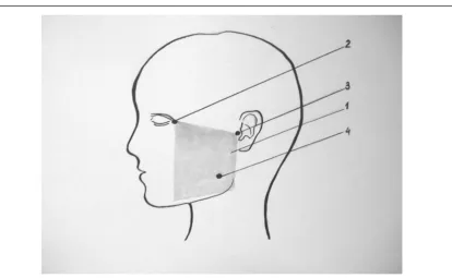
for this primary survey on the lateral sur-face of the patient’s sur-face the transparent plate (x-ray or thick plastic film) was imposed strict-ly to the landmarks: the middle of the tragus of the ear and the outer edge (corner) of the orbit. motor point marked by the dye was transferred on the transparent plate (fig. 3).
Fig. 2. Tomogram of the temporomandibular joint. A – the top of the articular tubercle; B – the lower edge of the external auditory canal; d – diameter of the head of the lower jaw; D1 – is the width of the joint space in the anterior; D2 – width of the joint space in the anterior-upper part; D3 – is the width of the joint space at the posterior e -upper part; D4 – is the width of the joint space in the
40
Medical sciences
fig. 3. diagram of motor points of the actual chewing muscles to hold myotonometry: where, 1 is a
transparent plate; 2 – the lateral edge of the orbit; 3 – the tragus of the ear; 4 – motor point of
actually chewing muscles
During the second study we put the film, respectively to benchmarks and through the hole marked motor point on the skin of the pa-tient. this method allowed to carry out identi-cal measurements.
Results of research and their discussion
To illustrate the efficiency of complex treatment here is an extract from a case history no. 102 of patient p. 68 years with complaints of difficulty in chewing and aesthetic disad-vantage (fig. 4).
the distal position of the mandible is de-termined by photostations. the results showed that the height of the upper part of the face (n – sn) did not meet the lower face (sn – gn). the severity of nasolabial folds indicates a decline in the height of the gnathic part.
during the examination of the oral cavity of the patient a decrease in the height of gnathic part, the presence of defects of dentition on the upper and lower jaws, cross-bite were objec-tively noted. tomography of the temporoman-dibular joints determined the distal position of the head of mandible, which allowed to make up a diagnosis of decompensated vertically distal form of increased abrasion of the front teeth of the upper jaw (fig. 5).
during the examination decrease in the height of the gnathic part, moderately
droop-ing corners of the mouth, intensive pattern of the nasolabial and chin folds was revealed. in the mouth cavity: increased abrasion of ante-rior and posteante-rior teeth of the upper jaw, the 2nd degree decompensated form, the upper and lower jaws included bilateral defects of denti-tion in lateral parts.
the patient underwent teleroentgeno-graphic study and imaging of the temporoman-dibular joints.
measurement of parameters of the teeth and dental arches was carried out on plaster models of the jaws and directly in the mouth cavity. occlusal relationships were evaluated, which showed non-physiological distribution of contact points, their asymmetry. in addition, the patient demonstrated the violation of the occlusal plane.
41
Medical sciences
А B
Fig. 4. Face photos patient P. 68 years before treatment. A – profile, B – in front
fig. 5. the ratio of the dentition of the patient p., 68 years
42
Medical sciences
x-ray examination of the temporoman-dibular joints identified the impairment of the normal topographical relations of elements of this articulation. the articular heads of the mandible on the left and on the right were shifted back, the expansion of the joint space in the anterior and narrowing in the back dpts was noted. the angle of the slope of the artic-ular tubercle, the size of which differed from the normal values on both sides was of impor-tance (fig. 7).
according to the computer program “software complex for determining the op-timal height of the bite in patients with creased dental abrasion (tmJ2015)” the in-crease in height of gnathic part 4.6 mm was recommended.
the patient was proposed a preliminary or-thopedic treatment of uncoupling the bite plate with the increase of vertical dimension 4.6 mm, to normalize position of the heads of the lower jaw shifted forward by 1.5 mm.
А B
fig. 7. tomogram of the temporomandibular joints patient p. 68 years before treatment. a – right joint, b – joint left
fig. 8. preparation of the oral cavity of the patient p. 68 years for prosthetics
43
Medical sciences
fig. 10. face photos of the patient p. 6 years after orthopedic treatment
44
Medical sciences
two weeks later, the tone of the masticatory muscles of the patient returned to normal, and after 3 months (after preparation of the abut-ment teeth) the final prosthetic metal-ceramic bridge prostheses with increase of the gnathic part of the person 4.6 mm was performed.
the effectiveness of the mastication in-creased from 20.1 + 0.7 percent (preliminary phase) to 78.2 + 1,2 % after orthopedic treat-ment. time of chewing: before treatment – 52.9 + 1,4 sec. after the orthopedic treatment – 23.4 + 0.5 sec. (figure. 8, 9, 10, 11, 12, 13).
a number of diseases, including high at-trition of natural teeth cause spatial variations in the chewing- vocal apparatus that affect the chewing muscles and tmJ, which are in their scope of activity and rest, and going beyond these limits leads to disorders followed by
de-compensation of supporting apparatus of the teeth. an aggravating factor can be parafunc-tion of chewing muscles, diseases of the tmJ and as a consequence some structural changes. It is therefore essential to define the parameters of changes in the position of the elements of tmJ from the value of the normalization of the height of gnathic part of the person’s face.
in orthopedic treatment of such patients the primary is clinical method, based on the doctor’s experience, accompanied by labora-tory methods, in particular the twg research and imaging of the tmJ that help to clarify medical tactics, previously developed during the clinical examination of patients. long-term results have confirmed the effectiveness of its construction in orthopedic treatment of such patients.
А В
Fig. 12. Side telerentgenogram of patient P. 68 years. A – pre-treatment. B – after the final orthopedic
treatment
А В
45
Medical sciences
imaging of the tmJ allows to obtain a cor-rect mapping of the true state of tmJ elements and their intra relationships and to clarify the features and regularities of changes during the process of increase of the height of gnathic part of the person’s face.
Conclusions
according to the results obtained studies, you can determine the amount of dissociation of dentition in patients with increased dental abrasion, taking into account the possibility of staged or simultaneous orthopedic treatment.
the proposed computer program allows to simulate the correct position of the ele-ments of the temporomandibular joints taking into account the normalization of the height of gnatichesky part of the person at patients with different clinical forms of increased abrasion of teeth.
the proposed method of assessing the quality of prosthetics allows to check the cor-rectness of the chosen tactics of orthopedic treatment of patients with increased dental abrasion.
The author confirms that the submitted data does not contain conflict of interests. GRATI-tudE the work was prepared with the sup-port of the ministry of education and science of the russian federation.
References
1. attin t. brushing abrasion of softaned and remineralised dentin: an in situ study / t. attin, s. siegel, w. bucalla // caries res. 2004. n. 38. p. 62-66.
2. azzopardi a. the measurement and prevention of ero-sion and abraero-sion / a. azzopardi, d.w. bartlett, t.f. watson // J. dent. 2001. – v. 29. – p. 395-400.
3. bondermarki l. Extraoral vs intraoral appliance for dis-tal Movement of Maxil1ary First Molars: А Randomized Con -trol1ed / 1. bondermarki, 1. Karlsson // angle orthodontist. – 2005. – № 5. – Р. 699–706.
4. de boever, J. a., adriaens p. a. occlusal relationship in patients with pain-dysfunction symptoms in the temporomandibu-lar joints. // J Oral Rehabil. – 1983. – Jan. – Vol. 10, № 1. – P. 1-7. 5. ilbuldu E., cakmak a., disci r., aydin r. comparison of laser, dry needling, and placebo laser treatments in myofascial pain syndrome. / // photomedicine and laser surgery. – 2004. – Aug. – Vol. 22, № 4. – P. 306-311.
6. fishchev b.s., lepilin a.v., sevastyanov a.v.,orlov v. i., balakhnichev d. n. the results of treatment of patients with defects of dentition in combination with cross bite with the use of computer modeling. // stomatology of children’s age and pre-vention. – 2015. – t. xiv. – no. 3 (46). – p. 55-58.
7. fishchev s.b., a.v. lepilin, d.n. balakhnichev et al. The Certificate of official registration program for computer no. 2007613744 // software complex to determine the opti-mal height of the bite in patients with increased dental abrasion (tmJ2015 test), state registered in the register of computer programs on 4 september 2015.
8. fishchev s.b., a.v. lepilin, d.n. balakhnichev et al. Method of assessing the quality of prosthetics / The patent for the meeting №2617229 Registered in the state to register of in -ventions on 24 april, 2017
9. fishchev b.s., lepilin a.v., sevastyanov a.v.,orlov v.i., balakhnichev d.n. the results of treatment of patients with defects of dentition in combination with cross bite with the use of computer modeling. // stomatology of children’s age and pre-vention. – 2015. – t. xiv. – no. 3 (46). – p. 55-58.
10. Jacobson А. Retrospective cephalometric investigation of the effects of soldered transpalatal arches оп the maxillary first molars during orthodontic treatment involving extraction of maxillary first bicuspids / А. Jacobson // American Joumal Of Orthodontics add Dentofacial Orthopedics. – 2006. – № 1. – Р. 81.
11. Katsoulis J., nikitovic s. g., spreng s. (et al.). pros-thetic rehabilitation and treatment outcome of partially edentu-lous pftients with severe tooth wear: 3-years results // J. dent. – 2011. – Vol. 39, № 10. – P. 662-71.
12. latino c. support of undermined occlusal enamel provided by restorative materials / c. latino, K. troendle, J.b. summit // Quintessence int. 2001. – vol. 32. – p. 287-292.
13. leinfelder K.f. analysis of adhesive systems of the 5th generation / F.K. Leinfelder// Стоматология. 2001. – № 3. – Р. 23-24.
14. leloup g. meta-analytical review of factors involved in dentin adherence / g. leloup, w. d’hoore, d. bouter // J. dent. Res. 2001. – V. 80. – № 7. – P.1605-1614.
15. Mercado J. Jefferson skeletal classification system (Jscs) and how it helps in extraction and non-extraction orth-odontic cases. // Int. J. Orthod. Milwaukee., 2007. – № 18(4). – Р. 31-34.
16. mok t.b. dental erosion: in vitro mode of wine asses-sor’s erosion // austral. dent. J. 2001. – n. 46(4). – p. 263-268.
17. Proffit W.R., Fields H.W. Contemporary Orthodontics, 4rd Edition. mosby. – 2007. – 751 p.
18. pullinger a.g., seligman d.a. multifactorial analysis of differences in temporomandibular joint hard tissue anatomic relationships between disk displacement with and without re-duction in women. / J. of prosthetic dentistry, 2001, V. 86, № 4, p. 407-419.