In Depth Study for Developing Efficient Direct
Somatic Embryogenesis of Jatropha Curcas L.:
Morphology and Ultrastructure
Totik Sri Mariani
1), Mirna Zuirda
1), Hiroshi Miyake
2)1)
School of Life Sciences and Technology, Bandung Institute of Technology, Gan esha 10, Bandung 40132, Indonesia
2)
Graduate School of Bioagricultural Sciences, Nagoya University, Nagoya 464-8601, Japan
Corresponding author : Totik Sri Mariani. Fax number : 62-22-7271569
E-mail : [email protected]
Abstract-- This is the first report on direct somatic
embryogenesis of Jatropha curcas L. For in depth study, morphological observation was performed by inverted
microscopy whereas ultrastructural observation was
conducted by transmission electron microscopy. The somatic
embryos derived from single cells, which divided, developed
into embryo proper with suspensor, ECS (Embryogenic cell
suspension), proembryo, globular embryo, heart and torpedo
stage embryo.The shoot and plantlet developed when the ECS
was plated on embryo induction medium I, embryo induction
medium II and embryo germination medium, consecutively.
The highest germination percentage was 63.3% on
IG4-gamborg medium supplemented with 3 ppm GA3 and 1
ppm IAA. The highest shoot development was on IG4-gamborg
(60%) and the highest plantlet development was on
IG4-glutamin (6.7%). The reason of low percentage of plantlet
development was revealed by observing the ultrastructural
features of the embryogenic cells and the somatic embryos.
Character of the embryogenic cells and somatic embryo was
forming deposit material in the vacuole. The deposit material
inhibited cellular metabolism of the cells.
Index Term-- Jatropha curcas, direct somatic embryogenesis in liquid medium, deposit material
I. INT RODUCT ION
Current day a wareness of the depletion of tradit ional energy
resources has evoked an intense interest in alternative
sources of energy. This has influenced trends in plant
biotechnology, resulting in attempts to use cell and plant
tissue for imp rove ment and rapid propagation of plants
producing oil with fuel application possibility. Jatropha
curcas (L.) is one plant that has oil capable to substitute
engine oil (biodiesel) (Nigam et al., 2005).
The genus Jatropha belongs to Euphorbiaceae is one of
the promising drought tolerant perennial species. The plant
is very hardy and can tole rate high te mperature and salinity.
The seeds contain semi-drying o il, an effic ient substitutes
for diesel engines. (Bhasubutra and Sutiponpeibun, 1982;
Ra ina, 1987). Hundred thousand hectares of land in Nus a
Tenggara Barat and Nusa Tenggara Timu r, Indonesia will be
planted by Jatropha curcas. Therefore , rapid and mass
propagation of Jatropha curcas using somatic
embryogenesis method is needed.
Sardana et al. (2000) carried out plant
regeneration fro m so matic e mb ryo of Jatropha curcas.
They used solid media for so matic e mb ryogenesis. They
induced proembryos fro m green, co mpact, and slow
growing e mbryogenic callus although their frequency was
rather limited. Liquid mediu m has several advantages such
as save place, labour, t ime , cost of mediu m and materia l. In
20 ml of liquid mediu m, Mariani et al. (2004) yie lded ± 100
globular somatic e mb ryo of Lithospermum erythrorhizon.
Therefore, in 1 liter of liquid mediu m, we yie lded ± 5000
more somat ic e mb ryos were p roduced in liquid mediu m
than in solid mediu m. In addit ion, the quality of somatic
embryo in liquid media was also improved rather than in
solid med ia. (Gupta et al., 2003). Accordingly, in this
present study we reported direct somatic e mb ryogenesis
fro m single ce ll of Jatropha curcas in liquid media.
Moreover, for in depth study, we reported observation of the
somatic e mbryos of J. curcas by transmission electron
microscopy.
II. MAT ERIAL AND MET HODS
Material : Hypocotyl fro m germinating seed of Jatropha
curcas
Method :
1. Optimizat ion of e mb ryogenic callus induction in
Jatropha curcas
Seeds were germinated on mo istened tissue paper.
Hypocotyls from the germinating seeds were used as
e xplant for somatic e mbryogenesis. The hypocotyl was cut
and placed onto the Embryogenic Ca llus Induction Medium
(ECIM) to induce friable e mb ryogenic callus format ion.
ECIM consisted of Murashige & Skoog Basal mediu m
supplemented with co mbination of 2,4-D (9 x 10⁻⁶ M, 1.35 x
10-5 M, 1,8 x 10⁻⁵ M) and BAP (0 M, 4,4 x 10⁻⁶ M), 2,4-D (9
x 10-6 M, 1.35 x 10⁻⁵ M, 1,8 x 10⁻⁵ M) and Kinetin (0 M, 4,4
x 10-6 M), IAA (1,14 x 10⁻⁵ M, 1.71 x 10⁻⁵ M, 2,28 x 10⁻⁵ M)
and BAP (0 M, 5,7 x 10⁻⁶ M).
2. Cells suspension
The e mbryogenic callus was transferred to the ce ll
suspension medium (CSM ) to induce cells format ion. The
combination o f 2,4-D and kinetin concentration was based
on the best embryogenic ca llus induction mediu m. The best
embryogenic ca llus induction mediu m was 1,35 x 10⁻⁵ M
2,4-D and 4.4.10-6 M kinetin. Therefore , the co mposition
med iu m for CSM was 6,75 x 10⁻⁶ M 2,4-D and 4,4 x 10⁻⁷ M
Kinetin.
3. Embryogenic cell suspension
The cell suspension in CSM was transferred to
embryogenic ce ll suspension mediu m (ECSM ). The
composition of ECSM was MS med iu m supplemented with
1,35 x 10⁻⁵ M 2,4-D and 4,4 x 10⁻⁷ M Kinetin. In this
medium KNO3 was changed by 6 gL-1 K3citrat.
4. Plantlet development
The embryogenic cells was subcultured to embryo
induction mediu m I (EDMI) and e mbryo induction mediu m
II (EDM II) befo re transferred to e mbryo germination
med iu m (EGM). The composition of EDM I was half
strength macronutrient of MS, mic ronutrient of MS, 10 ppm
ascorbic acid, 20 pp m citric acid, 25 pp m adenine sulfat,
100 p m g lutamine, 2 pp m kinetin and 1,5 pp m IBA.The
composition of EDM II was the same as EDM I.
supplemented with 3 ppm IAA and 3 ppm BAP. The
composition of EGM was listed in Tabel 1.
TABLE I
PLANT GROWTH REGULATOR VARIATION IN EMBRYO GERMINATION MEDIUM
GA3
(ppm)
IAA
(ppm) 0
Gamborg Glutamin
2 3 2 3
0 control
1 IG3-gamborg IG4-gamborg IG3-glutamin IG4-glutamin
IG-gluta min mediu m consisted of ½ MS mac ronutrient+
MS micronutrient+ 10 pp m ascorbic acid+20 pp m cit ric
acid+25 pp m adenine sulphate+ 100 pp m gluta mine
vitamin. Five time repletion was performed on each
medium variation.
5. Transmission electron microscopy
Ce lls, e mbryogenic cells and the somatic e mbryos were
prefixed in 0.1 M cacodylate buffer pH 7.2 containing 5%
glutaraldehyde for 24 h, rinsed 3 t imes in the same buffe r,
postfixed in 2% os miu m tetro xide in cacodylate buffer for
12 h, rinsed in the same buffer once and distilled water
twice, and gradually dehydrated in an alcohol series, a ll at 4
degree C. The samp les then were in filtrated and e mbedded
with Spurr's resin at room temperature. The e mbedded
samples we re poly me rized in an oven at 70 degree C for 24
h. Ultrathin sections were made with an u ltra microto me at a
thickness of 70-90 n m. These sections were stained using
aquaeous 2% uranyl acetate for 30 min, and with lead
citrate for 10 min. Then, they were observed by TEM.
III. RESULT S AND DISCUSSION
1. Optimizat ion of e mb ryogenic callus induction in
Jatropha curcas
Fig. 1. Friable embryogenic callus of Jatropha curcas L.
I . MS Basal mediu m with the addit ion of 2,4-D and BAP
combination
II. M S Basal med iu m with the addition of 2,4-D and
Kinetin combination
2,4-D
(M)
Kin (M)
9 x 10⁻⁶ 1.35 x 10⁻⁵ 1,8 x 10⁻⁵
0
Ca llus (+),
White Yellow,
Friable (+),
Wet
Ca llus (+),
White Yellow,
Friable (+),
Wet
Ca llus (+),
White Yellow,
Friable (+),
Wet
4,4 x 10⁻⁶
Ca llus (++),
White Yellow,
Friable (++),
Wet
Ca llus (++),
White Yellow,
Friable (+++),
Wet
Ca llus (++),
White Yellow,
Friable (++),
Wet 2,4-D (M) BAP (M)
9 x 10⁻⁶ 1.35 x 10⁻⁵ 1,8 x 10⁻⁵
0 Ca llus(+),White
Yellow
Ca llus(+),White
Yellow
Ca llus(+),White
Yellow
4,4 x
III. MS Basal mediu m with the addition of IAA and BAP
combination
IAA
(M)
BAP
(M)
1,14 x 10⁻⁵ 1.71 x 10⁻⁵ 2,28 x 10⁻⁵
0
Ca llus(+),
White Ye llo w,
Dry, Root(+)
Ca llus(+),
White Ye llow,
Dry, Root(+)
Ca llus(+),
White Ye llow,
Dry, Root(+)
5,7 x
10-6
Ca llus(+),
White Ye llo w,
Dry, Root(+)
Ca llus(+),
White Ye llow,
Dry ,Root(+)
Ca llus(+),
White Ye llow,
Dry ,Root(+)
Note:
+
: a few
++
: moderate
+++ : abundant
Conclusion: The best embryogenic callus medium was
basal medium JC supplemented with 1,35 x 10
-5M
2,4-D and 4.4.10
-6M kinetin.
Hypocotyl explant cultured on callus induction mediu m
supplemented with comb ination of 2,4-D and BAP
produced compact white yellow non embryogenic callus.
This kind of callus was not suitable for cell suspension
initiat ion. Hypocotyl e xplant cultured on callus induction
med iu m supplemented with co mbination of IAA and BAP
produced rooted dry white yellow ca llus. This kind of ca llus
was also not suitable for ce ll suspension establishment.
White yellow, friable and wet e mbryogenic callus was
induced from hypocotyl exp lant on callus induction
med iu m supplemented with co mbination o f 2,4 -D and
kinetin. The best embryogenic callus was induced on callus
induction mediu m supplemented with 1.35. 10-5 M 2,4-D
and 4.4.10-6 M kinetin.
Cells in nodules at the surface of embryogenic
callus was undegoing high prolife ration cell and formed
me riste matic ce ll agregate, which have the potency became
embryogenic ce lls in the suitable med iu m. (Filho and
Hattori, 1997). Touchet et al (1991) found that nodular
embryogenic callus consisted of single embryogenic cells.
The aggregates were co mposed of typical meristematic cells
containing soluble proteins and sometimes starch granules.
They had a round prominent nucleus, and a dense
cytoplasm with s mall vacuoles. These embryogenic masses
were d ividing active ly. To check the e mbryogenicity of the
callus, it was inoculated into cell suspension medium.
2. Somatic embryogenesis and embryogenic cell
suspension
A
B
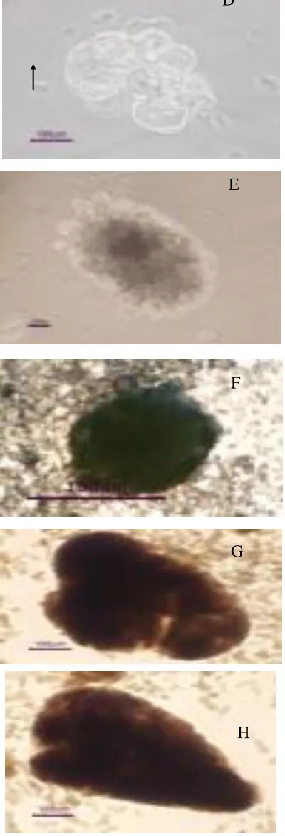
C
Fig. 2. A-H. Single cells (A); Dividing cells (B); Embryo proper with
suspensor (C); ECS (D); Proembryo (E); Globular somatic embryo (F);
Heart stage (G); Torpedo stage (H) of Jatropha curcas L. Note : S =
Suspensor, EP = Embryo proper
White yellow friab le e mbryogenic calli were inoculated into
cell suspension medium supplemented with 6,75 x 10⁻⁶ M
2,4-D and 4,4 x 10⁻⁷ M Kinetin. In this med iu m, single
embryogenic cells we re produced (Fig. 2 A). The single
embryogenic ce ll is round and small, has dense cytoplasm,
large nucleus, little vacuole and thin cell wa ll. This single
embryogenic ce ll d ivided within 5 days of culture as shown
in Fig. 2 B. Fig. 2 C. shows embryo proper with suspensor.
This e mbryo proper with suspensor often occurred in the
cell suspension mediu m 7 days of culture. The e xistence of
the suspensor gave the evidence that the embryo derived
fro m a single ce ll. Mariani et al. (2002) reported that at an
early stage, suspensors were observed on the elongated rice
somatic e mbryo, because the somatic e mb ryo was
unicellu lar origin. Un icellula r o rig in means that the somatic
embryo was derived fro m a single ce ll. Suspensor in
somatic e mbryo was also found in Phaseolus vulgaris
(Puspitawati 1997), Vigna radiata (Puspitawati, 1997;
Fitriani, 2002) Allium sativum (Nu rwendah, 2002),
Lithospermum erythrorhizon (Ramayanti, 2003; Marian i et
al., 2004), and Elaeis guineensis (Wardjo, 2006). Therea fter,
the embryo proper divided further and formed
embryogenic cells suspension (ECS ) after 10 days of
culture (Fig. 2 D.). The ECS grew into proembryo (Fig. 2
E) and globular somatic e mbryo (Fig. 2 F) a fter 14 days
of culture. Heart stage (Fig. 2 G) and torpedo stage somatic
embryo (Fig. 2 H) were developed in e mb ryo development
medium after 14 days of culture.
According to our result, the so matic e mbryo of
Jatropha curcas L. underwent direct somatic
embryogenesis derived fro m single ce ll. This is coincide
with Noerhadi (1988) that direct somatic e mbryogenesis
was the formation of e mbryos from single ce ll. This is
advantegous because it can reduce somaclonal variation. In
addition, this advantage will be important in applying the
somatic e mbryo in p lant genetic transformation (Mariani et
al., 2002).
After it was understood that the cells of Jatropha
curcas enable to undergo somatic e mbryogenesis,
D
E
G
embryogenic cell suspension (ECS) was established. The
ECS was established within 2 wee ks of culture in
embryogenic ce ll suspension mediu m (ECSM ) (Fig. 3). The
composition of ECSM was JC mediu m supplemented with
1,35 x 10⁻⁵ M 2,4-D and 4,4 x 10⁻⁷ M Kinetin. The
population of e mbryogenic cells in ECS was very uniform
(Fig. 4).
Fig. 3. Embryogenic cell suspension Jatropha curcas L.
Fig. 4. Population of embryogenic cells of Jatropha curcas L. under
inverted microscope
The ECS (Emb ryogenic cell suspension) is useful for
protoplast isolation, mic ropropagation and as materia l for
genetic transformation.
3. Plantlet development of Jatropha curcas L.
The plantlet developed fro m the e mb ryogenic cells of
Jatropha curcas L. after the ECS was cultured on embryo
induction mediu m I, e mb ryo induction med iu m II and
embryo germination mediu m, consecutively. Fig. 5.
showed the plantlet of Jatropha curcas L.
Fig. 5. Plantlet of Jatropha curcas on MS medium
supplemented with 3ppm GA3 and 1ppm IAA
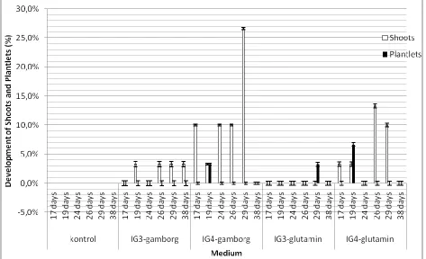
Germination of e mbryos we re showed in fig. 6. The highest
germination percentage was 63.3% on IG4-ga mborg
med iu m. There was no germination on control mediu m. In
germinated embryos, shoot and plantlet were still mixed.
Therefore, observation on shoots and plantlet were
performed and showed in fig. 7.
Fig. 6. Percentage of embryos germination on various embryo germination
medium
The highest shoot development was on IG
4-gamborg
(60%) and the highest plantlet development was on
IG
4-glutamin (6.7%). It indicated that 1 ppm IAA and
shoot and plantlet. Glutamin helped the development
of plantlet on IG3-glutamin and Ig4-glutamin media.
Purnamaningsih
(2002)
reported
that
glutamine enhanced the development of embryo into
torpedo and cotyledone stage. Others substances in
glutamine medium such as citric acid as potassium
chelating agent could increase somatic embryos
amount and ascorbic acid as antioxidant could convert
somatic embryos into plantlet (Herman, 2002).
Fig. 7. Development of shoots and plantlets on various embryo germination medium
4. Ultrastructural features
The percentage of plantlet development of
Jatropha curcas
L. was low (6.7%). Therefore, we
observed the ultrastructural features of the cells,
embryogenic cells and the somatic embryos by
transmission electron microscopy. Fig 8. shows the
ultrastructure features of cells suspension of
Jatropha
curcas
L. It was clear that the vacuole was clean. Fig.
9 showed the ultrastructural features of embryogenic
cells. It was prominent that the vacuoles contain many
deposit material. Fig. 10. shows the ultrastructural
features of somatic embryos. It indicated that the
deposit material these deposits seem to form inside the
Fig. 9. Ultratsructural features of the embryogenic cells of
Jatropha curcas L.
Fig. 10. Ultrastructural features of the somatic embryo of
Jatropha curcas L.
Based on the observation of the ultrastructural features of
the embryogenic cells and the somatic e mbryos, it was
understood why the percentage of plantlet develop ment was
low. It was due to the deposit material in the vacuoles. The
deposit material inhibited cellula r metabolis m of the cells.
Therefore, only 6.7% of p lantlet was developed fro m the
embryogenic cells of Jatropha curcas L.
CONCLUSION
In conclusion, this is the first report of direct somatic
embryogenesis of Jatropha curcas fro m single ce lls.
Somat ic e mbryos of J. curcas L. have been established in
suitable liquid mediu m. The somatic e mb ryos derived from
single cell. Th is is advantageous because it can reduce
somaclonal variation. In addit ion, this advantage will be
important in applying the somatic e mbryo in plant genetic
transformation. Plantlet of J . curcas developed fro m
embryogenic ce ll suspension plated in solid med iu m.
Character of the e mbryogenic ce lls and so matic e mbryo is
forming deposit material in the vacuole. It is suggested that
KNO3 should not be changed by 6 gL-1 K3citrat because it
formed deposit material in the vacuole of cell.
ACKNOWLEDGEMENT
Asahi Glass Foundation greatly facilitated this study and
are gratefully appreciated.
REFERENCES
[1] Nigam, N., Saxena, O.P and Braganza, V.. Trends in Plant
Tissue Culture and Biotechnology. 2005. Ed. Agrobios,
India.
[2] Basubutra,R. and Sutiponpeibum, S. Renewable energy. Review
Journal. 1982(4): 56-70.
[3] Sardana, J., Batra, A., Ali, D.J. Phytomorphology. 2000. 50
(3/4) 239-42.
[4] Mariani, T.S., Ramayanti, O., Yazaki, K. and H. Miyake.
Annales Bogorienses. 2004. 9(2):72-79.
[5] Gupta, P.K., Timmis, R. and Carlson W. Abstract of SIVB
conference. 2003.
[6] Filho, J.C.B and Hattori, K. R Bras Fisiol.Veg. 1997. 9 (3):
185-188.
[7] Touchet (de) B, Duval Y and Pannerier C. Plant Cell Reports.
[8] Mariani, T.S., Miyake, H. and Y. Takeoka. Jurnal Matematika
dan Sains. 2002. 7(2): 53-56
[9] Puspitawati, R.P. Tesis Sarjana ITB 1997. Bandung
[10] Fitriani, N. Thesis Pascasarjana ITB. 2002. Bandung
[11] Nurwendah, I. Skripsi Sarjana ITB 2002. Bandung
[12] Ramayanti, O. Skripsi Sarjana ITB. 2003. Bandung.
[13] Mariani, T.S., Ramayanti, O., Yazaki, K. and H. Miyake.
Annales Bogorienses. 2004. 9(2):72-79.
[14] Wardjo, N.G. .Skripsi Sarjana ITB. 2005. Bandung.
[15] Noerhadi, E. Seminar Bioteknologi Tanaman 1988: 4
[16] Purnamaningsih, R. Buletin AgroBio. 2002. 5(2):51-58
[17]Herman, Edwin B.. Recent Advances in Plant tissue Culture VII.
Regeneration and Micropropagation : Techniques, Media and