International Journal of Engineering Inventions
e-ISSN: 2278-7461, p-ISSN: 2319-6491
Volume 5 Issue 8 [Sep2016] PP: 18-29
Silk in Biomedical Engineering: A Review
Zubir, N.
1, Pushpanathan, K.
21
Department of Biology, Centre for Foundation Studies (CFS), International Islamic University Malaysia (IIUM), JalanUniversiti, 46350 Petaling Jaya, Selangor
2Department of Biomedical Engineering, Faculty of Engineering, University of Malaya, 50603 Kuala Lumpur,
Malaysia
Abstract:-The astounding biomaterial features of silk for example its high mechanical strength, biocompatibility and degradability have led to plethoric study on the silk properties. Tissue engineering and regenerative medicine are the fields that have been widely explored using silk scaffolds due to their mechanical stability. The mechanical stability of the scaffold is vital for the cells to adhere, expand, divide, proliferate and differentiate. Various types of connective tissues for examples tendon, ligament, bone and cartilage have been used. Apart from that, the scaffold should also display remarkable biocompatibility feature, should be porous with high surface area, should match the target tissue and reproducible. Another important aspect of the scaffold that needs significant consideration is the usage of nanoscale structure for the cells to be able to recognize and elicit response. Hence, nanofibres is the most suitable format of the artificial matrices to be considered in the tissue engineering technique. Inexpensive and simple approach pertaining to nanofibres approach is electrospinning method which capable to be scaled up and to produce 3 dimensional, porous structure with high surface area. Vast studies on the tissue engineering pertaining to silk and the silk regenerative properties have been associated with the impressive mechanical strength of the silk.
Keywords: biocompatibility, biomaterial, cell growth, sericine, silk fibroin
I.
INTRODUCTION
The use of silk in medicine has spanned for centuries as a suture material [1]. In the field of regenerative medicine, silk is used as a scaffold in engineering connective tissues: tendon, ligament, bone and cartilage. Its outstanding biomaterial properties has attracted numerous and extensive research to study its potential use. Silk is tough but light weight and biodegradable. The silk’s morphologies and its biocompatibility are much of desire for it elicits less or no immune response in hosts [2].
Silks are produced by silkworms, spiders, fleas, mites and scorpion. These animals use silk for preying, protection and even reproduction [3, 4]. Silks are a protein polymer of large molecular weight, hydrophobic and thermally stable at extreme temperatures. Its properties vary depending on the source, surrounding temperature and the way they are harvested. For example: compared to cocoon silks, immobilized silkworms silks which were artificially extracted are far better. Silks from forced silking technique, compared to silks collected from freely walking spiders are significantly different in their respective properties [3, 5].
Assessment of biodegradability of silk scaffold is of most importance, for silk is highly regarded as non-degradable material. Silk fibroin (SF, extracted from B. Mori) takes over 60 days for in vivo degradation. For a material to be absorbable, it needs to lose most of its tensile strength within 60 days in vivo. However, dragline spider silk with its rapid degradation when implanted in the skin could be possibly to be used as a scaffold [6, 7, 8]. Silk protein occurs in crystalline β sheets structure, composed of mainly fibroin and sericin. The repetitive sequence in fibroin polymer could contribute to the structure of cocoon, nest and web formation while the crystalline β sheets arrangement plays a huge role on the degradability of silk [9, 10, 11, 12].
Research has been carried out using silk fibroin in investigating the adhesion, proliferation and differentiation of stem cell in vivo, in addition to the in vivo tissue repair [13]. Controlling variables that affect spider silk assembly and processing in vitro as well in vivo, such as solvent, pH, water, salt and protein concentration has proved to be of many difficulties. However, focusing on degradability assessment, in vitro system setting provide simpler and cheaper alternative [5].
At the molecular level, tissue regeneration is highly dependent on the extracellular matrix (ECM). The cells need a support platform for proliferation and differentiation into tissues. Therefore for the scaffold to mimic the ECM, it should have great biocompatibility, porous to allow oxygen and nutrition diffusion, with large surface area, match the target tissue and reproducible [14].
environment can also be adjusted [15, 16, 17, 18, 19]. Isolated mesenchymal stem cells (MSCs) from adult bone marrow was used in in vitro replication of cartilage with highly porous 3D silk scaffold. The MSCs was found out to be attached, proliferated and differentiated in the silk scaffold [20]. A study using electrospun silk matrices scaffold and human bone marrow stromal cell (BMCs) also found out that the BMCs adhere, differentiate and proliferate on the silk matrices [21]. In another study, human keratinocytes and fibroblast cultures was observed to adhere and spreading type I collagen triggered by electrospun SF nanofibers which give large surface area for cell attachment within the 3D feature [22].
Further discoveries on how connective tissues regenerate could be applied in peripheral nerve regeneration by using silk as the scaffold. Sericin which is a major component of silk was tested in vivo for peripheral nerve regeneration, with the same mechanical strength of porous conduit found in peripheral nerve tissue. Investigation of rat sciatic nerve repair was conducted, using fabricated sericine/silicone double conduit to achieve maximum functional recovery as the single conduit could only achieve marginal recovery [23].
II.
PROPERTIES OF SILK
The fibrous silk protein is mainly composed of fibroin and sericin. By referring to Figure 1, Silk Fibroin (SF) consists of layers of antiparallel ß crystalline or pleated structures which rich in amino acid Alanine (Ala) and Glycine (Gly). The ß crystalline structure contributes to its high mechanical strength and had much influence on the degradability property of silk as the biomaterial [9, 10, 11, 12].
Figure 1. Silk Fibroin (SF) [24]
Table 1: Comparison between the mechanical properties of silk and man-madefibres[26]
Material
Strength, max/GPa
Extensibility , max
Toughness/ MJm-3
B. mori silk 0.6 0.18 70
A.
diadematus 1.1 0.27 160
MA silk A.
diadematus 0.5 2.70 150
Flag silk
Nylon 0.95 0.18 80
Kevlar49TM 3.6 0.027 50
High-tensile
steel 1.5 0.008 6
III
PROTEIN PROPERTIES AND CHARACTERISTICS
During metamorphosis, silkworms produce cocoons which are polyamino acid-based to protect themselves whereas spiders produce spidroins [26]. Both polyamino acid and spidroins are both made up of amino acids [27].Polyamino acids are synthesised when DNA is used as a blueprint in the mRNA production through a process of transcription. The mRNA is then used as a blueprint by the ribosome for the production of polypeptide which are sequence specific where the process is known as translation. The translation process is basically the translation of information stored in polynucleic acid which is the genetic code into polyamino acid [28]. The primary structure of the material is the sequence of residues in the polypeptide which exhibits different functional groups. These functional groups are grouped into polar, non-polar, aromatic, anionic or cationic [29]. Secondary structures such as - helices, -sheets and –turns which are determined by supramolecular interactions - hydrogen bonding between amide bonds or p interactions between aromatics groups [30, 31].
Formation of -helices occurs when hydrogen bonds between hydrogen atom attached to the nitrogen atom of an amide and the carbonyl atom of the fourth on the amino-terminal side of the peptide bond causes polymer to coil into an axis. The - helix stimulated by ion pair formation between oppositely charged residues which are 3 to 4 amino acids apart. It is also stimulated by the p-interactions between the aromatic amino acid which are spaced as the former [32]. As for the -sheets formation, hydrogen bonding between the chains causes the polypeptide chain to take a form of zigzag. -sheets formation is possible with the amino acids with small chains such as glycine and alanine. The formation of -sheets would not be possible with amino acids with larger chains [33].
The 3-D arrangement of the polypeptide chains is totally dependent cross-links which are either covalent or non-covalent between the different regions within the chain while the primary structure of polypeptide is responsible for the regions which are either locally disorganised or organised – which is the tertiary structure [34]. Quaternary structure occurs when different polypeptide chains interacts with each other. The process ‘folding’ takes place after polymerisation which is aided by other proteins [35].
IV
TYPES OF SILKS
Silkworm silk
structural strength of silk as a whole. The molar ratio of these three proteins – H-chain: L-chain: P25 is 6:6:1 [36, 37, 38].
Spider silk
Spider silks consists of many silks which are made up of different compositions of proteins and has different mechanical properties from each other.
Major Ampullate silk (MA silk)
This type of silk has a very high tensile strength and has moderate elasticity with molecular weight more than 300kDa. MA silk has a structure similar to silk produced by silkworm. This silk has a diameter between 1 to 20mm which depends entirely on the species of the spiders. Two major protein components of this type of silk are Major Ampullate Spidroins 1 and 2 which mainly consists of glycine, alanine and proline [39, 40].
Minor Ampullate silk
Minor ampullate silk have similar mechanical properties as Major Ampullate silk. It has a molecular weight of more than 250 kDa. The major protein components are Minor ampullate spidroins 1 and 2 which are similar to Major Ampullate silk in terms of composition but with major differences – there are no presence of proline and amount of glutamate is reduced [41,42].
Flagelliform silk
Flagelliform silk are highly elastics and has a molecular weight of 500 kDa. This type of silk is made up of one major protein which contains higher amount of proline and valine and reduced amount of alanine [43].
Pyriform silk
This silk has low amount of small non-polar amino acids and higher amount of polar and charged amino acids – which are significant for cross-linking [44].
Aggregate silk
This type of silk consists of glycoproteins and small hygroscopic peptides. It has small amounts of non-polar amino acids while proline quantity is significantly higher. Cross-linking and hydration of the fibres are mainly contributed by polar and charged amino acids [45].
Silk Processing
Manipulating factors such as solvent, pH, water, concentration of salt or concentration of protein which affects the assembly and processing of spider silk both in vivo and in vitro is one of the obstacles faced by researchers. However, this system provides a much simpler and cheaper conditions in vitro as more emphasis is given towards evaluating the degradability [5]. Tissue regeneration is dependent on extracellular matrix (ECM). Scaffolds are fundamental to tissue regeneration. The constructs act as mechanical support for the cells and allowing the cells to divide, proliferate and differentiate into tissues. Therefore, scaffolds need to mimic natural ECM by having favourable characteristics such as biocompatibility, porosity and reproducibility [14]. Using nanoscale structures in scaffolds would allow the cells to recognize and stimulate response. Therefore, nanotechnology integrated in fabrication of scaffolds would be a great advancement in tissue regeneration. Electrospinning method would be an inexpensive and simple approach in obtaining the nanofibres and has the potential to be scaled up.
Apart from the factors mentioned earlier, parameters such as substrates, solutions and environment can be manipulated [15, 16, 17, 18, 19]. Environmental factors such as pH, water content, concentration of protein, salt, and charge can have an impact on the processing and assembly of spider silk in vivo and in vitro. Interchain interactions for example are increased with enhanced calcium concentration and low water content [46].
Hydrogels which are developed from the recombinant spider silk exhibit high elastic modulus and were discovered to be stable for several weeks. Polymer hydrogel have been researched in tissue engineering for the development of porous yet stable scaffolds. Nanofiber-based hydrogels were produced from engineered and recombinant spider silk proteins. The silk nanofibers are stable semi-flexible polymers which assemble into hydrogel networks.
successful in generating spider silk although the quantity of silk produced is not satisfactory. These genetically engineered spider silk proteins which have been formed into films can be chemically modified [47].
V
APPLICATION AND RESEARCH ON SILK: PAST AND PRESENT
Silk, in the form of sutures, has been used for centuries [1]. In the 19th century, Louis Pasteur who has made a significant contribution by saving the silk industry in France from bacterial infection and indirectly triggered the development of silk in the western countries. In 1901, Dr.IshiwataShigetane successfully discovered and identified the bacteria strain which was affecting the silkworm. The bacteria strain is Bacillus thuringiensis. Only in 19th century, metal wires were replaced with silk for sutures by the surgeons [48].
Numerous research have been conducted on silk due to its superior and remarkable biomaterial properties for example high strength, toughness, lightweight, biocompatibility, and degradability [49]. The main component of the silk which is silk fibroin (SF) has been used to explore the adhesion, proliferation and differentiation of stem cell in vivo apart from tissue repair [13].
The electrospun SF nanofibers demonstrated a high level of surface area with the three dimensional feature for the attachment of cells. The feature was observed to have stimulated the cell adhesion and spreading of type I collagen [22]. In 2004, the potential use of the electrospun silk matrices was investigated using the Human bone barrow stromal cells (BMSCs). The aqueous regenerated silkworm silk solutions was used to prepare the silk matrices [21]. The electrospun silk fibroin-based fibers with average diameter 700-750 nm (Fig 2) has shown to promote the adhesion, spreading and proliferation of BMSCs. The ability of the electrospun silk matrices to support the attachment, spreading and growth of the BMSCs in vitro due to its mechanical strength apart from its biocompatibility and biodegradable properties. (Fig 2)
Figure 2. Scanning electron micrographs of BMSCs growing on electrospun mats and native silk fibroin matrices after 1, 7 and 14 days (scale bar: 5mm) [21]
The fabrication of silk fibroin (SF) nanofiber nonwovens for cell culture of normal human keratinocytes and fibroblasts was carried out. They adopted the electrospinning method in their study to explore the cell attachment and spreading. The morphology and microstructure of as-spun and chemically treated SF nanofibers were studied using scanning electron microscopy (SEM) and mercury porosimetry. The cytocompatibility and cell behaviour onto the electrospun SF nanofibers were also studied [22].
In 2005, adult mesenchymal stem cells (MSCs) were isolated from adult whole bone marrow to study on their attachment, spreading and proliferation on the highly porous three dimensional silk scaffold [20]. The study has shown that MSCs attached, proliferated and differentiated in the silk scaffold.
In another study, scaffolds which range from twisted fibre architectures composed of silk fibres to complex three-dimensional braided structures composed of poly (L-lactic acid) fibres were explored. Among the objectives of these tissue-engineered are the application of growth factors to promote ligament tissue regeneration while providing mechanical properties similar to natural ligament constructs apart from applying approaches such as the use of porous scaffolds as well as the use of cells [50].
The limited ability of the self-repair capacity is experienced by adult cartilage tissue, especially in the case of severe damages for examples developmental abnormalities, trauma, or aging-related degeneration. Adult mesenchymal stem cells (MSCs) were observed to have the ability to divide and differentiate into bone, cartilage, and fat cells. The spatial cell arrangement and distribution in the MSCs-silk scaffold constructs mimic those in native articular cartilage tissue after 3 weeks of cultivation [20].
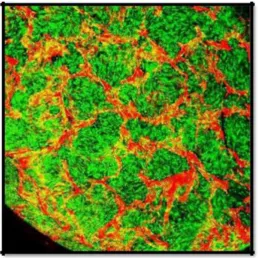
demonstrated that the electrospun silk fibroin-based scaffolds supported hMSC proliferation, growth and differentiation [15]. Films, fibres, nets, meshes, membranes, yarns, and sponges are various formats of silk fibroin (SF) which have been widely explored. They have been observed to support stem cell adhesion, proliferation, and differentiation in vitro apart from to trigger tissue repair in vivo. The silk-based biomaterials have been shown to be promising scaffolds for tissue engineering of bone, ligament, cartilage, stem cells as well as skin. By referring to figure 3, the three-dimensional distribution of human bone marrow derived mesenchymal stem cells was observed in porous aqueous derived silk fibroin scaffolds at week 3 [13].
In 2008, increased amount of primineralized silk fibroin using Hydroxyapatite (HAP) and BMP-2 was used [51]. The increased in osteoconductive and biomimetic growth of calcium phosphate were shown on silk fibroin. Enhanced outcome for bone tissue engineering was demonstrated in the study. ‘
Figure 3. Cells were stained using 2 mMcalcein AM (green for live cells) and 4 mMEthD-1 (red for mostly silk fibroin and also dead cells) (Molecular Probes, Eugene, OR) for 30-45 min at room temperature and evaluated using a Bio-Rad MRC 1024 confocal microscope with Lasersharp 2000 software [13].
The exploration on the nerve regeneration was triggered by the plethoric information and study on the regeneration of connective tissues. Silk has been identified as the potential biomaterial to regenerate damaged peripheral nerves for instance, sensory neuron, motor neuron or sciatic nerves. Another major component of silk, sericin, has been studied in vivo for the peripheral nerve regeneration. The recovery of a rat sciatic nerve was studied using the double conduits, sericine/silicone. It has been observed that the mechanical strength of conduit porous matched the peripheral nerve tissue. The double conduit was developed to attain maximum functional recovery. This is because the single conduit could only reach marginal recovery [52].
Figure 4. Architecture of nanofibers and culture of Schwann cells over nanofibrousscaffolds. (A) SF
nanofibrous scaffold and (B) nanocomposite (GNP-SF) nanofibrous scaffold. (E) SF nanofibrous scaffold after 5 days of culturing Schwann cells and (F) GNP-SF nanocomposite nanofibrous scaffold after 5 days of
culturing Schwann cells (G) [53]
Composite silk fibres with improved overall mechanical properties can be manufactured through the creation of transgenic silkworms which encoded chimeric silkworm or spider silk proteins. The piggyBac vectors were used in the process of forming the transgenic silkworms [54].
drug delivery and also for controlled release of DNA for gene therapy [58]. It was found that human MSC seeded on silk hydrogel increased from150 ± 30 cells/mm2 to 622 ± 87 cells/mm2[59]. Silk fibroin have been explored in bone tissue engineering [60]. Metabolic activity and proliferation of ATDC5 cells seeded onnon- and cross-linked Eri fibroin silk sponges showed significant increment until 14 days of culture [61].
VI
ADVANTAGES OF SILK
Comprehensive research have been carried out over the past few years to formulate a biomaterial which is similar to the human body in terms of physical and mechanical properties. It is even more challenging to create a material which is both biocompatible and non-toxic to the human body. In the field of medicine, natural material is preferred compared to their synthetic counterparts. This is due to its outstanding mechanical properties. Silk’s tensile strength is between 0.5 to 0.6GPa. Elongation of silk can reach up to 40% of its initial length [62]. All of these contribute to the toughness of silk [63]. Silk has remarkable strength to density ratio due to its strength and light-weight [64]. Another superior feature of silk is it can resist high level of stress and strain [62]. Mechanical properties is totally dependent on the type of silk [64]. Silk’s excellent mechanical properties can be utilized in the biodegradability area of biomaterial.
Apart from excellent mechanical properties, silk has also good biocompatibility. Lewis rats exhibited mild immune response when 3D silk scaffold were implanted in them [65, 66]. Silk has also been used in the production of ligament in tissue engineering. The pig model showed no signs of adverse effect and the in-vivo culture did not malfunction [67]. Silk’s biocompatibility is far more superior compared to collagen and PLA as it induced lower immune response [9]. In addition, silk has lower inflammatory response compared to polystyrene and poly (2-hydroxyethyl methacrylate).
Silk is both biodegradable and bio-resorbable [68]. Biodegradable is defined as the loss of mass of the material where the fragments can be moved from the implantation area but bot completely from the body [69]. On the other hand, bio-resorbable is the ability to completely metabolise the by-products due to the degradation. Implantation of water-based scaffold in Lewis rats degraded within a few weeks and was completely metabolised within a year [66]. The by-products from the degradation imposed no harmful effects. Systems which uses silk can be prepared using water based solution which requires only mild conditions for manufacturing. These systems only require room temperature, neutral pH and without the need of high force shear. These conditions are very favourable for loading sensitive drugs onto silk implants [70]. These conditions also increases the ability of biosensor to bio-integrate in vivo [62]. By using water vapour annealing, mechanical stretching and ultrasonic treatment, resistance towards dissolution, thermal and enzymatic degradation can be induced in silk [63,71].
In addition to that, silk also exhibits excellent optical transparency in the visible range which is very beneficial to the biosensor field [55, 72]. Silk is economically viable as large amount of silk are produced for the textile industry.
VII
LIMITATIONS OF SILK
The disadvantage of the degumming process is that the process alters the mechanical properties of the silk which is not favourable [73, 74]. The long term effect of silk are still questionable. More comprehensive research needs to be carried out in order to observe the long term effect of silk in human body. A major issue in bio-implants is the generation of particulate debris which may induce inflammation [75]. Fractions from silk are able to prompt immune response [76]. Apart from that, enzymatic reaction on silk which degrades the silk could result in reduction of cyto-compatibility feature [77]. On the other hand, many researches which were carried out proved otherwise [78, 79]. Degradation of silk may result in amyloidogenesis which may subsequently result in the degradation of surrounding tissue [80].
VIII
FUTURE WORK
Silk has been extensively researched in numerous field specifically in medical field. For instance, silk is integrated in the design of a microchip which would aid in eliminating infections [81]. This microchip is created as such that it is dissolvable. It is made up of magnesium on a film of silk. The microchip is turned on with the assistance of wireless connection and the resistor in the microchip heats up. The heat would in turn eliminate the infections. Silk is also being developed as printable inkjet [89]. This would reform the future of wound healing. Wound dressing with drugs can be printed out according to the size and shape of the wound. The ink doubles as a contamination detector. Silk balls are being created as a treatment to arthritis. The silk balls are injected into arthritic joints as lubricants. Silk is being developed as vaccines which requires no refrigeration [82].
IX
CONCLUSION
dimensional (3D) and porous silk scaffold adopting electrospinning method. The method produces nanofiber structure which mimic the extracellular matrix (ECM) of the cells. Nonetheless, more research is required for the silk to advance in the field of biomedical engineering.
X.
CONFLICT OF INTERESTS
The authors declare that there is no conflict of interest regarding the publication of this article. None of the authors has a financial relationship with a commercial entity that has an interest in the subject of this paper,
REFERENCE
[1] Thilagavathi, G., &Viju, S. (2015). 11 - Silk as a suture material. In A. Basu (Ed.), Advances in Silk Science and Technology (pp. 219-232): Woodhead Publishing.
[2] Das, S., Bora, U., Borthakur, B., &Kundu, S. (2014). Applications of silk biomaterials in tissue engineering and regenerative medicine. Silk biomaterials for tissue engineering and regenerative medicine. Woodhead Publishing Limited, 41-77.
[3] Hakimi, O., Knight, D. P., Vollrath, F., &Vadgama, P. (2007). Spider and mulberry silkworm silks as compatible biomaterials. Composites Part B: Engineering, 38(3), 324-337. doi: 10.1016/j.compositesb.2006.06.012
[4] Guinea, G. V., Elices, M., Pérez-Rigueiro, J., & Plaza, G. R. (2015). 10 - Structure and properties of spider and silkworm silk for tissue scaffolds*. In A. Basu (Ed.), Advances in Silk Science and Technology (pp. 185-217): Woodhead Publishing.
[5] Mortimer, B., & Holland, C. (2015). 12 - The use of spider silk as a biomaterial. In A. Basu (Ed.), Advances in Silk Science and Technology (pp. 233-260): Woodhead Publishing.
[6] Zhao, J., Zhang, Z., Wang, S., Sun, X., Zhang, X., Chen, J., . . . Jiang, X. (2009). Apatite-coated silk fibroin scaffolds to healing mandibular border defects in canines. Bone, 45(3), 517-527. doi: 10.1016/j.bone.2009.05.026
[7] Yan, L., Correia, C., Correia, J. S., Caridade, S., Fernandes, E., Mano, J., . . . Reis, R. (2012). Development of macro/micro porous silk fibroin scaffolds with nano-sized calcium phosphate particles for bone tissue engineering. Journal of tissue engineering and regenerative medicine, 6(Suppl. 1), 181. [8] Teuschl, A. H., Schuh, C. M., Halbweis, R., Marton, G., Pajer, K., Hopf, R., . . .Redl, H. (2015). Silk
Fibroin for Peripheral Nerve Regeneration: A Novel Preparation Method Improved Mechanical Characteristics and Supports Regeneration in Rat Sciatic Nerves. Tissue Engineering Part A, 21, S36-S36. [9] Vepari, C., & Kaplan, D. L. (2007). Silk as a Biomaterial. ProgPolymSci, 32(8-9), 991-1007. doi:
10.1016/j.progpolymsci.2007.05.013
[10] Shi, P., & Goh, J. C. H. (2012). Self-assembled silk fibroin particles: Tunable size and appearance. Powder Technology, 215-216, 85-90. doi: 10.1016/j.powtec.2011.09.012
[11] Ratner, B. D. (2013). The Biomaterials Literature. 1491-1493. doi: 10.1016/b978-0-08-087780-8.00146-7 [12] Abdel-Naby, W., & Lawrence, B. D. (2015). 9 - Processing of silk biomaterials. In A. Basu (Ed.),
Advances in Silk Science and Technology (pp. 171-183): Woodhead Publishing.
[13] Wang, Y., Kim, H. J., Vunjak-Novakovic, G., & Kaplan, D. L. (2006). Stem cell-based tissue engineering with silk biomaterials. Biomaterials, 27(36), 6064-6082. doi: 10.1016/j.biomaterials.2006.07.008
[14] Wenk, E., Meinel, A. J., Wildy, S., Merkle, H. P., &Meinel, L. (2009). Microporous silk fibroin scaffolds embedding PLGA microparticles for controlled growth factor delivery in tissue engineering. Biomaterials, 30(13), 2571-2581. doi: 10.1016/j.biomaterials.2008.12.073
[15] Li, C., Vepari, C., Jin, H. J., Kim, H. J., & Kaplan, D. L. (2006). Electrospun silk-BMP-2 scaffolds for bone tissue engineering. Biomaterials, 27(16), 3115-3124. doi: 10.1016/j.biomaterials.2006.01.022 [16] Wang, X., Hu, X., Daley, A., Rabotyagova, O., Cebe, P., & Kaplan, D. L. (2007). Nanolayer biomaterial
coatings of silk fibroin for controlled release. J Control Release, 121(3), 190-199. doi: 10.1016/j.jconrel.2007.06.006
[17] Zhang, X., Reagan, M. R., & Kaplan, D. L. (2009). Electrospun silk biomaterial scaffolds for regenerative medicine. Adv Drug Deliv Rev, 61(12), 988-1006. doi: 10.1016/j.addr.2009.07.005
[18] Feng, Z., Rong, L., Zuo, B. Q., & Qin, J. Z. (2010, 18-20 June 2010). Electrospun Silk Fibroin Nanofiber Tubes for Peripheral Nerve Regeneration. Paper presented at the Bioinformatics and Biomedical Engineering (iCBBE), 2010 4th International Conference on.
[19] Liu, H., Li, X., Zhou, G., Fan, H., & Fan, Y. (2011). Electrospun sulfated silk fibroin nanofibrous scaffolds for vascular tissue engineering. Biomaterials, 32(15), 3784-3793.
[21] Jin, H. (2004). Human bone marrow stromal cell responses on electrospun silk fibroin mats. Biomaterials, 25(6), 1039-1047. doi: 10.1016/s0142-9612(03)00609-4
[22] Min, B., Lee, G., Kim, S., Nam, Y., Lee, T., & Park, W. (2004). Electrospinning of silk fibroin nanofibers and its effect on the adhesion and spreading of normal human keratinocytes and fibroblasts in vitro. Biomaterials, 25(7-8), 1289-1297. http://dx.doi.org/10.1016/j.biomaterials.2003.08.045
[23] Xie, H., Yang, W., Chen, J., Zhang, J., Lu, X., Zhao, X., . . . Wang, Z. (2015). A Silk Sericin/Silicone Nerve Guidance Conduit Promotes Regeneration of a Transected Sciatic Nerve. Advanced healthcare materials.
[24] Permanent Waving Is Biochemical Engineering - Glucose Phosphate. (2016). Barnardhealth.us. Retrieved 2 April 2016, from http://www.barnardhealth.us/glucose-phosphate/permanent-waving-is-biochemical-engineering.html
[25] Eisoldt, L., Smith, A., &Scheibel, T. (2011). Decoding the secrets of spider silk. Materials Today, 14(3), 80-86. http://dx.doi.org/10.1016/s1369-7021(11)70057-8
[26] Hardy, J., Römer, L., &Scheibel, T. (2008). Polymeric materials based on silk proteins. Polymer, 49(20), 4309-4327. http://dx.doi.org/10.1016/j.polymer.2008.08.006
[27] Kawamoto, K., Kawamoto, T., Shiba, H., &Hosono, K. (2013). A histochemical study of the posterior silk glands of Bombyxmori during metamorphosis from larvae to pupae using frozen sections. Biotechnic&Histochemistry, 89(2), 145-152. http://dx.doi.org/10.3109/10520295.2013.830777
[28] Heijenoort, J. (2001). Formation of the glycan chains in the synthesis of bacterial peptidoglycan.Glycobiology, 11(3), 25R36R.http://dx.doi.org/10.1093/glycob/11.3.25r
[29] Millard, M., &Masri, M. (1974). Detection and analysis of protein functional group modification by x-ray photoelectron spectrometry. Analytical Chemistry, 46(12), 1820-1822. http://dx.doi.org/10.1021/ac60348a039
[30] Eisenberg, D. (2003). The discovery of the  -helix and  -sheet, the principal structural features of proteins. Proceedings Of The National Academy Of Sciences, 100(20), 11207-11210. http://dx.doi.org/10.1073/pnas.2034522100
[31] Sakai, F., Yang, G., Weiss, M., Liu, Y., Chen, G., & Jiang, M. (2014). Protein crystalline frameworks with controllable interpenetration directed by dual supramolecular interactions. Nature Communications, 5, 4634. http://dx.doi.org/10.1038/ncomms5634
[32] Doshi, U., & Muñoz, V. (2004). The Principles of α-Helix Formation: Explaining Complex Kinetics with Nucleation−Elongation Theory. The Journal of Physical Chemistry B, 108(24), 8497-8506. http://dx.doi.org/10.1021/jp049896a
[33] Fukushima, Y. (1996). Synthesis and Highly Stable β-Sheet Formation of Sequential Alternating Amphiphilic Polypeptides. Polym J, 28(2), 113-120. http://dx.doi.org/10.1295/polymj.28.113
[34] Alberts, B. (2002). Molecular biology of the cell. New York: Garland Science.
[35] Zhang, S., Pan, Q., Zhang, H., Zhang, Y., & Wang, H. (2003). Classification of protein quaternary structure with support vector machine. Bioinformatics, 19(18), 2390-2396. http://dx.doi.org/10.1093/bioinformatics/btg331
[36] Inoue, S., Tanaka, K., Arisaka, F., Kimura, S., Ohtomo, K., & Mizuno, S. (2000). Silk Fibroin of BombyxmoriIs Secreted, Assembling a High Molecular Mass Elementary Unit Consisting of H-chain, L-chain, and P25, with a 6:6:1 Molar Ratio. Journal Of Biological Chemistry, 275(51), 40517-40528. http://dx.doi.org/10.1074/jbc.m006897200
[37] Sirichaisit, J., Brookes, V., Young, R., &Vollrath, F. (2003). Analysis of Structure/Property Relationships in Silkworm ( Bombyxmori ) and Spider Dragline ( Nephila edulis ) Silks Using Raman Spectroscopy. Biomacromolecules, 4(2), 387-394. http://dx.doi.org/10.1021/bm0256956
[38] Sehnal, F., &Žurovec, M. (2004). Construction of Silk Fiber Core in Lepidoptera. Biomacromolecules, 5(3), 666-674. http://dx.doi.org/10.1021/bm0344046
[39] Boutry, C., Řezáč M., &Blackledge, T. (2011). Plasticity in Major Ampullate Silk Production in Relation
to Spider Phylogeny and Ecology. Plos ONE, 6(7), e22467.
http://dx.doi.org/10.1371/journal.pone.0022467
[40] Madurga, R., Plaza, G., Blackledge, T., Guinea, G., Elices, M., &Pérez-Rigueiro, J. (2016). Material properties of evolutionary diverse spider silks described by variation in a single structural parameter. Sci. Rep., 6, 18991. http://dx.doi.org/10.1038/srep18991
[41] Chen, G., Liu, X., Zhang, Y., Lin, S., Yang, Z., & Johansson, J. et al. (2012). Full-Length Minor
Ampullate Spidroin Gene Sequence. Plos ONE, 7(12), e52293.
[42] Guinea, G., Elices, M., Plaza, G., Perea, G., Daza, R., &Riekel, C. et al. (2012). Minor Ampullate Silks from Nephila and Argiope Spiders: Tensile Properties and Microstructural Characterization. Biomacromolecules, 13(7), 2087-2098. http://dx.doi.org/10.1021/bm3004644
[43] Adrianos, S., Teulã, F., Hinman, M., Jones, J., Weber, W., Yarger, J., & Lewis, R. (2013).Nephilaclavipes Flagelliform Silk-Like GGX Motifs Contribute to Extensibility and Spacer Motifs Contribute to Strength in Synthetic Spider Silk Fibers. Biomacromolecules, 14(6), 1751-1760.
http://dx.doi.org/10.1021/bm400125w
[44] Perry, D., Bittencourt, D., Siltberg-Liberles, J., Rech, E., & Lewis, R. (2010). Piriform Spider Silk Sequences Reveal Unique Repetitive Elements. Biomacromolecules, 11(11), 3000-3006. http://dx.doi.org/10.1021/bm1007585
[45] Vollrath, F. (2006). Spider Silk: Thousands of Nano-Filaments and Dollops of Sticky Glue. Current Biology, 16(21), R925-R927. http://dx.doi.org/10.1016/j.cub.2006.09.050
[46] Foo, C. W. P., Bini, E., Hensman, J., Knight, D., Lewis, R., & Kaplan, D. (2006). Role of pH and charge on silk protein assembly in insects and spiders. Applied Physics A, 82(2), 223-233.
[47] Huemmerich, D., Slotta, U., &Scheibel, T. (2006). Processing and modification of films made from recombinant spider silk proteins. Applied Physics A, 82(2), 219-222.
[48] Kundu, B., Kurland, N., Bano, S., Patra, C., Engel, F., Yadavalli, V., &Kundu, S. (2014). Silk proteins for biomedical applications: Bioengineering perspectives. Progress In Polymer Science, 39(2), 251-267. http://dx.doi.org/10.1016/j.progpolymsci.2013.09.002
[49] Das, S., Bora, U., Borthakur, B., &Kundu, S. (2014). Applications of silk biomaterials in tissue engineering and regenerative medicine. Silk biomaterials for tissue engineering and regenerative medicine. Woodhead Publishing Limited, 41-77.
[50] Laurencin, C. T., & Freeman, J. W. (2005). Ligament tissue engineering: an evolutionary materials science approach. Biomaterials, 26(36), 7530-7536. doi: 10.1016/j.biomaterials.2005.05.073
[51] Kim, H., Kim, U., Kim, H., Li, C., Wada, M., Leisk, G., & Kaplan, D. (2008). Bone tissue engineering with premineralized silk scaffolds. Bone, 42(6), 1226-1234. http://dx.doi.org/10.1016/j.bone.2008.02.007 [52] Xie, H., Yang, W., Chen, J., Zhang, J., Lu, X., Zhao, X., . . . Wang, Z. (2015). A Silk Sericin/Silicone
Nerve Guidance Conduit Promotes Regeneration of a Transected Sciatic Nerve. Advanced healthcare materials.
[53] Das, S., Sharma, M., Saharia, D., Sarma, K. K., Sarma, M. G., Borthakur, B. B., & Bora, U. (2015). Data in support of in vivo studies of silk based gold nano-composite conduits for functional peripheral nerve regeneration. Data in Brief, 4, 315-321.
[54] Teulé, F., Miao, Y.-G., Sohn, B.-H., Kim, Y.-S., Hull, J. J., Fraser, M. J., . . . Jarvis, D. L. (2012). Silkworms transformed with chimeric silkworm/spider silk genes spin composite silk fibers with improved mechanical properties. Proceedings of the National Academy of Sciences, 109(3), 923-928. [55] Liu, S., Dong, C., Lu, G., Lu, Q., Li, Z., Kaplan, D. L., & Zhu, H. (2013). Bilayered vascular grafts based
on silk proteins. Actabiomaterialia, 9(11), 8991-9003.
[56] Huang, L., Li, C., Yuan, W., & Shi, G. (2013). Strong composite films with layered structures prepared by casting silk fibroin–graphene oxide hydrogels. Nanoscale, 5(9), 3780-3786.
[57] Yang, M., Shuai, Y., He, W., Min, S., & Zhu, L. (2012). Preparation of porous scaffolds from silk fibroin extracted from the silk gland of Bombyxmori (B. mori). International journal of molecular sciences, 13(6), 7762-7775.
[58] Kapoor, S. &Kundu, S. (2016). Silk protein-based hydrogels: Promising advanced materials for biomedical applications. ActaBiomaterialia, 31, 17-32. http://dx.doi.org/10.1016/j.actbio.2015.11.034 [59] Floren, M., Bonani, W., Dharmarajan, A., Motta, A., Migliaresi, C., & Tan, W. (2016). Human
mesenchymal stem cells cultured on silk hydrogels with variable stiffness and growth factor differentiate into mature smooth muscle cell phenotype. ActaBiomaterialia, 31, 156-166. http://dx.doi.org/10.1016/j.actbio.2015.11.051
[60] Melke, J., Midha, S., Ghosh, S., Ito, K., & Hofmann, S. (2016). Silk fibroin as biomaterial for bone tissue engineering. ActaBiomaterialia, 31, 1-16. http://dx.doi.org/10.1016/j.actbio.2015.09.005
[61] [61] Silva, S., Oliveira, N., Oliveira, M., da Costa, D., Naskar, D., & Mano, J. et al. (2016). Fabrication and characterization of Eri silk fibers-based sponges for biomedical application. ActaBiomaterialia, 32, 178-189. http://dx.doi.org/10.1016/j.actbio.2016.01.003
[62] Rajkhowa, R., Gupta, V., & Kothari, V. (2000). Tensile stress, strain and recovery behavior of Indian silk fibers and their structural dependence. Journal of Applied Polymer Science, 77(11), 2418-2429. http://dx.doi.org/10.1002/1097-4628(20000912)77:11<2418::aid-app10>3.3.co;2-h
[64] Ko, F., Kawabata, S., Inoue, M., Niwa, M., Fossey, S., & Song, J. (2001). Engineering Properties of Spider Silk. MRS Proc., 702. http://dx.doi.org/10.1557/proc-702-u1.4.1
[65] Cheung, H., Lau, K., Ho, M., & Mosallam, A. (2009). Study on the Mechanical Properties of Different Silkworm Silk Fibers. Journal of Composite Materials, 43(22), 2521-2531. http://dx.doi.org/10.1177/0021998309345347
[66] Zafar, M., & Al-Samadani, K. (2014). Potential use of natural silk for bio-dental applications. Journal OfTaibah University Medical Sciences, 9(3), 171-177. http://dx.doi.org/10.1016/j.jtumed.2014.01.003 [67] Wang, Y., Rudym, D., Walsh, A., Abrahamsen, L., Kim, H., & Kim, H. et al. (2008). In vivo degradation
of three-dimensional silk fibroin scaffolds. Biomaterials, 29(24-25), 3415-3428. http://dx.doi.org/10.1016/j.biomaterials.2008.05.002
[68] Fan, H., Liu, H., Toh, S., & Goh, J. (2009). Anterior cruciate ligament regeneration using mesenchymal stem cells and silk scaffold in large animal model. Biomaterials, 30(28), 4967-4977. http://dx.doi.org/10.1016/j.biomaterials.2009.05.048
[69] Ho, M., Lau, K., Wang, H., & Bhattacharyya, D. (2011). Characteristics of a silk fibre reinforced biodegradable plastic. Composites Part B: Engineering, 42(2), 117-122. http://dx.doi.org/10.1016/j.compositesb.2010.10.007
[70] Vert, M., Li, S., Spenlehauer, G., & Guerin, P. (1992). Bioresorbability and biocompatibility of aliphatic polyesters. J Mater Sci: Mater Med, 3(6), 432-446. http://dx.doi.org/10.1007/bf00701240
[71] Karageorgiou, V., Meinel, L., Hofmann, S., Malhotra, A., Volloch, V., & Kaplan, D. (2004). Bone morphogenetic protein-2 decorated silk fibroin films induce osteogenic differentiation of human bone marrow stromal cells. Journal Of Biomedical Materials Research, 71A(3), 528-537. http://dx.doi.org/10.1002/jbm.a.30186
[72] Wang, X., Yucel, T., Lu, Q., Hu, X., & Kaplan, D. (2010). Silk nanospheres and microspheres from silk/pva blend films for drug delivery. Biomaterials, 31(6), 1025-1035. http://dx.doi.org/10.1016/j.biomaterials.2009.11.002
[73] Omenetto, F., & Kaplan, D. (2008). A new route for silk. Nature Photonics, 2(11), 641-643. http://dx.doi.org/10.1038/nphoton.2008.207
[74] Perez-Rigueiro, J., Elices, M., Llorca, J., &Viney, C. (2002). Effect of degumming on the tensile properties of silkworm (Bombyxmori) silk fiber. Journal of Applied Polymer Science, 84(7), 1431-1437. http://dx.doi.org/10.1002/app.10366
[75] Jiang, P., Liu, H., Wang, C., Wu, L., Huang, J., &Guo, C. (2006). Tensile behavior and morphology of differently degummed silkworm (Bombyxmori) cocoon silk fibres. Materials Letters, 60(7), 919-925. http://dx.doi.org/10.1016/j.matlet.2005.10.056
[76] Purdue, P., Koulouvaris, P., Nestor, B., &Sculco, T. (2006). The Central Role of Wear Debris in PeriprostheticOsteolysis. HSS Journal, 2(2), 102-113. http://dx.doi.org/10.1007/s11420-006-9003-6 [77] Altman, G., Diaz, F., Jakuba, C., Calabro, T., Horan, R., & Chen, J. et al. (2003). Silk-based biomaterials.
Biomaterials, 24(3), 401-416. http://dx.doi.org/10.1016/s0142-9612(02)00353-8
[78] Minoura, N., Aiba, S., Higuchi, M., Gotoh, Y., Tsukada, M., & Imai, Y. (1995). Attachment and Growth of Fibroblast Cells on Silk Fibroin. Biochemical And Biophysical Research Communications, 208(2), 511-516. http://dx.doi.org/10.1006/bbrc.1995.1368
[79] Baran, E., Tuzlakoğlu, K., Mano, J., & Reis, R. (2012). Enzymatic degradation behavior and cytocompatibility of silk fibroin–starch–chitosan conjugate membranes. Materials Science And Engineering: C, 32(6), 1314-1322. http://dx.doi.org/10.1016/j.msec.2012.02.015
[80] Serban, M., Panilaitis, B., & Kaplan, D. (2011). Silk fibroin and polyethylene glycol-based biocompatible tissue adhesives. Journal Of Biomedical Materials Research Part A, 98A(4), 567-575. http://dx.doi.org/10.1002/jbm.a.33149
[81] Kearns, V., MacIntosh, A., Crawford, A., & Hatton, P. (2008). Silk-based Biomaterials for Tissue Engineering. In Tissue Engineering (1st ed.). Sheffield: Centre for Biomaterials & Tissue Engineering, University of Sheffield.
[82] Sheena McKenzie, f. (2015). Silk 'microchip' heals you from the inside - CNN.com. CNN. Retrieved 5 November 2015, from http://edition.cnn.com/2015/06/19/tech/silk-microchip-science-healing-alternative-uses/
![Figure 1. Silk Fibroin (SF) [24]](https://thumb-us.123doks.com/thumbv2/123dok_us/1364585.1645895/2.595.189.416.306.409/figure-silk-fibroin-sf.webp)
![Table 1: Comparison between the mechanical properties of silk and man-madefibres [26] Strength, ExtensibilityToughness/](https://thumb-us.123doks.com/thumbv2/123dok_us/1364585.1645895/3.595.116.467.109.370/table-comparison-mechanical-properties-silk-madefibres-strength-extensibilitytoughness.webp)
![Figure 2. Scanning electron micrographs of BMSCs growing on electrospun mats and native silk fibroin matrices after 1, 7 and 14 days (scale bar: 5mm) [21]](https://thumb-us.123doks.com/thumbv2/123dok_us/1364585.1645895/5.595.202.434.333.469/figure-scanning-electron-micrographs-growing-electrospun-fibroin-matrices.webp)