Original Research Article.
Clinical and Radiological Assessment of Intraarticular Displaced Calcaneum
Fractures Operated with Locking Branched Calcaneal Plate:
A Prospective Study
Jairam D Jagiasi
1, Amit Joshi
2, Manikanta Babu
3*, Mihir R Patel
4, Amol Bochare
5, Manan Vora
61Professor & HOD, 2Senior Registrar, 3*PG Resident, 4Assistant Professor, 5SMO, 6Junior Resident, Department of Orthopaedics, Dr. RN Cooper Hospital and HBTMC, Mumbai, Maharashtra, India. ABSTRACT
Background: Calcaneal fractures are common, but are difficult to manage. Immediate concern is soft tissue problems, while long term concern is pain as sequelae of subtalar arthritis. A consensus has not been reached in the management of calcaneal fractures.
Objective: This study aims to evaluate clinical and radiological outcomes of the patients managed with open reduction and internal fixation with Calcaneal Locking Plates for the displaced intra-articular calcaneal fractures presenting in the Department of Orthopaedics at Dr. R.N. Cooper muncipal general hospital, Juhu, Mumbai.
Method: This was a prospective study, conducted on
displaced intra articular calcaneal fractures from January 2016 through January 2018. The patients underwent open reduction and internal fixation with Locking Branched Calcaneal Plates through the extensile lateral approach. Post-operatively, ankle was mobilized after two weeks. Weight bearing was started after 12 weeks. Patients were evaluated clinically with Maryland foot score and radiologically with measurements of
Boehler’s and Gissane angle.
Results: Twenty cases of calcaneal fractures managed with open reduction and internal fixation with Locking Branched Calcaneal Plates were available for final evaluation. Sixteen of the enrolled patients were males in their third decade of life. On average, calcaneal fractures were operated on seven days after the injury. Sanders Type II were seen in 75% of the cases and Sanders Type III were in 25%. Mean follow-up duration was 12 months. The average Maryland foot score was 77.27.
Fifteen cases (75%) had good, four cases (20%) had fair, and one case (5%) had poor outcome score. There was statistically
significant improvement in Boehler’s and Gissane angle across
all enrolled patients.
Conclusion: Displaced intra-articular calcaneal fractures treated operatively with open reduction and internal fixation with Locking Branched Calcaneal Plates through the extended lateral approach, with proper planning of operation and surgical techniques in soft tissue handling, results in good clinical as well as radiological outcomes.
Key Words: Boehler’s Angle, Gissane Angle, Calcaneum,
Locking Branched Calcaneal Plates, Maryland Foot Score, ORIF.
*Correspondence to:
Dr. Manikanta Babu, PG Resident,
Department of Orthopaedics, Dr. RN Cooper Hospital and HBTMC, Mumbai, Maharashtra, India. Article History:
Received: 29-04-2018, Revised: 21-05-2018, Accepted: 07-06-2018
Access this article online
Website: www.ijmrp.com
Quick Response code
DOI:
10.21276/ijmrp.2018.4.4.004
INTRODUCTION
Calcaneal fractures, which account for 2% of total fractures, are one of the most common fractures of the foot.1-7 They include 60%
of tarsal bone fractures.10% of fractures are bilateral and 75% are intra articular.10% of fractures are associated with vertebrae fractures. Mechanism of injury in majority of patients is axial loading i.e. fall from height. Other mechanisms are brake pedal injuries and high velocity trauma. Current development in imaging technology has allowed better understanding of this complex fracture pathology. Sanders classification of intra articular Calcaneum fractures is being used widely because of its correlation with prognosis and planning for surgery. Treating
Calcaneum fractures is a challenge for orthopaedic surgeon. Treatment options ranges from non-operative to operative methods. There is controversy whether to manage these fractures conservatively or open reduction and internal fixation (ORIF). ORIF with Locking Branched Calcaneal Plate (LBCP) has better radiological and functional outcomes when they are reviewed over the long-term.8,9 The drawbacks to ORIF with LBCP are the
Conservative management reduces the likelihood of soft tissue complications but has drawbacks such as: increased risk of chronic pain, gait abnormality, prolonged recovery, short and wide heel, sub-fibular impingement, disparity in height and increased morbidity which can be minimized by ORIF.11
The available literatures provide insufficient evidence regarding best management strategy for these fractures. The paucity of literature about the operative management from our part of the world has made it difficult to follow evidence based practice for our patients. The current study evaluates clinical and radiological outcome of patients with intra-articular displaced calcaneal fractures managed with ORIF with LBCP.
MATERIALS AND METHODS
A prospective quantitative descriptive cohort study was carried out over 2 years, from January 2016 to January 2018, in all consecutive cases of closed displaced intra-articular fractures of calcaneum in skeletally mature patients that presented to the Department of Orthopaedics at Dr. R.N. Cooper muncipal general hospital, Juhu, Mumbai.. Out of total 30 cases of calcaneal fractures that were managed during the study period, 25 cases were eligible for the study and 20 cases were available for final follow up. Open calcaneal fractures, patients unwilling for the operative management, skeletally immature patients, cases managed conservatively were excluded from the study. Written informed consent was obtained from all patients to be managed with ORIF with LBCP. The data were recorded in proforma including: epidemiological information, fracture details from X-rays and CT scans, preoperative, perioperative, and postoperative details including wound condition.Fractures classified using sanders classification. Routine investigations carried out to get fitness for surgery. Patients underwent Open reduction and internal fixation with locking branched calcaneal plate through extensile lateral approach under spinal anaesthesia. Post-operative physiotherapy followed according to protocol. Patients will be followed up at 6 weekly interval until fracture union. Clinical evaluation done by using Maryland foot scoring system. Radiological evaluation done by using Boehlers angle and Critical angle of Gissane on Lateral radiographs of calcaneum. Functional outcome was recorded at every followup after 12 wks of surgery.
Procedure Details
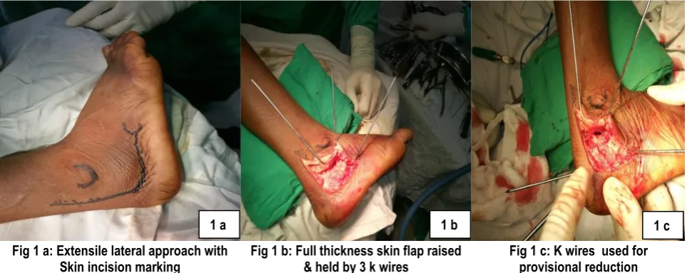
The operation was carried out with the patient in lateral decubitus position with the affected side up and using extensile lateral incision with elevation of single full thickness skin flap up from the bone. Instead of using tooth forceps, tissue was reflected with a penfield and held with the use of three 2 mm Kirschner wires at the lateral surface of the talus. On the bulged lateral wall of the fractured calcaneum, a window was opened by elevating the bone flap, through which the depressed fragment was elevated, anatomically reducing the articular surface of the calcaneum. The reduction was confirmed by visualization through talo-calcaneal joint and also by lateral view of calcaneum in C arm. The bone flap was then turned down, to close the window. The reduction was temporarily held in place with a K wire that was passed from heel through the calcaneum to the talus. An appropriate sized branched calcaneum locking plate was placed on the lateral wall of the calcaneum and initially fixed with non-locking cortical screws, in order to generate good lag effect allowing proper buttressing of the plate onto the lateral wall of calcaneum. Then, the non-locking cortical screws were replaced by the locking ones ensuring the stable fixation of the plate. The wound was then irrigated and closed in two layers: three to four polygalactin sutures for subcutaneous tissues and tension free polypropylene sutures for skin over an eight French closed suction drain. The ankle and foot were padded well and splinted with below knee slab in neutral position.
Post-operatively ankle was elevated over a pillow, intravenous cefuroxime 750mg q8h was given for 48 hours followed by oral cefuroxime for five days. Wound was inspected and drain was removed on the second postoperative day. Five to seven days post-operatively, once the wrinkle sign appeared, ankle range of motion exercises was begun.
The patients were discharged with advice of suture removal on fourteenth postoperative day. Patients were advised to continue B/K slab for total of six weeks post-operation with intermittent range of motion exercises. Weight bearing was started on 12th week post operation. At the final follow up, the functional outcome was assessed with Maryland foot score and the radiological
outcome by Boehler’s Tuber and Gissane angles on lateral view of ankle X-ray.
Fig 1 a: Extensile lateral approach with
Skin incision marking Fig 1 b: Full thickness skin flap raised & held by 3 k wires Fig 1 c: K wires used for provisional reduction
Fig 1 d: LBCP is used to buttress the
lateral aspect of calcaneum Fig 1 e,f: Intraop reduction under c-arm AP and Axial views showing maintained boehlers angle and gissane angle
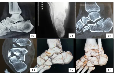
Fig 2 (a-f): Preoperative X-rays and CT Scans
Fig 3 (a,b): Postoperative xrays at 12 months followup
1 d 1 e 1 f
2 b 2 c
2 d 2 e 2 f
2 a
Table 1: Clinical Outcome
Functional Outcome Number (%)
Maryland footscore (Mean ± SD) 77.27 ± 8.04
Excellent (90–100) 0
Good (75–89) 15 (75)
Fair (50–74) 4 (25)
Poor (<50) 1 (5)
Overall 20(100)
Table 2: Radiological outcome and relation of Boehler’s and Gissane angle with functional outcome
Maryland foot score
Boehler’s Angle Good Fair and Poor Over all
Pre-OP 0.3 1 0.45
Post-Op 29.3 21 27.4
Gissane Angle
Pre-Op 147.7 148.6 147.6
Post-Op 116.1 136 121
Table 3: Demographic information
Age Mean : 32 years ± 8 years
Range ( 17 to 60 years)
Gender Male : Female =16:4
(Male : 4 times more)
Laterality Right : Left = 11:9
Injury to Operation duration
Mean : 7 days ± 4.8 days Range : (1 to 19) days Operation to Discharge
duration
Mean : 5.6 days ± 2.5 days Range : (2 to 12) days
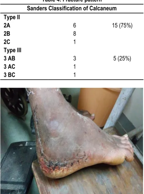
Table 4: Fracture pattern Sanders Classification of Calcaneum Type II
2A 6 15 (75%)
2B 8
2C 1
Type III
3 AB 3 5 (25%)
3 AC 1
3 BC 1
Fig 4: Wound condition on suture removal postop day 14
RESULTS
Twenty cases of calcaneal fractures managed with ORIF with LBCP were available for final evaluation. Clinical outcome, as measured using the Maryland foot score, showed majority of the patients had a good outcome. Radiological assessment comparing pre-operative to post-operative improvement in
Boehler’s and Critical angle of Gissane showed significant
improvement after ORIF with LBCP.
The average age of cohort members in this study was 32 years. Male preponderance was observed with male to female ratio of 4:1. The majority of the cases presented with isolated injuries however, some fractures occurred in patients along with multiple traumatic injuries (spine injuries being most common) that were managed appropriately. After arrival in the Emergency Department or Orthopedic Outpatient Department, each patient underwent an X-ray and a CT scan of the calcaneus. Fifteen patients had Sanders Type II fractures and five patients had Sanders Type III fractures. However, there were no Sanders I and IV type fractures in the current study. In average, the final follow-up was 12 months post-operation with a range of 6 to 18 months. Three patients developed local complications. Two patients had superficial wound infections with sero-sanguineous discharge, but negative culture. The infections improved with repeated dressing and local wound care. One patient had superficial skin necrosis which was treated with debridement. Seven patients had longer hospital stays (more than five days): three of those patients had local complications; other three had associated fractures such as tibia, femur, or spine from which they were recovering and one patient had no significant medical problems, but stayed longer despite advice of discharge
DISCUSSION
Recent trend shift from conservative to operative management of displaced calcaneal fractures has been observed.12-14 In our
department, until early 2016, we were managing these cases predominantly with closed reduction with pins and plaster casts or screws. With availability of equipment such as locking plates and improvements in technical skill, especially in the handling of the soft tissue, we have seen a gradual shift to operative management in recent years.
The majority of the patients in this study were males in third decade of life and spine injuries were the most common associated injuries. This trend was reported by other similar studies.15,16 Most males in this age group are engaged in
agricultural or industrial work and are at risk for these types of injuries.
al. from India, operation was performed within mean 9.2 days with a range of 2 to 19 days and a mean of 6.5 days with the range of 2 to 14 days respectively.15-18
The patients enrolled in this study predominantly had Sanders type II and few had Sander III calcaneal fractures. Kumar et al cited a Sanders type II as the most common fracture type with 47% of cases. However, Joshi et al. had majority (41.93%) of fractures, of Sander III. Sanders classification, a CT based classification, is a little bit cumbersome however it has the advantage of assisting in formulation of a surgical plan.16,19,20
Though some studies suggest there is no significant difference in clinical outcome of operative versus conservative management, there are some recent literatures which suggest that the outcomes are more favourable with operative management.21 Joshi et al.
had an average Maryland foot score of 83.68 in their study of surgically treated calcaneal fractures and 61.76% had good results.16 In another study evaluating functional outcome by the
same scoring system for calcaneal fractures and managed with locking plates, Kumar et al. showed gradual improvement of the score over the time from the first follow-up visit at 6 weeks post-operation with 93% of cases graded fair and 7% graded poor, to the final follow-up visit at one year with 97% of cases graded as excellent.20 Similarly, Zwipp et al., Lakhey et al., and Jain et al.
had shown excellent to good results with operative management, though they used different scoring system for the functional outcome measurement, namely, Merle d’Aubigne score, Modified Rowe’s Score, and American Orthopaedics Foot and Ankle score
respectively.15,17,18 In this study, Maryland foot score was used and
the average score in the final follow up was 72.7 with a range of 60 to 80. We did not have any patients with an excellent result and one patient had poor result, however there were significant number of good results (77.3 %). Our findings are consistent with the literatures showing better results with operative management.15-18 We expect that with time, the average Maryland
foot score is expected to improve further with continuous physiotherapy.
We have observed a statistically significant overall improvement in
the preoperative versus postoperative Boehler’s angle as well as
in Gissane angle. We have observed a positive correlation
between the amount of restoration of Boehler’s and Gissane
angles in relation to better functional outcome score. Patients with
Good clinical outcome had better restored Boehler’s and Gissane
angles compared to the patients with Fair and Poor outcome. A similar trend is found in a study by Kulkarni et al. and Makki et al. however direct comparison of those findings to ours is challenging since they used the Creighton-Nebraska score for assessing the functional outcome.22,23 Meena et al. in a meta-analysis of
randomized controlled trials of operative versus non-operative treatment for calcaneal fractures, favours operative management and concludes that functional outcome is better and pain is less when post-operative Boehler’s angle is restored and anatomic
reduction is achieved.8
Despite many advantages, operative management is not without some risks. The main complications those appear in the literature are wound dehiscence, necrosis and infections. We had three patients with wound problems but with conservative management their wound condition improved. Joshi et al. had 8.8% of their cohort with wound issues, Zwipp et al. had 13.2%, Jain et al. had 19.2% and Lakhey et al. had 25% of the cases with wound related
problems.15-18 Other notable complications that the literature
mentions are broadening and varus of heel, subtalar impingement, and subtalar arthritis. This may be because we had smaller cohort size and relatively shorter time for final follow-up, in average 12 months. Complication rates as high as 54% have been reported in patients with operative management, hence the literature suggests proper patient selection is important. Patients with comorbidities like peripheral vascular disease, diabetes mellitus, and tobacco smoking are not good candidates for operative management.
The measurement tool for functional outcome (Maryland foot score) has not been translated and validated in the local language and context. This could have had an impact in our observations. Other limitations in our study were relatively shorter average final follow-up time and single centre study of single cohort only.
CONCLUSION
Open reduction and internal fixation with locking branched calcaneal plates through the extended lateral approach results in a good clinical and radiological outcome in the management of displaced intra-articular calcaneal fractures. Proper patient selection, planning of operation, and proper surgical techniques, especially in regard to soft tissue handling are necessary in order to avoid wound complications and ensure the better outcomes for the patients.
REFERENCES
1. Rodríguez SR, Garduño RB, Raygoza CO. Surgical treatment of calcaneal fractures with a special titanium AO plate. Acta Orthopedica Mexicana. 2004;18(Suppl. 1):34-8.
2. Fitzgibbons TC, McMullen ST, Mormino MA. Fractures and dislocations of the calcaneum. In: Bucholz RW and Heckman JD
Eds. Rockwood and Green’s Factures in adults, Vol.3, 5th ed.
Philadelphia: Lippincott Williams & Wilkins, 2001: 2133-2179. 3. Sanders R. Displaced intra-articular fractures of the calcaneus. J Bone Joint Surg Am. 2000;82(2):225-50.
4. Giachino AA, Uhthoff HK. Intra-articular fractusres of the calcaneus. J Bone Joint Surg Am. 1989;71(5):784-7.
5. Paley D, Hall H. Intra-articular fractures of the calcaneus. A critical analysis of results and prognostic factors. J Bone Joint Surg Am. 1993;75(3):342-54.
6. Sanders R. Intra-articular fractures of the calcaneus: present state of the art. J Orthop Trauma. 1992;6(2):252-65.
7. Simpson LA, Schulak DA, Spiegel PG. Intraarticular fractures of the calcaneus: a review. Contemp Orthop. 1983;6(1):19-29. 8. Meena S, Gangary SK, Sharma P. Review article: Operative versus non-operative treatment for displaced intra-arrticular calcaneal fracture: a meta-analysis of randomized controlled trials. J Orthopedic Surg. 2016;24(3):411-6.
9. Leung KS, Yuen KM, Chan WS. Operative treatment of displaced intra-articular fractures of the calcaneum: middle-term results. J Bone Joint Surg Br. 1993;75:196-201.
10. Rammelt S, Zwipp H. Fractures of the calcaneus: current treatment strategies. Acta Chir Orthop Traumatol Cech 2014;81:177-96.
13. Brauer CA, Manns BJ, Ko M, Donaldson C, Buckley R. An economic evaluation of operative compared with nonoperative management of displaced intra-articular calcaneal fractures. J Bone Joint Surg. 2005;87:2471-9.
14. Lim EV, Leung JP. Complications of intra-articular calcaneal fracture. Clin Orthop Relat Res. 2001;391:7-16.
15. Jain S, Jain AK, Kumar I. Outcome of open reduction and internal fixation of intraarticular calcaneal fracture fixed with locking calcaneal plate. Chinese J of Traumatology. 2013;16(6):355-60.
16. Joshi J, Gupta A, Menon H, Patel M, Lakhani D. Functional outcome of surgically treated sanders types II, III, IV calcaneal Fractures: An observational study. IJSS Journal of Surgery. 2015;2:1-7.
17. Zwipp H, Rammelt S, Barthel S. Calcaneal fractures – open reduction and internal fixation (ORIF). Injury, Int. J. Care Injured. 2004;35:46-54.
18. Lakhey S, Manandhar RR, Pradhan RL, Pandey BK, Sharma S, Rijal KP. Functional outcome of operatively treated displaced intra-articular calcaneal fractures using two parallel contoured reconstruction plates. KUMJ. 2010;8(1):12-7.
19. Sanders R, Fortin P, Dipasquale T, WallingA. Operative treatment in 120 displaced intraarticular calcaneal fractures results using a prognostic
Computed Tomography scan classification. Clin Res. 1993;290:87-95.
20. Kumar S, Krishna LG, Singh D, Arora S. Evaluation of functional outcome and complications of locking calcaneum
plate for fracture calcaneum. J clinical Orthop and Trauma. 2015;6:147-52.
21. Buckley R, Tough S, McCormack R, Plate G, Petrie D, Galpin R. Operative compared with nonoperative treatment of displaced intra-articular calcaneal fracture – A prospective, randomized, controlled multicentral trial. J Bone Joint Surg Am. 2002;84(10):1733-44.
22. Kulkarni HG, Gaonkar KL, Patel NS. Plating for intra-articular calcaneal fractures. Is it an overkill? J Clinical Orthop and Trauma. 2015; 6: 153-9.
23. Makki D, Alnajjar HM, Walkay S, Ramkumar U, Watson AJ. Allen PW. Osteosynthesis of displaced intra-articular fractures of the calcaneum. J Bone Joint Surg Br. 2010;92-B693-700.
[
Source of Support: Nil.
Conflict of Interest: None Declared.
Copyright: © the author(s) and publisher. IJMRP is an official publication of Ibn Sina Academy of Medieval Medicine & Sciences, registered in 2001 under Indian Trusts Act, 1882. This is an open access article distributed under the terms of the Creative Commons Attribution Non-commercial License, which permits unrestricted non-commercial use, distribution, and reproduction in any medium, provided the original work is properly cited.