Logistic Regression Machine Learning Algorithm
On Mri Brain Image For Fast And Accurate
Diagnosis
Shaik Sofia Saba,D Sreelakshmi, P Sampath kumar, K Sai kumar, shaik Rushdiya saba
Abstract: In medical field fast and accurate diagnosis has required for treatment, but present technologies do not have such facility. So need to implement an efficient diagnosis application for effective treatment. In this work LR (Logistic Regression) based Machine Learning (ML) method for classification and global threshold segmentation for pre-processing. At 1st stage image acquisition and de-noising has been performed, 2nd stage classification and regression has attained with ML techniques. In this MRI brain Image computer aided automatic detection system for brain disorders and tumors has been proposed. Computational methods of this work are threshold segmentation followed by LR-ML classifier. This experiment consists of 120 brain images of 15-normal and 105- abnormal from real time MRI brain database. The performance metrics like accuracy on training and testing images is 99.46% has been obtain. This technique is compared to recently published techniques conclude that LR-ML with Th- segmentation has Fast-real and accurate brain diagnosis system
Index Terms: MRI- brain Image, LR-ML, Th-segmentation, MIT-data set, and accuracy.
—————————— ——————————
1.
INTRODUCTION
This Research introduces a technique that uses a ML (machine learning algorithms), with global threshold based segmentation MR (magnetic resonance) brain pictures into a number of tissue classes. The network utilizes various patch sizes and various decision trees to obtain multi-scale data about each segment to guarantee the technique obtain precise segmentation details as well as spatial consistency. The technique does not depend on explicit characteristics, but learns to recognize information based on training data that is essential for classification. Only one anatomical MR picture is required for the technique. Using this method not only acquires clear image data but also obtain the De-noise image. This investigation is process on medical image De-noising with the logistic regression (LR Machine-learning) Machine learning technique. Brain disorders or tumours are significant major causes of brain mis-functionality. A tumour is a small tissue that grows out of control with respect to brain. Most of the countries faces brain disorders, around 10 billion people died because of brain tumours [1]. MRI brain image here MRI scan for tumor detection is performed. Segmentation and classification done but needs accuracy, So, brain tumour or disorders are detect in an early stage, therefore this problem has overcome. In this investigation magnetic resonance image with respect to brain has collected from real time diagnosis canters. By using image processing and computer design an application for tumour detection is implemented [3].
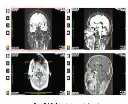
Fig. 1.MRI brain from dataset
Fig.1 explains about Computer aided design from
Fig. 2.conventional model on MR scan
2.
LITERATURE SURVEY
K.Selvanayaki et.al 2010[1] explained about MRI brain tumour detection system with automatic cad detection, in this work computer aided design has implemented with the
__________________________________
• S. Sofia saba is currently working as an Assistant Professor in G.pullaiah college of engineering and technology, Kurnool, AP, India, E-mail: [email protected]
• D. Sreelakshmi is currently working as an Assistant Professor in Narayana Engg College, Nellore, India. E-mail: [email protected]
• P. Sampath kumar is currently working as an Associate Professor in Mallareddy institute of technology, Hyderabad, India.
7077
help of segmentation on MR brain images. H. Fujita et.al 2008[2] Investigated on emerging cad system with three wings has discussed. The country of japan health care monitoring system is used this computer brain diagnosis method. But, the accuracy is need to improve. H. Arimura et.al 2012 [3] explains about MRI image analysis with machine learning technique. In this MRI medical brain image with intelligence diagnosis analysis has been improved. But, needs to improve true positive rate. E.S. El-Dahshan.et.al 2010 [8] In this research hybrid MRI brain image with signal processing techniques have used as classification and denoising methods used as a pre-processor. This technique has limitations like time of imaging, accuracy etc. Y. Zhang.et.al 2011 [9]
PROBLEM IDENTIFICATION
1. Denoising on MRI brain image 2. Risk free decision on processed data 3. Less Efficiency and PSNR in existed method
2.1 Problem solution
1. LR Machine-learning
2.2 Parameters estimated: 1. MSE
2. PSNR 3. CC 4. SSIM 5. CNR
In this research hybrid export MRI brain system has been implemented for fast and accurate system. But, the system has limitation at classification and noising. So, need to design a new application. S. Chaplot.et.al 2006 [12] Wavelet transform based MRI brain image system has implemented with the help of support vector machine. In this neural networks control action has been done based on bio-medical signal processing application.
3.
METHODOLOGY
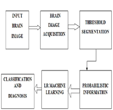
Fig. 3. Proposed block diagram
In the third step threshold based segmentation has been performed. In this segmentation threshold value has been fixed by averaging grey and white pixels of MRI brain images.
Fig. 4.median filters flow chart.
In this input brain image block requires MRI brain images from dataset. This dataset consist of 100 MRI brain images combination of normal and abnormal MRI brain scan images. In the second step image acquisition has performed based on histogram equalization and denoising methods by using adaptive median filter. This adaptive median filter consist of Yi,j as weighted MR image, Wmax is the weight maximum, Si,jmin,w, Si,jmed,w, Si,jmax,w has been computed for denoising.
__________________________________________ Algorithm1
_______________________________________________ ______
Step 1: Input image Step 2: RGB to grey image Step 3: Import local dataset
Step 4: Calculate the threshold value
Step 5: Compare with The result. If Th is less than the present pixel value, it has been decided as background, else object.
Step 6: Stop the process.
Algorithm: 2 – Classification by using Logistic Regression
By using algorithm 2 classifications of brain images has estimated. This mechanism is very useful for fast and accurate brain diagnosis system.
TABLE:1 STATISTICAL SYSTEM DATA
MRI brain image DE-NOISE RATIO
Sample -12 10.19
Sample -17 9.971
Sample -13 10.19
Sample -24 10.13
Sample -27 9.12
Sample -24 10.27
Sample -34 10.17
TABLE:2F0 AND Y-F0FUNTION
X Y F0 Y-F0
12 10.19 10.00586 0.18414
17 9.971 10.00586 -0.03486
13 10.19 10.00586 0.18414
24 10.13 10.00586 0.12414
27 9.12 10.00586 -0.88586
24 10.27 10.00586 0.26414
34 10.17 10.00586 0.16414
7079 Fig. 6. F0 vs Y-F0.
Above discussions explains that statistical estimation of X, in this X value is decided with mean of 3 elements i.e until 12. The values taken from sample numeric are like five,
seven, twelve, and twenty three, twenty five, twenty nine, thirty seven.
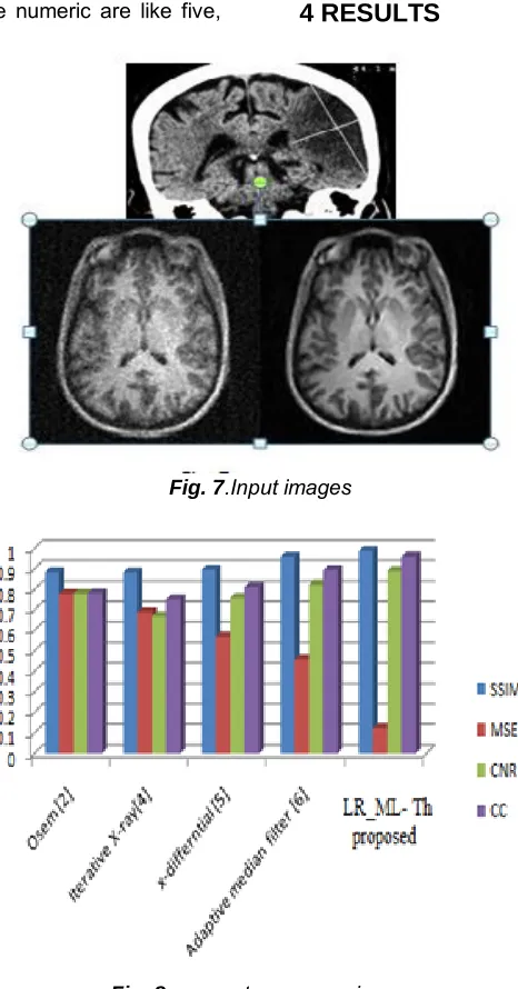
4 RESULTS
Fig. 7.Input images
Table :3 comparison of results
5 CONCLUSION
In this investigation, proposed LR-ML threshold based MRI brain diagnosis is implemented, this work consists of accurate tumour detection with fast and accurate processPast work is implanted with conventional methods so need more time to give the diagnosis of brain. So in this Research machine learning based that model is attained for fast and accurate diagnosis of brain, quality assessment parameters gives the robust accuracy and throughput. The PSNR is improved by 9.321%, SSIM improved by 2.1%, MSE=85.3%, CNR= 7.1%, CC= 7.7% has been improved, this is good improvement compared to survey methods. This investigation is very useful for brain tumour detection at fast and accurate diagnosis.
REFERENCES
[1]. K.Selvanayaki, M. Karnan, CAD System for Automatic Detection of Brain Tumor through Magnetic Resonance Image-A Review, International Journal of Engineering Science and Technology, vol.2 (2010), pp. 5890-5901.
[2]. H. Fujita, Y. Uchiyama, T. Nakagawa, D. Fukuoka, Y. Hatanaka, T. Hara, G. N. Lee, Y. Hayashi, Y. Ikedo, X. Gao, X. Zhou, Computer-aided diagnosis: The emerging of three CAD systems induced by Japanese health care needs, Computer Methods and Programs in Biomedicine, vol. 92, No. 3 (2008) , pp. 238-248.
[3]. H. Arimura, C. Tokunaga, Y. Yamashita, J. Kuwazuru, Magnetic Resonance Image Analysis f
[4]. r Brain CAD Systems with Machine Learning, Machine Learning in Computer-Aided Diagnosis: Medical Imaging Intelligence and Analysis. IGI Global (2012), pp. 258-296.
[5]. J. M. Kinser, J. L. Johnson, Stabilized Input with a Feedback Pulse-Coupled Neural Network, Optical Engineering, vol. 35 (1996), pp. 2158-2161. [6]. N. Marshkole, B. K. Singh, A.S Thoke, Texture
and Shape based Classification of Brain Tumors using Linear Vector Quantization, International Journal of Computer Applications, vol. 30, No.11 (2011).
[7]. T. Lindblad, J. M. Kinser, Image processing using pulse-coupled neural networks, Springer, Reading (2005).
[8]. Y. Zhang, S. Wang, L. Wu, A Novel Method for Magnetic Resonance Brain Image Classification Based on Adaptive Chaotic PSO, Progress In Electromagnetics Research, vol. 109 (2010), pp. 325-343.
[9]. E.S. El-Dahshan, T. Hosny, A.B. M. Salem, Hybrid intelligent techniques for MRI brain images classification, Digital Signal Processing, vol. 20 (2010), pp. 433-441.
[10].Y. Zhang, Z. Dong, L. Wu, S. Wang, A hybrid method for MRI brain image classification, Expert Systems with Application, vol. 38 (2011), pp. 10049–10053.
[11].M. Jafari and S. Kasaei. Automatic Brain Tissue Detection in MRI Images Using Seeded Region Growing Segmentation and Neural Network Classification. Australian Journal of Basic & Applied Sciences, vol. 5 (2011), pp. 1066-1079.
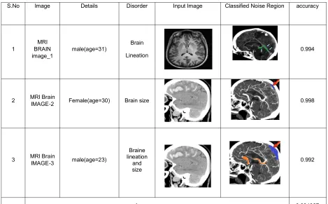
S.No Image Details Disorder Input Image Classified Noise Region accuracy
1
MRI BRAIN image_1
male(age=31) Brain Lineation
0.994
2 MRI Brain
IMAGE-2 Female(age=30) Brain size 0.998
3 MRI Brain IMAGE-3 male(age=23)
Braine lineation
and size
0.992
7081
[12]. Harvard Medical School, Web: http://med.harvard.edu/ AANLIB/
[13]. S. Chaplot, L.M. Patnaik, N.R. Jagannathan,Classification of magnetic resonance brain images using wavelets as input to support vector machine and neural network, Biomedical Signal Processing and Control, vol. 1(2006), pp. 86–92.
[14]. Inthiyaz, S., Sk, H. A., Konduri, P. S., Inani, A., Risthitha, N., Dhiraj, V., ... & Saikumar, K. (2020). A novel approach of MRI-CT Image fusion using CWT for finding Disease location. International Journal of Research in Pharmaceutical Sciences, 11(1), 497-506.
[15]. Shameem, S., RamaKrishna, T. V., Sahithi, M., Rohitha, B., Keerthana, J., Sk, H. A., ... & Saikumar, K. (2020). Design of MEMS-based Microfludic Channel to Detect Cancer Cells in Blood. International Journal of Research in Pharmaceutical Sciences, 11(1), 561-566.
[16]. Saikumar, K., & Rajesh, V. (2020). Coronary blockage of artery for Heart diagnosis with DT Artificial Intelligence Algorithm. International Journal of Research in Pharmaceutical Sciences, 11(1), 471-479.
[17]. Ahammad, Sk, V. Rajesh, K. Saikumar, Sridevi Jalakam, and G. N. S. Kumar. "Statistical analysis of spinal cord injury severity detection on high dimensional MRI data." International Journal of Electrical & Computer Engineering (2088-8708) 9 (2019).
[18]. Sai Kumar Kayam, K.Raghava Rao, Srinivasakumar Ch, Gurram narender, A Sreenivasa rao, Sk Hasane Ahammad. ―Efficient 1024-Point Low Power Radix-22 FFT Processor with MFFMD.‖ International Journal of Innovative Technology and Exploring Engineering (IJITEE) ISSN: 2278-3075, Volume-8 Issue-7, May, 2019. [19]. Saikumar, K., Rajesh, V., Ramya, N., Ahammad,
S. H., & Kumar, G. N. S. (2019). A deep learning process for spine and heart segmentation using pixel-based convolutional networks. Journal of International Pharmaceutical Research, 46(1), 278-282.