CANDIDIASIS AMONG PREGNANT WOMEN: A REVIEW
9
0
0
Full text
(2) 16. Chandrashekhar Unakal. et al., Int. J. Pharm. & H. Care Res., Vol.–02 (01) 2014 [15 - 23]. guillermondii, and Candida kyfeare commonly identified in pregnant women(2,5,6,7). Vaginal Candidiasis is common and frequent disease among women during their child bearing age; women with this age group face at least one episode of vaginal Candidiasis in their lifetime(8). Number of gravida and stage of trimester have been associated with development of vaginal Candidiasis. Studies showed that women in their 3rd trimester and multigravidae women had the highest rate of Candidiasis occurrence(2, 8). Pregnant women are more susceptible to many diseases including Candidiasis due to immune suppression and hormonal imbalance; progesterone and estrogen are the known hormones that elevate during pregnancy. These two hormones suppress the normal functioning of the body’s immune system(8). Candidaspecies are normally found in human body but there are factors that determine the organism to be pathogen to the host (human body). Virulence factors of the organism and/or predisposing factors of the host determine whether the organism remains as a commensal or become pathogen and causes disease(8, 9). A number of predisposing factors to Candidiasis have been identified by different studies. Some of these factors include, HIV AIDS, steroids and some cancer medications(1), hot weather and tight clothing, obesity, pregnancy(8), diabetes, birth control pills and overuse of antibiotics(9). Now a day Candidiasis among pregnant women is increasing due to several factors; so understanding of these factors will help the prevention, control and management of the disease among pregnant women. Therefore, the aim of this review was to document updates about the burden of the disease in pregnant women, the predominant Candida species involved, hormonal relations with pathogenesis and the virulence factor for the development of disease. Virulence factors of Candida Candida is common flora of human body in healthy individual however Candida expresses a variety of virulence factors that contribute to its pathogenesis for persistent infection and tissue damage of the host when immunity is debilitated(10,11). Major virulence factors of Candida are its ability to adapt to a variety of. habitats of the body (oropharyngea, gastro intestine and female genitalia), adherence to host cells, the ability to switch between the yeast form and filamentous (pseudo hyphae formation), biofilm formation and production of hydrolytic enzymes such as proteinases, phospholipases, lipases and other factors play a major role in successful colonization and subsequent infection of Candida.(4, 10, 11, 12, 13) Ability to adapt different anatomical site Candida species colonize and cause disease in different anatomical sites including skin, oral cavity and esophagus, gastrointestinal tract, vagina and vascular system by using different virulence factors. For example Candida albican expresses PHR1 in blood stream or in tissue to adapt the neutral PH while it expresses RPH2 in the vagina to optimize and survive at acidic pH(12). Adherence of Candida to host surfaces (adhesion) The ability of Candida to adhere to host surfaces is a prerequisite for both successful commensal carriage as well as persistence during active infection(11). Candida has the ability to adhere to several host cell types, including epithelial, endothelial and phagocytic cells. Among the different type of adhesions expressed by Candida albicans, agglutinin-like sequence which is consisted of several glycosylated proteins is very important for successful adhesion(10, 11, 12). Hyphal formation The ability to switch between the yeast form and pseudo hyphal form is one of the virulence factor of Candida species(12). Among the different species of Candida, Candida albicans and Candida dubliniensis are associated with the generation of hyphae(13). Hyphae are believed to play an important role in tissue and biomaterial invasion. Study reported that, species that do form hyphae have high ability to invade tissue and are resistant to phagocytosis(4). Hence, hyphal formation is considered to be significant to the pathogenicity of Candida. Biofilm formation Biofilms can be defined as microbial communities or aggregation of microorganisms that are often (but not necessarily) attached to a solid surface. Candida strains that have the ability to form biofilms are more virulent than others; this has. www.ijphr.com.
(3) 17. Chandrashekhar Unakal. et al., Int. J. Pharm. & H. Care Res., Vol.–02 (01) 2014 [15 - 23]. been associated with increased expression of virulence factors as well as reduced susceptibility to antimicrobial agents(4). Production of hydrolytic enzymes Candida produces several hydrolytic enzymes including secreted aspartyl proteinases, phospholipases, lipases, phosphomonoesterase and hexosaminidase. These enzymes have the capacity to degrade human proteins and help the organism to invade the human body easily hence are associated with tissue invasion (4,11,12). Pathogenesis The genus Candida has diverse species that are common residents of soil and mucosal surfaces of human gastrointestinal tract, genito-urinary tract and the mouth(14). But there are a number of factors that favors the organism to be pathogen to the host; the factors may be virulence factor of the organism or predisposing factor of the host(9). The common predisposing factors of the host are: broad spectrum antibiotic treatment, chronic corticosteroid therapy, diabetes, pregnancy, organ transplantation, Human Immunodeficiency Virus(15), oral contraceptive pills and contraception devices (diaphragm, vaginal contraceptive sponge, intrauterine device)(16). All these factors have their own impact on host either by directly suppressing the host immunity, inhibit the normal flora of the body that competes pathogen or by disturbing the normal physiology of the human body which results in progress of disease condition(15,16). During pregnancy the level of estrogen and progesterone increase. This results in immune supersession for example; progesterone has suppressive effect on neutrophils(8). As studies indicated, neutrophils have important role to defend the host from Candida infection by oxidative and non-oxidative killing mechanisms and also neutrophils are the only effector cells that can inhibit the development of Candida from yeast-tohyphae using oxidative burst mechanisms this is very crucial to prevent invasive Candida formation(10). But due to high production of progesterone during pregnancy, the normal functioning of neutrophils is impaired which in turn increases Candida colonization and infection. In addition high production of estrogen during pregnancy has its own impact on immunoglobulin secretion in the vagina which results in mucosal. surface defense failure immunoglobulins(8).. due. to. lack. of. Generally, the main reason for increased vaginal Candidiasis in pregnant women is due to elevation of reproductive hormones during pregnancy. Study revealed that vaginal Candidiasis is very rare in post-menopausal women and girls(17). But high levels of reproductive hormones during pregnancy provide an increased amount of glycogen in the vagina which enhances growth of Candida species because glycogen serves as a carbon source for Candida growth(17, 18). In addition estrogen enhances adherence of yeast to vagina by reducing immunoglobulin secretion and fungal inhibition activity of epithelial cells(8). Clinical manifestation Candidiasis shows different signs and symptoms depending on the site of infection. For example Candida infection in the skin will manifest patches on skin, itchy of skin, scabs and pustules may be seen around the edge of the rash. These clinical manifestations are commonly seen around groin, the folds of the buttocks, between the breasts, toes, or fingers. Candidiasis in oral cavity causes curdlike white patches inside the mouth, on the tongue and palate and around the lips. Clinical manifestations of Candidiasis in pregnant women are vaginal pruritis, thick crud/cheese like vaginal discharge, itching, redness, burning, swelling and pain during walking and urination(8, 9, 19). Candidiasis clinical presentation based on anatomical site of infection is presented here below: Oropharyngeal Candidiasis Oropharyngeal Candidiasis commonly called oral Candidiasis or oral thrush is an opportunistic yeast infection of the mouth, pharynx, tongue, and buccal mucosa. Oral Candidiasis is the predominant opportunistic infection in HIV infected patients and Candida albicans is the most common species identified from Oropharyngeal Candidiasis(20). Oropharyngeal Candidiasis can present in one of four forms: Pseudomembranous Candidiasis, Erythematous Candidiasis, Hyperplastic Candidiasis, and Angular cheilitis are commonly observed clinical manifestation of oral Candidiasis(11).. www.ijphr.com.
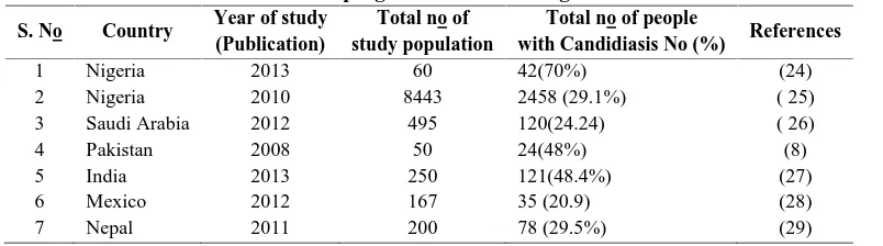
(4) 18. Chandrashekhar Unakal. et al., Int. J. Pharm. & H. Care Res., Vol.–02 (01) 2014 [15 - 23]. Candidemia Candidemia or systemic Candidiasis is the infection of blood by Candida species. Invasive Candidiasis or candidemia is the most frequent cause of morbidity and mortality in hospitalized patients(15). Invasive Candidiasis is increasing in the past two decades associates with medical advances such as invasive medical devices, organ transplantation, intravascular catheters, intensive cancer chemotherapy, broad spectrum antimicrobial therapy; mechanical ventilation and prolonged hospital stay are the common risk factors in addition to HIV/AIDS. Study reported isolation of Candida albicans, Candida glabrata, Candida parapsilosis, Candida tropicalis, and Candida Krusei from patients with systemic Candidiasis. Invasive Candidiasis is the fourth leading cause of nosocomial bloodstream infection in the United States(10,15). Vaginal Candidiasis Vaginal Candidiasis is the most common type of Candidiasis in pregnant women .The clinical manifestation of the disease is characterized by vaginal pruritis, thick crud/cheese like vaginal discharge, itching, redness, burning, swelling and pain during walking and urination(8, 9). These clinical signs and symptoms, though not specific, are important for the diagnosis of vaginal Candidiasis. Pregnant women with such clinical manifestations should be treated as early as possible because Candidiasis is reported as a risk factor for preterm birth(21). Vaginal Candidiasis can be classified as either uncomplicated or complicated. According to Center for Disease prevention and Control (CDC), approximately 10%–20% of women with vaginal Candidiasis will have complicated type that necessitates diagnostic and therapeutic considerations. Uncomplicated type of Candidiasis is sporadic or infrequent, mild-tomoderate vulvo vaginal Candidiasis and mostly. caused by Candida albican and this may occur in immuno competent women while complicated form of vaginal Candidiasis is severe form and recurrent infection is common. In complicated form of Candidiasis, non albican Candida species are common and the disease is commonly seen in diabetic, debilitated or immunosuppressed women. The outcome of complicated vaginal Candidiasis is psychosexual problems (e.g. feeling unclean or not wanting to have sex) and depression(22). Epidemiology Vaginal Candidiasis is the most common disease in pregnant women all over the world. The risk of carriage to develop disease in pregnant women is two times higher than non-pregnant women particularly during the third trimester due to changes in the levels of reproductive hormones and deposition of glycogen in the vagina(23,24). Studies show that Candidiasis is the most common cause of vaginitis; in United States it is the second leading cause of viginitis next to bacterial vaginosis and it is also common in Europe(23). Study from Spain reported 28% prevalence of Candida species among pregnant women. Of these, Candida albicans was the major isolate 90.4% followed by Candida glabrata 6.3%; Candida parapsilosis 1.1% and Candida kefyr 1.1 %(7). Other studies around the world also reveled high prevalence of the disease; Sarajevo 46.8%(17), Cuba 42.3%(23) and Kenya 42.7%(2). Most studies reported Candida albican as the most predominant cause of vaginal Candidiasis in pregnant women which accounts 8095% of Candida infection(2, 5, 11, 17,18). From non albican group, Candida glabratais the leading cause of vaginal Candidiasis in pregnant women(17). In Ethiopia however, epidemiological studies of Candidiasis are limited to HIV patients and diabetic patients. Hence, literatures about the epidemiology of Candidiasis among pregnant women are lacking.. Table No. 01: Prevalence of Candidiasis in pregnant women among different countries around the world S. No. Country. 1 2 3 4 5 6 7. Nigeria Nigeria Saudi Arabia Pakistan India Mexico Nepal. Year of study (Publication) 2013 2010 2012 2008 2013 2012 2011. Total no of study population 60 8443 495 50 250 167 200. www.ijphr.com. Total no of people with Candidiasis No (%) 42(70%) 2458 (29.1%) 120(24.24) 24(48%) 121(48.4%) 35 (20.9) 78 (29.5%). References (24) ( 25) ( 26) (8) (27) (28) (29).
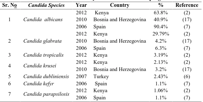
(5) Chandrashekhar Unakal. et al., Int. J. Pharm. & H. Care Res., Vol.–02 (01) 2014 [15 - 23]. During this review, there were few studies on Candidiasis in Jimma(30), Addis Ababa(31) and. 19. Gondar(32) among HIV patients and among diabetic patient in Gondar(33) as well.. Table No. 02. Common Candida species identified from pregnant women Sr. No. Candida Species. 1. Candida albicans. 2. Candida glabrata. 3. Candida tropicalis. 4. Candida krusei. 5 6. Candida dubliniensis Candida kefyr. 7. Candida parapsilosis. Year 2012 2010 2006 2012 2010 2006 2012 2012 2010 2007 2006 2012 2006. Diagnosis of Candidiasis Candidiasis can be diagnosed based on the clinical feature of the disease. Although the signs and symptoms of Candidiasis are relatively nonspecific, clinical manifestations of Candida infection serve as a diagnostic approach of the disease(34, 35,36). Microscopic examination of normal saline or 10% potassium hydroxide wet mount preparation and gram stained preparation from vaginal discharge of clinically positive pregnant women are basic tests for the diagnosis of Candidiasis(8, 18, 36). Microscopic examination identifies only the presence of Candida but do not isolate the species. Hence, further identification methods are necessary to identify the species of Candida such as culture using Sabourand dextrose agar (SDA) which can support growth of Candida and suppresses the growth of many bacteria due to its low pH and antibiotic content(2,6). It is also possible to use CHROM agar to detect mixed cultures(17). CHROM agar is a fungal culture media which can be used to identify species based on the reaction between different species of Candida and chromogenic substrate after incubation at 37 ˚C; different Candida species produce different type of colonies with different color(17). Germ tube test is also another type of Candida species identification method which helps to identify hyphae former Candida species such as Candida albican and Candida dubliniensis; hyphae formers produce hyphae when incubated at 35 ˚C in serum for 2-4 hour(6, 17). Moreover, yeast assimilation test is the safest identification method of non albicans species. This test is based on the ability of yeast to assimilate organic compounds(17).. Country Kenya Bosnia and Herzegovina Spain Kenya Bosnia and Herzegovina Spain Kenya Kenya Bosnia and Herzegovina Turkey Spain Kenya Spain. % 63.8% 40.9% 90.4% 29.79% 4.2% 6.3% 3.19% 2.13% 3.2% 2.43% 1.1% 1.06% 1.1%. Reference (2) (17) (7) (2) (17) (7) (2) (2) (17) (6) (7) (2) (7). Diagnosis of invasive Candidiasis is not simple because clinical presentation of invasive Candidiasis is not specific and up to 50% blood cultures can be negative or may only become positive late in the infection. Hence invasive Candidiasis can be diagnosed using different methods such as antibody detection, though it is not sensitive in immune compromised individuals and nonspecific to superficial colonization. Detection of antigens such as extracellularly secreted aspartyl proteinase which is produced by Candida albicans and some other Candida species are target for diagnosis of invasive Candidiasis. Histo pathological examination of tissue sections is also one of the most reliable methods of establishing a diagnosis of systemic fungal infection(37). Rapid test kits are also available for the diagnosis of Candida; for example Immunologic Latex Agglutination Test is the one used for Candida diagnosis(23). Now days there are also more sensitive and specific molecular techniques such as polymerase chain reaction (PCR) and 18S rRNA gene clone library methods which allow identification of drug resistant Candida in addition to species identification. The basic principle of PCR is amplifying of the target DNA in high amount with in different temperature ranges(28, 33,38). Vulvovaginal Candidiasis diagnosis and treatment algorism indicated physical examination for abnormal pelvic findings as a primary step. When the patient has abnormal pelvic finding direct microscopy will be considered. Those positive for. www.ijphr.com.
(6) 20. Chandrashekhar Unakal. et al., Int. J. Pharm. & H. Care Res., Vol.–02 (01) 2014 [15 - 23]. direct microscopy plus pH value of discharge <4.5 and no excess of white blood cells in the discharge, atimycotic treatment will be initiated. On the other hand in the presence of positive microscopic examination, if pH of discharge is >4.5 and excess of white blood cells exist in the discharge, mixed infection is considered. When the patient is negative for microscopic examination and the pH of the discharge is <4.5 with no excess of white blood cells in the absence of trichomonads infection and clue cells, while culture considered antimycotic treatment will be initiated for the patient(18). Despite the availability of different diagnostic modalities, in Ethiopia however, Candidiasis is being diagnosed clinically and through microscopic identification of the yeast in clinical samples. Therefore, evaluation of different diagnostic modalities and selection of the best Candidasis diagnostic method among pregnant women in developing countries like Ethiopia has a paramount significance for proper management of the disease. Treatment of Candidiasis in pregnant women Antifungal drug choice is determined by several factors including the patient’s medical history, specific symptoms of Candidiasis, severity of infection, predicted compliance with application method, and drug sensitivity pattern of the isolated Candida species(35). There are several types of antifungal agents commercially available for the treatment of Candidiasis which include the following: imidazole antifungals (eg, butoconazole, clotrimazole, miconazole), triazole antifungals (eg, fluconazole, terconazole), and polyene antifungals (eg, nystatin). Study conducted in 1997 recommend topical antifungal agents for the treatment of Candidiasis in pregnant women while it discourages use of oral antifungal agents because of fetal complications(35). To the contrary, prospective and observational studies proves that using these antifungal drugs during pregnancy is not associated with increased risk of major malformations although there was case report due to high dose of oral antifungal agents(39). Other study reported that even though it is possible to use many type of topical antifungal agents, topical imidazole appears to be more effective than nystatin for treating symptomatic vaginal Candidiasis in pregnancy(40). Different studies. agree on seven days antifungal topical therapy as the best for treating Candidiasis in pregnant women rather than shorter duration since short term therapy is associated with treatment failures(39,40). Treating of Candida in pregnant women has significant role to reduce preterm birth in addition to maintaining mothers’ health. Study showed that Candida colonization is a risk factor for preterm birth and infant mortality; hence treating of pregnant women reduces preterm birth(2, 41). Retrospective analysis of data in Hungary revealed that 34-64% of preterm birth was prevented by applying vaginal clotrimazole treatment of pregnant women who had vaginal Candidiasis(42). Antifungal susceptibility testing can be performed using different methods such as broth microdilution method this can be performed as follows : prepare inoculums suspension adjusted to a 0.5 McFarland Standard, dilute the working suspension into 1:100 dilution, add 10µl suspension with 10µl sterile normal saline then mix suspension and pour into an inoculation tray in Tryptose Soya Broth, then shake the microtiter plate on a plate shaker for 30 seconds to ensure even distribution of the inoculums, incubate at 37°C for 48 hours, take out the microtiter plate from the incubator and shake for 5 minutes. Finally the minimum inhibitory concentrations will be determined by reading the optical density of each well at 530 nm spectrophotometrically(43, 44). The second method is using modified disc diffusion method, by adding 2% glucose and methylene blue to Mueller-Hinton agar to accelerate growth of Candida and inhibit the growth of bacteria. The test can be performed as follows: prepare colony from an overnight subculture on SDA suspension in sterile normal saline and vortex (mix) which should be comparable to 0.5 McFarland Standard, then dilute 1:2 with sterile saline solution, then inoculate the Mueller-Hinton agar with moistened cotton swab then allow to dry for around 15 mint, place antifungal discs on surface of Mueller-Hinton agar then incubate the plates at 37oC for 24 hours finally measure the zone of inhibition using ruler(45). Empiric antifungal therapy to treat vaginal Candidiasis could act as risk factor for increased resistance of Candida species to antifungal agents; since all Candida species do not have equal susceptibility pattern. For example antifungal susceptibility studies done in Kenya and Brazil. www.ijphr.com.
(7) Chandrashekhar Unakal. et al., Int. J. Pharm. & H. Care Res., Vol.–02 (01) 2014 [15 - 23]. reveled that Candida krusie was resistant to ketoconazole(2,44). Hence during treatment of Candidiasis antifungal susceptibility test is necessary because all Candida species may not be susceptible to different antifungal drugs. Since drug resistance pattern of Candida species is variable to the different types of drugs used for the treatment of Candidiasis, susceptibility testing is very important to select appropriate antifungal agents. Study conducted to determine antifungal susceptibility of different Candida species revealed that Candida albicans, Candida kefyr and Candida parapsilosis were susceptible to fluconazole, ketoconazole, itraconazole and nystatin while 1 in 6 Candida glabrata isolates showed resistance to azole drugs(7). Other study conducted to determine the susceptibility patternof Candida species result reported that Candida albican was susceptibility to Clotrimazole while resistance to Fluconazole, Ketoconazole, and Econazole was detected. Most of other Candida species show resistance against azoles specially among most isolates of Candida glabrata was resistant to Ketoconazole, Clotrimazole, Fluconazole and Econazole(44,45).. drug sensitivity pattern of Candidaspecies isolated because some Candid species are resistant to one or more antifungal agents. During this review, there were no literatures that show epidemiology of Candidiasis among pregnant women in Ethiopia. Therefore, epidemiological studies regarding Candidiasis among pregnant women have paramount significance.. References 1.. 2.. 3.. 4.. Conclusion Pregnant women are more vulnerable to vaginal Candidiasis due to reproductive hormone elevation which suppresses the normal activity of the body’s immune function and enhances the growth of Candida by increasing glycogen level in the vagina. Although Candidiasis has different clinical presentations vaginal Candidiasis is the predominant one in pregnant women. Candidiasis can be diagnosed using a combination of different methods such as clinical sign and symptom, microscope, culture, serology (antigen & antibody detection), germ tube test, sugar assimilation and PCR. Antifungal susceptibility testing can be performed using broth micro-dilution or modified disc diffusion Mueller-Hinton agar method. Empirical antifungal therapy is a risk for drug resistanceamong Candida species. Most of non albican Candida species are resistant to azole drugs. Seven days treatment is necessary to cure pregnant women and effective treatment reduces the risk of preterm birth.. 21. 5.. 6.. 7.. 8.. 9.. Recommendations Vaginal Candidiasis is common in pregnant women therefore early diagnosis and treatment should be practiced. Antifungal treatment should be based on. www.ijphr.com. Shaheen MA, Taha M. Species identification of candidia isoltes obtained from oral lesions of Hospitalized and Non Hospitalized patients with oral Candidiasis. Egyptian Dermatology online Journal 2006; 2(1):1-11. Chengo NM. Isolation, identification and susceptibility profile of Candida species to antifungal agents in pregnant women in Thika District Hospital, Kenya. Available at http://irlibrary.ku.ac.ke/handle/123456789/3973. Jacqueline M A, Bettina CF. Candida infection of the genitourinary tract. Clinical Microbiology Review2010; 23(2):253-273. David WW, Tomoari K, Sonia S, Sladjana M, Michael A OL. Candida biofilms and oral candidosis: treatment and prevention. Periodontology 2011; 55:250–265. Ibrahim SM, Mohammed B, Yahaya M, Audu BM, Ibrahim HA. Prevalence of vaginal Candidiasis among pregnant women with abnormal vaginal discharge in Maiduguri. Nigerian Journal of Medicine 2013; 22(2):138-42. Us E, Cengiz, SA. Prevalence and phenotypic evaluation of Candida dubliniensis in pregnant women with vulvovaginalcandidosis in a university hospital in Ankara. Mycosis 2007; 50: 13–20. García HM, García SD, Copolillo EF, Cora EM, Barata AD, Vay CA, Et.al.Prevalence of vaginal Candidiasis in pregnant women, Identification of yeasts and susceptibility to antifungal agents.Revista Argentina de Microbiologica2006; 38 (1):9-12. Aaleeha A, Rubeena H, Sadia I and Tahir M.Vulvovaginal Candidiasis in pregnancy.Biomedical Journal 2008;24:54-56. Alli JAO, Okonko IO, Odu NN, Kolade AF, Nwanze JC. Detection and prevalence of Candida isolates among patients in Ibadan, Southwestern Nigeria.Journal of Microbiology and Biotechnology2011; 1(3):176-184..
(8) 22. Chandrashekhar Unakal. et al., Int. J. Pharm. & H. Care Res., Vol.–02 (01) 2014 [15 - 23]. 10. Michail SL, Mihai GN .Candida and Host Determinants of Susceptibility to Invasive Candidiasis. PLOS. 2013; 9 (1):1-5. 11. David W, Michael L. Pathogenesis and treatment of oral candidosis. Journal of Oral Microbiology 2011; 3:4. 12. Yun-Liang Y. virulence factor of Candida species. Journal of Microbiology immunology Infect.2003; 36:223-228. 13. Jackson AP, Gamble JA, Yeomans T, Moran GP, Saunders D,Harris D, et al. Comparative genomics of the fungal pathogens Candida dubliniensis and Candida albicans. Genome Research2009; 19: 2231-44. 14. Lucy AN.Diversity of Pathogenic Candida Species Colonizing Women with and without Candida Vaginitis in Dares Salaam Region, Tanzania.Journal of Biology and Life Science2013; 4(1):153. 15. Mukta NC, Prabha A, Rajeev A, Ashok KS. Study of risk factors and prevalence of invasive Candidiasis in a tertiary care hospital. Indian Society of Critical Care Medicine2007; 11(2):67-73. 16. Dariane CP, Luana THB, Luciane NC and Alexandre MF. A six year epidemiological survey of vulvovaginal Candidiasis in cytopathology reports in the state of Rio grandedosul, Brazil. Abr.2012; 41 (2): 163168. 17. Babic M, Hukic M.Candida albicans and nonalbicans species as etiological agent of vaginitis in pregnant and non-pregnant women. Bosnian Journal of Basic Medical Science 2010; 10(1):89-97. 18. Jack DS. Vulvovaginalcandidosis. Lancet 2007; 369: 1961–71. 19. Okonkwo NJ. Prevalence of Vaginal Candidiasis among Pregnant Women in Nnewi Town of Anambra State, Nigeria. International Multi-Disciplinary Journal2010; 4(4) 539-548. 20. Sow PG, Coume M, Gaye, Dia A, Traore AT I. Prevalence and Distribution of Candida Species in HIV Infected Persons on Antiretroviral Therapy in Dakar.International Journal of Modern Biology and Medicine2012; 1(3): 145-155. 21. Christine LR, Jonathan MM, Kristen RR, Warwick BG, Judy MS, George Ket al.Protocol for a randomised controlled trial of treatment of asymptomatic Candidiasis for the. 22.. 23.. 24.. 25.. 26.. 27.. 28.. 29.. 30.. www.ijphr.com. prevention of preterm birth. BMC Pregnancy and Childbirth 2011;11(19):11-19. MeReC B. An update on vulvovaginal Candidiasis (thrush). National prescribing center2004;14 (4):13-16. Octavio FL, Dra M, Isela L. Prevalence of Candida albicans and Trichomonasvaginalis in Pregnant Women in Havana City by an Immunologic Latex Agglutination Test.Medscape General Medicine 2004; 6(4): 50. Oyewole OA, Okoliegbe IN, Alkhalil S, Isah P. Prevalence of Vaginal Candidiasis among Pregnant Women Attending Federal University of Technology, Minna, Nigeria, Bosso Clinic. Research Journal of Pharmaceutical, Biological and Chemical Sciences 2013; 4(1):113-120. Jombo GTA., Opajobi SO, Egah DZ, Banwat EB, Denen PA. Symptomatic vulvovaginal Candidiasis and genital colonization by Candida species in Nigeria. Journal of Public Health Epidemiology. 2010; 2(6):147-151. . Raid AA, Talat AE, Yazeed AA, ZiabZakey A. Prevalence and comparison for detection methods of Candida species in vaginal specimens from pregnant and non pregnant Saudi women. African Journal of Microbiology Research 2013; 7(1): 56-65. Deepa B, Subbannayya K, Sunil R, Rao TV. Clinico- mycological profile of vaginal Candidiasis in a tertiary care hospital in Kerala. International Journal of Research in Biological Science. 2013; 3(1): 55-59. Saraín MAR, Peggy EAG, Víctor MVV, Beatriz XC, Yolanda CP. A molecular epidemiological study of Prevalence of Candida spp. in women in the City of Tuxtla Gutierrez, Chiapas. International Biotechnology Color Journal2012; 2(2): 6- 14. Shrestha S, Tuladhar NR, Basnyat S, Acharya GP, Shrestha P , and Kumar P. Prevalence of vaginitis among pregnant women attending Paropakar Maternity and Women’s Hospital, Thapathali, Kathmandu, Nepal. Nepal Medical College Journal2011; 13(4): 293-296. Lissane S. Socio-demographic and clinical profile of AIDS patients in Jimma referral Hospital, Southwest Ethiopia. Ethiop. J. Heath Dev. 2004; 18(3): 204-205..
(9) Chandrashekhar Unakal. et al., Int. J. Pharm. & H. Care Res., Vol.–02 (01) 2014 [15 - 23]. 31. Guteta S, Feleke Y, Fekade D, Neway M, Diro E. Prevalence of oral and perioral manifestations in HIV positive adults at TikurAnbessa Teaching Hospital Addis Ababa, Ethiopia. Ethiop Med J. 2008; 46(4):349-57. 32. Debasu D, Gizachew Y, Desalegn W, Belay A. Common opportunistic infections and their CD4 cell correlates among HIV-infected patients attending at antiretroviral therapy clinic of Gondar University Hospital, Northwest Ethiopia.BMC Research Notes 2013, 6:534. 33. Gizachew Y, Daniel A, Yimtubezinash W, Chandrashekhar U.Prevalence of Candiduria in Diabetic Patients Attending Gondar University Hospital, Gondar, Ethiopia. Iranian Journal of Kidney Diseases 2013; 7:102-7. 34. Wenjin Q, Yifu S. Epidemiological study on vaginal Candida glabrata isolated from pregnant women .Scandinavian Journal of Infectious Disease2006; 38(1):49-54. 35. Sebastian F, Joseph A, Nancy B, Kelly E, Mark GM, Susan MM Et.al Treatment Considerations in Vulvovaginal Candidiasis. The Female Patient 1997; 22:1-17. 36. Gondo F, da Silva M.G, Polettini J, Tristao AR, Peracoli JC, Witkin SS Et.alVaginal Flora Alterations and Clinical Symptoms in Low-Risk Pregnant Women. Gynecologic and obstetrics investigation2011 (3); 71:158–162. 37. Arjuna NB, Christine JM. Laboratory Diagnosis of Invasive Candidiasis. The Journal of Microbiology 2005; 43(1): 65-84.. 23. 38. Zheng NN, Guo XC, Lv W, Chen XX, Feng GF. Characterization of the vaginal fungal flora in pregnant diabetic women by 18S rRNAsequencing. European Journal of Clinical Microbiology & Infectious Diseases2013; 32(8):1031-40. 39. Derrick S, Adrienne E. Vaginal yeast infections during pregnancy. Can Fam Physician 2009; 55 (3) 255-256. 40. Young G, Jewell D. Topical treatment for vaginal Candidiasis (thrush) in pregnancy. TheCochrane Collaboration 2013; 2:1-14. 41. Christine L.R, Kristen R, George K, Jonathan MM. Treatment of asymptomatic vaginal Candidiasis in pregnancy to prevent preterm birth. BMC Pregnancy and Childbirth 2011; 11:18:1-6. 42. Czeizel AE, Fladung B, Vargha P. Preterm birth reduction after clotrimazole treatment during pregnancy. European Journal of Obstetrics, Gynecology, and Reproductive Biology 2004; 116(2):157-63. 43. Andargachew M, AfeworkK, Belay A, Beyene M, Aschalew G,Martha A.Frequent detection of ‘azole’ resistant Candida species among late presenting AIDS patients in northwest Ethiopia.BMC Infectious Diseases 2013; 11(82):5. 44. Luciana B D, Márcia de Souza CM, Maria WS, José MF, Rosane CH. Vulvovaginal Candidiasis in MatoGrosso, Brazil: pregnancy status, causative species and drugs tests. Brazilian Journal of Microbiology20011; 42(4):3. 45. Rehab S, AL-Maliki, Zouhair I, AL-Ani. Antifungal resistance of Candida species isolated from Iraqi women infected with vulvovaginal Candidiasis. QMJ 2011;7 (11): 117-127.. www.ijphr.com.
(10)
Figure
Related documents
Speaker; Left vs. Right), whether the probe name matched the sentential referent (Name.. Match; Match vs. Mismatch), the point at which the probe was played in the sentence.
Between September 2002 and December 2002, a subgroup Bb with 25 patients was randomly compared with patients receiving classical vaginal hysterectomy with general anesthetic
three-dimensional cell culture and anticancer drug activity evaluation in a microfluidic chip. Lee, S.-A., Kang, E., Ju, J., Kim, D.-S., and Lee, S.-H., Spheroid-based
According to the results and the importance of barbering job and requirement to regarding occupational health to maintain the health security and society's health by
In such cases, the expert is in fact faced with the same problem as the one which consists of the identification of a very short handwritten text, one or two
Efficacy of Patrapinda Sweda and Matra Basti (Combined Therapy) in the Management of Sandhivata (Osteo Arthritis). Source of support: Nil, Conflict of interest:
Our comparison of patients’ self-reported history of CRC screening and physicians’ reports showed dependable agreement in the types of tests and concordance in the
9 shows scattering parameters of the proposed filter with surface mount capacitor using ADS simulator, which are very close to the scattering parameters derived from CST Microwave
