83
IMPLICATION OF PROSTATE SPECIFIC ANTIGEN IN BENIGN PROSTATIC
HYPERPLASIA
SINGH SK
1, PRABHU GGL
21
Department of Surgery, BP Koirala Institute of Health Sciences, Dharan, Nepal 2
Department of Urology, Kasturba Gandhi Medical College, Mangalore, Karnataka, India
ABSTRACT
Background: Prostate-specific antigen (PSA) is the prostatic tumour marker of choice. Because PSA is not prostate cancer specific and prostate cancer develops in man at an age when the prevalence of benign prostatic hyperplasia is high, several parameters have been developed and investigated to enhance the sensitivity and specificity of the PSA test. The parameters included are PSA density (serum PSA concentration divided by the size of the prostate gland), PSA velocity (change in serum PSA over time), age specific reference ranges for serum PSA, percent free PSA, Digital rectal examination.
Aim and Objectives: The aim of this study is to find out the implication of prostate specific antigen in benign prostatic hyperplasia.
Materials and Methods: In this research, a correlative study of total serum PSA with digital rectal examination, prostatic volume, Transabdominal Ultrasonography, urine analysis with culture and sensitivity and histopathological examination of the patients with prostatic hyperplasia was done in 100 patients.
Here our exclusion criterion was the patients with carcinoma of prostate. PSA was estimated in fasting blood sample and was collected when patient is without Foleys catheter or with catheter (24 hours after catheterization). It was estimated by Ciba Corning Automated Chemiluminescence System (ACS) using both monoclonal and polyclonal anti-PSA antibody titers.
Results and conclusion: Age specific rise in Serum prostate specific antigen level was found. The prostatic volume was highly significant in correlation with Serum prostate specific antigen. Digital Rectal Examination when compared with other parameters was found not to be significant in predicting the prostatic disease.
The Total Serum PSA level done in all the cases does not show significance and none of the patients had malignancy even with Serum PSA> 4ng/ml. When prostate specific antigen density was calculated and compared with biopsy report positively, it was significant and was helpful in predicting the prostatic diseases and prognosis. With this we conclude that clinical correlation with Digital Rectal Examination and Transabdominal Ultrasound is very much helpful in predicting malignancy than totally relying on Serum PSA. We found that PSA is neither organ specific chemical nor disease specific.
Keywords: Benign prostatic hyperplasia, Transabdominal ultrasonography, Total Serum PSA, Digital rectal examination
INTRODUCTION
Prostate is an accessory gland of male reproductive system, secretion of which adds to the bulk of seminal fluid. It is firm due to dense fibromuscular stroma in which the complex glandular acini are embedded. Prostatic secretion includes: Water and Electrolytes (Calcium, Potassium, Sodium chloride, Phosphate, Bicarbonates), Zinc- mean content in prostate (692mg/100gm) of dry weight, Polyols i.e. sorbitol and
inositol, nitrogenous bases i.e. choline and its derivatives and spermine and spermidine that distinguish human semen from other body fluids, lipids, Prostaglandins, citric acid –maintains an osmotic equilibrium of semen, proteins, fibrinolysin and fibrinogenase–maintains the semen in liquid state during ejaculation, proteolytic enzymes, phosphatase, glucoronidase, prostate specific antigen (PSA). Prostate gland remains relatively small throughout childhood and begins to grow at puberty under the influence of testosterone. The gland is almost stationary by the age of 20yrs and remains at this size upto the age of approximately 50yrs. At this time in some men it begins to involute along with the decreased production of testosterone by the testis. However, benign *Corresponding author:
Email: [email protected]
84
prostatic adenoma frequently develops in the prostate in older men.Benign Prostatic Hyperplasia (BPH) is the commonest benign tumor in the ageing male and is universal after the age of 80 yrs. It is symptomatic in 25% of the total male population and in 50% of those aged 80 years and more, however the incidence is rising [1]. The cellular features of BPH are:- Cell clusters flat and cohesive, honeycombing prominent, myoepithelial cells present, detached single epithelial cells with normal nuclei, Nuclei- round, oval and equal in size, Mitosis-absent. Other cells include inflammatory cells, corpora amylacea, squamous cells and epithelial contaminants from the rectal mucosa and cells of the seminal epithelium. Prostatic secretion includes: Water and Electrolytes (Calcium, Potassium, Sodium chloride, Phosphate, Bicarbonates), Zinc- mean content in prostate (692mg/100gm) of dry weight, Polyols i.e. sorbitol and inositol, nitrogenous bases i.e. choline and its derivatives and spermine and spermidine that distinguish human semen from other body fluids, lipids, Prostaglandins, citric acid –maintains an osmotic equilibrium of semen, proteins, fibrinolysin and fibrinogenase–maintains the semen in liquid statduring ejaculation, proteolytic enzymes, phosphatase, glucoronidase, prostate specific antigen (PSA). Prostate gland remains relatively small throughout childhood and begins to grow at puberty under the influence of testosterone. The gland is almost stationary by the age of 20yrs and remains at this size upto the age of approximately 50yrs.
At this time in some men it begins to involute along with the decreased production of testosterone by the testis. However, benign prostatic adenoma frequently develops in the prostate in older men.Benign Prostatic Hyperplasia (BPH) is the commonest benign tumor in the ageing male and is universal after the age of 80 yrs. It is symptomatic in 25% of the total male population and in 50% of those aged 80 years and more, however the incidence is rising [1]. The cellular features of BPH are:- Cell clusters flat and cohesive, honeycombing prominent, myoepithelial cells present, detached single epithelial cells with normal nuclei, Nuclei- round, oval and equal in size, Mitosis-absent. Other cells include inflammatory cells, corpora amylacea, squamous cells and epithelial contaminants from the rectal mucosa and cells of the seminal epithelium. Prostatitis is another disorder associated with prostate gland. In
acute prostatitis, the prostate is enlarged, soft and spongy. Congestion and boggy suppuration of the entire gland occurs. There are minute, disseminated abscesses seen as well, coalescent focal areas of necrosis and diffuse edema of the gland. Chronic prostatitis is characterized by the presence of numerous aggregates of lymphocytes, plasma cells, macrophages and neutrophils within the substance of the prostate.
85
glandular ducts secretes PSA and effectively prevents the escape of the protease into the general circulation. However, small amounts of PSA leak into the circulation and this increase in prostate disease. The ratio between the PSA concentration in seminal plasma(0.5-3g/L) and in serum (<4g/1) is approximately 106:1 in healthy men [7]. Gann et al [10] have shown that men with a baseline serum PSA between 2-4ng/ml are more than 12 times more likely to be diagnosed with prostate cancer during next 10 years than men with PSA level less than 1ng/ml. Catalona et al [11] found pathologically organ- volume in cubic centimeters) has been suggested as one possible method to improve the specificity of serum PSA testing. Seamen and associates [12] reported that PSA density was significantly higher in men with prostate cancer than in those without prostate cancer, despite having similar mean serum PSA levels. Smith and associates [13] showed that a cut-off of 0.09 yielded a sensitivity of 90% and a specificity of 40% for men in an early detection study in which serum PSA levels are between 4-10ng/ml. Using a PSA slope (PSA velocity) cut off of 0.75 ng/ml per year would have yielded a specificity of 90% for prostate cancer detection, and this was confirmed by Smith and Catalona [14]. Using age-specific reference 4-10ng/ml, f-PSA/t-PSA ratio significantly differentiated between those with benign and malignant conditions. Catalona and associates [16] stated that using a percent f-PSA cut-off of 20% or less, 38% of biopsies could have been eliminated and more than 90% of cancers still detected. Epstein et al [17] stated that serum PSA levels, PSA-density, and needle biopsy pathologic findings are accurate predictors of tumour extent. It may be reasonable to follow up some patients whose tumours are most likely insignificant with serial PSA measurements and repeated biopsies. In this study, we have employed the serum PSA level (in ng/ml), digital rectal examination, ultrasonologically measured prostate volume and correlated the values and analyzed with histopathology reports.
The aim of this study is to find out the implication of prostate specific antigen in benign prostatic hyperplasia.
MATERIALS AND METHODS
In this research, a correlative study of serum PSA with digital rectal examination, prostatic volume and histopathological examination of the patients with prostatic hyperplasia was done. This study was done in the inpatients of K.M.C Hospital, Mangalore, Karnataka, India. We studied 100 patients in total who came with various complain and were examined clinically thoroughly with main stress on digital rectal
examination (DRE) to access the nature of the prostate. Then all these patients were subjected to various investigations which included the Total Serum PSA, Transabdominal Ultrasonography, urine analysis with culture and sensitivity along with trans-rectal biopsy of prostate wherever clinically indicated. Here our exclusion criteria was the patients with carcinoma pf prostate. Then the patients were subjected to the indicated management and followed up. We also treated the co-morbid associated diseases in these patients.The main component of this study, the total Serum PSA was estimated once in each patient due to financial constraints. PSA was estimated in fasting blood sample and was collected when patient is without Foleys catheter or with catheter (24 hrs after catheterization)
It was estimated by Ciba Corning Automated Chemiluminescence System (ACS) using both monoclonal and polyclonal anti-PSA antibody titers. These values are then correlated with clinical examination, prostate volume and histopathological diagnosis of the prostatic hypertrophy.
RESULTS
This is a retrospective random study of 100 cases admitted at the K.M.C Hospital, Mangalore. These patients are mostly from South Kanara District and other neighboring Districts of Kerala. Majority of the patients belong to lower and middle socio-economic strata of mixed occupations.
AGE INCIDENCE
Benign Prostatic Hyperplasia manifested clinically between the age of 39 years and 95 years in this series. The maximum incidence however was between 60-70 years. Of the 100 cases studied, 44 belongs to this group giving a percentage of 44%. Mean age in our study was 68 years. It is detailed in table 1.
URINE CULTURE
This was done in all the patients irrespective of symptoms and sign of urinary tract infection (UTI). From our study E. Coli was the commonest organism causing urinary tract infection, which was 10%. In our study 78% of our patients did not have any growth on culture. The detail of urine culture is shown in table 2.
SERUM PROSTATE SPECIFIC ANTIGEN (PSA)
86
4ng/ml. The PSA level in our study population is tabulated in table 3.TRANSABDOMINAL ULTRASONOGRAM
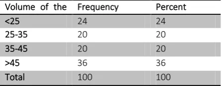
In this series we have done transabdominal ultrasonogram to estimate the volume of the prostate gland. It was calculated by the Ellipsoid formula: V= Length × Height × Width × 0.52 Most of the patients had Grade-III prostatic enlargement. Minimum USG volume was 12gms and maximum was 156gms. Mean weight in our study was 45gms. The ultrasonogram result is shown in detail in table 4.
DIGITAL RECTAL EXAMINATION
Digital rectal examination was one of the main clinical criteria where we found that most of the patients were having Grade-II prostatomegaly, prostate was firm in consistency, no nodule found and mucosa was free. The detail about grading of prostatomegaly is shown in table 5.All the patient underwent TURP in our study group. 17% of our patient underwent Transrectal
biopsy of prostate on clinical suspicion, however none of them had malignancy.
CORRELATIVE STUDY
Transabdominal Ultrasonography (TAUSG) Vs DRE This was done to know the significance of DRE in prostatomegaly when compared with the help of Transabdominal Ultrasonography. In our study it was found to be highly significant. Results are depicted in table 6.
RELATIONSHIP OF PSA AND DRE:
This was done to know whether there was any variation of Serum PSA level in clinically graded prostatomegaly. From our study it is found that PSA was not significantly raised and was not significant with digital rectal examination. The relationship is shown in table 7.
RELATIONSHIP OF USG VOLUME VS PSA
In our study, it was found that as the grade of
prostate increases, PSA value also increases. USG
volume is also highly significant in our study. The
correlation is shown in table 8.
Table 1 Serum Prostate Specific Antigen level in the study population
Serum PSA level
Frequency Percent
<4 17 17
4-10 42 42
10-25 33 33
≥25 8 8
Total 100 100
Table 2 volume of the prostate gland in the study population
Volume of the
prostate gland
Frequency Percent
<25 24 24
25-35 20 20
35-45 20 20
>45 36 36
Total 100 100
Table 3 Incidence of Benign Prostatic Hyperplasia according to age group
Age group Frequency Percent
<50 4 4.0
50 - 60 13 13.0
60 - 70 44 44.0
>70 39 39.0
Total 100 100%
Table 4 Organism causing the urinary tract infection in our study population according to the urine culture report
Culture isolates Frequency Percent
No growth 78 78
Acinetobactor species 1 1
Candida species Significant 2 2
Citobacter species Significant growth
1 1
E. Coli significant 10 10
Enterobacter significant 1 1
Klebsiella Significant 1 1
Mixed flora 2 2
Pseudomonas significant 4 4
87
DISCUSSION
In this series we have done comparative study of Serum Prostate Specific Antigen (PSA) concentration and prostatic volume with the help of DRE & TAUSG and correlated histopathology in the diagnosis of prostatic diseases. For this study we collected 100 cases from KMC Hospital, Mangalore. All the patient were admitted in the hospital. We have done retrospective random selection of patient. From these cases we collected the various parameters which were required for the study apart from routine data.
AGE
From our series of 100 cases who were diagnosed clinically as prostatic enlargement by Digital Rectal Examination (DRE), we found that prostatic hypertrophy can appear in the age group of 39−95. Of these, most cases i.e 44% were in the age group of 60-70 years. The mean age was 68 years.
The age of the patient when correlated with Serum PSA value, in our study showed significance. In our study there is significant rise in Serum PSA value as the age advances. Similar study done by Babaian RJ et al [18] in 1992, also showed there is significant rise in
Serum PSA value as the age advances. However, glandular dynamics among aging men, suggest that regular PSA monitoring could assist in the assessment of malignant potential and with decisions on
appropriate further management. Here our study showed that clinically DRE and TAUSG could be equally beneficial in diagnosing BPH.
TRANSABDOMINAL ULTRASONOGRAPHY:
In our series most of the patients had Grade-III prostatic enlargement. Minimum USG volume was 12gms and maximum was 156gms. Mean weight in our study was 45gms. In correlation there was progressive rise in Serum PSA as the weight of the prostate increases. In our study it was highly significant.
SERUM PROSTATE SPECIFIC ANTIGEN:
Cooner and associate reported that 20% of men with prostate cancer had normal (<4ng/ml) serum PSA level. Brawer et al found 32% of men with prostatic cancer had normal Serum PSA level. Catalona et al [11] found that pathologically organ confined cancers in 63% of men undergoing initial or serial PSA based screening. From our study, we found that other than Serum PSA level, clinical & radiological parameters are equally important in evaluating patient with prostatomegaly.
Table 8 Grade of prostatomegaly in the
study population
Grade
of
prostatomegaly
Frequency Percent
1
36
36
2
40
40
3
23
23
4
1
1
Total
100
100
Table 7 comparison of transabdominal
Ultrasonography with digital rectal
examination
N
mean±SD
P
value
1
36
28.25±11.44
<0.001
2
40
44.77±23.66
<0.001
3
23
70.06±32.48
<0.001
4
1
89.00±
<0.001
Table 5 Relationship of Prostate specific antigen with digital rectal examination
N
mean±SD
P value
1.00
36
9.70±16.05
>0.05
2.00
40
13.89±16.22
>0.05
3.00
24
16.69±21.91
>0.05
Table 6 : correlations of transabdominal Ultrasonography volume with Prostate specific antigen
PSA
USG vol
r
0.316
p
0.001 vhs
N
100
88
PROSTATE VOLUME AND PSA DENSITY
The measurement of prostatic volume using the Ellipsoid formula [19] is accurate and the detailed method employed in this study is reproducible in experienced hands. In our study, it was found that as the prostate volume increases PSA value also increases. P value = 0.001 in our study is highly significant.
CONCLUSION
In this study, of total Serum PSA was correlated to age, DRE, prostatic volume and histopathology.It was found that there is age specific rise in Serum PSA level. The prostatic volume was highly significant in correlation with Serum PSA where it showed progressive rise in level of Serum PSA as the volume increases. DRE when compared with other parameters (PSA, PSA-D) was found not to be significant in predicting the prostatic disease. The Total Serum PSA level done in all the cases does not show significance and none of the patients had malignancy even with Serum PSA>4ng/ml. When PSA –density was calculated and compared with biopsy report positively, it was significant and was helpful in predicting the prostatic diseases and prognosis. With this we conclude that clinical correlation with DRE and Transabdominal Ultrasound is very much helpful in predicting malignancy than totally relying on Serum PSA. We found that PSA is neither organ specific chemical nor disease specific. We can hope that further research will lead to new and enhanced application of PSA which will be accurately organ specific and disease specific tumor marker.
REFERENCES
1. Cockett ATK. Editorial Comment. The Journal of
Urology :1964. Available from:
http://dx.doi.org/10.1097/00005392-199606000-00042
2.Nelson WG. Commentary on Huggins and Hodges: “Studies on Prostatic Cancer.”Cancer Research ; 2016 Jan 14;76(2):186–7. Available from: http://dx.doi.org/10.1158/0008-5472.can-15-3172
3. Oesterling JE. Introduction and editorial comment.
Urology;38(1):2–3. Available from:
http://dx.doi.org/10.1016/0090-4295(91)80190-i
4.ABLIN RJ, SOANES WA, BRONSON P, WITEBSKY E. PRECIPITATING ANTIGENS OF THE NORMAL HUMAN
PROSTATE. Reproduction; 1970 Aug 1;22(3):573–4.
Available from:
http://dx.doi.org/10.1530/jrf.0.0220573
5. Hara M, Inoue T. Forensic immunological studies of human seminal plasma. Forensic Science; 1975
Jan;5(2):131–2. Available from:
http://dx.doi.org/10.1016/0300-9432(75)90320-9
6.Wang MC, Valenzuela LA, Murphy GP, Chu TM. Purification of a human prostate specific antigen. The Journal of Urology ; 2002 Feb;167(2):960–4. Available from: http://dx.doi.org/10.1016/s0022-5347(02)80311-1
7.Lilja H, Laurell C-B. Liquefaction of coagulated human semen. Scandinavian Journal of Clinical and Laboratory Investigation; 1984 Jan;44(5):447–52.
Available from:
http://dx.doi.org/10.3109/00365518409083836
8.LEE C, KEEFER M, ZHAO ZW, KROES R, BERG L, LIU X, et al. Demonstration of the Role of Prostate-Specific Antigen in Semen Liquefaction by Two-Dimensional Electrophoresis. Journal of Andrologyl; 1989 Nov
12;10(6):432–8. Available from:
http://dx.doi.org/10.1002/j.1939-4640.1989.tb00134.x
9. CHRISTENSSON A, LAURELL C-B, LILJA H. Enzymatic activity of prostate-specific antigen and its reactions with extracellular serine proteinase inhibitors. European Journal of Biochemistry ;194(3):755–63. Available from: http://dx.doi.org/10.1111/j.1432-1033.1990.tb19466.x
10.Gann PH. A Prospective Evaluation of Plasma Prostate-Specific Antigen for Detection of Prostatic Cancer. JAMA: The Journal of the American Medical Association; 1995 Jan 25;273(4):289. Available from: http://dx.doi.org/10.1001/jama.1995.03520280035036
11. Catalona WJ. Prostate cancer. Current Problems in
Surgery :395–461. Available from:
http://dx.doi.org/10.1016/0011-3840(90)90011-s
12. Luderer AA, Chen Y-T, Soriano TF, Kramp WJ, Carlson G, Cuny C, et al. Measurement of the proportion of free to total prostate-specific antigen improves diagnostic performance of prostate-specific antigen in the diagnostic gray zone of total prostate-specific antigen. Urology; 1995 Aug;46(2):187–94. Available from: http://dx.doi.org/10.1016/s0090-4295(99)80192-7
89
free PSA, PSA density, and age-specific PSA cutoffs for prostate cancer detection and staging. Urology; 2000Aug;56(2):255–60. Available from:
http://dx.doi.org/10.1016/s0090-4295(00)00637-3
14. Carter H, Kettermann A, Warlick C, Metter E, Landis P, Walsh P, et al. Expectant management of prostate cancer with curative intent: an update of the Johns Hopkins experience. International braz j uro; 2007
Dec;33(6). Available from:
http://dx.doi.org/10.1590/s1677-55382007000600032
15.Ochiai A, Fritsche HA, Babaian RJ. Influence of anthropometric measurements, age, and prostate volume on prostate-specific antigen levels in men with a low risk of prostate cancer; 2005 Oct;66(4):819–23.
Available from:
http://dx.doi.org/10.1016/j.urology.2005.04.040
16 Littrup PJ, Kane RA, Williams CR, Egglin TK, Lee F, Torp-Pedersen S, et al. Determination of prostate volume with transrectal US for cancer screening. Part I. Comparison with prostate-specific antigen assays. Radiology; 1991 Feb;178(2):537–42. Available from: http://dx.doi.org/10.1148/radiology.178.2.1702894
17. Herschman JD, Smith DS, Catalona WJ. Effect of ejaculation on serum total and free prostate-specific
antigen concentrations. Urology ; 1997 Aug;50(2):239– 43. Available from: http://dx.doi.org/10.1016/s0090-4295(97)00209-4
18. Smith JR, Freije D, Carpten JD, Gronberg H, Xu J, Isaacs SD, et al. Major Susceptibility Locus for Prostate Cancer on Chromosome 1 Suggested by a Genome-Wide Search. Science [Internet]. American Association for the Advancement of Science (AAAS); 1996 Nov 22;274(5291):1371–4. Available from:
http://dx.doi.org/10.1126/science.274.5291.1371