1535-9778/10/$12.00 doi:10.1128/EC.00271-09
Copyright © 2010, American Society for Microbiology. All Rights Reserved.
Isocitrate Dehydrogenase Is Important for Nitrosative Stress
Resistance in
Cryptococcus neoformans
, but Oxidative
Stress Resistance Is Not Dependent on
Glucose-6-Phosphate Dehydrogenase
䌤
Sarah M. Brown,
1,2Rajendra Upadhya,
3James D. Shoemaker,
2and Jennifer K. Lodge
3*
Division of Laboratory and Genomic Medicine, Department of Pathology, Washington University School of Medicine,
660 S. Euclid Avenue, St. Louis, Missouri 631101; Edward A. Doisy Department of Biochemistry and
Molecular Biology, St. Louis University School of Medicine, 1100 S. Grand Boulevard, St. Louis,
Missouri 631042; and Department of Molecular Microbiology, Washington University School of
Medicine, 660 S. Euclid Avenue, St. Louis, Missouri 631103
Received 16 September 2009/Accepted 2 April 2010
The opportunistic intracellular fungal pathogenCryptococcus neoformansdepends on many antioxidant and
denitrosylating proteins and pathways for virulence in the immunocompromised host. These include the
glutathione and thioredoxin pathways, thiol peroxidase, cytochromecperoxidase, and flavohemoglobin
deni-trosylase. All of these ultimately depend on NADPH for either catalytic activity or maintenance of a reduced, functional form. The need for NADPH during oxidative stress is well established in many systems, but a role in resistance to nitrosative stress has not been as well characterized. In this study we investigated the roles of
two sources of NADPH, glucose-6-phosphate dehydrogenase (Zwf1) and NADPⴙ-dependent isocitrate
dehy-drogenase (Idp1), in production of NADPH and resistance to oxidative and nitrosative stress. Deletion ofZWF1
inC. neoformansdid not result in an oxidative stress sensitivity phenotype or changes in the amount of NADPH
produced during oxidative stress compared to those for the wild type. Deletion ofIDP1resulted in greater
sensitivity to nitrosative stress than to oxidative stress. The amount of NADPH increased 2-fold over that in
the wild type during nitrosative stress, and yet theidp1⌬strain accumulated more mitochondrial damage than
the wild type during nitrosative stress. This is the first report of the importance of Idp1 and NADPH for nitrosative stress resistance.
The alveolar macrophage can produce microbicidal amounts of toxic reactive oxygen species (ROS) and reactive nitrogen species (RNS) following phagocytosis (27, 53). Despite this, the opportunistic fungal pathogenCryptococcus neoformansis able to inhabit and replicate within phagocytes of the mam-malian host and to exit these cells unharmed (1, 2, 40). The intracellular pathogenicity ofC. neoformansis most likely fa-cilitated by stress resistance mechanisms, including a number of antioxidant proteins and pathways involved in the detoxifi-cation of ROS and RNS. Specifically, these include the syn-thesis of mannitol, a free radical scavenger (9, 20); the small protein flavohemoglobin denitrosylase (Fhb1), which is essen-tial for resistance ofC. neoformansto nitrosative stress (10, 14, 32); and the glutathione and thioredoxin antioxidant systems, which are both important for stress resistance and virulence (42, 43, 45).
Even with different mechanisms of catalysis and/or cellular localization, one thing that these stress resistance proteins and pathways have in common is the requirement for NADPH as a cofactor. NADPH is used as an electron donor either in recy-cling of oxidized, inactive enzymes to reduced, active forms or directly in catalytic activity. For example, Fhb1 binds NADPH
during its catalytic activity and uses it directly as an electron donor for the reduction of NO䡠to NO3(21). Catalases, which
are highly conserved antioxidants that dismute H2O2 to
mo-lecular oxygen and water, consist of four units each with a molecule of NADPH bound in the core (18, 36, 59). The tripeptide glutathione (GSH) is oxidized to glutathione disul-fide (GSSG), a homodimer held together by a disuldisul-fide bridge, during its oxidative state. GSSG can be reduced back to GSH by glutathione reductase, an enzyme that requires NADPH for electrons used in reduction. Similarly, glutathione peroxidase and thiol peroxidase ultimately depend on NADPH for recy-cling from an oxidized, inactive form back to a reduced, active form (57).
NADPH is classically recognized as being produced by the highly conserved, cytosolic pentose phosphate pathway. This pathway has been shown to be important for reductive bio-chemistry during oxidative stress in many organisms. The pen-tose phosphate pathway is an essential factor in maintaining health of erythrocytes, cells that, due to their biological func-tion, have considerable risk for oxidative damage. Humans deficient in the pathway have hemolytic anemia, as their eryth-rocytes are unable to maintain sufficient pools of reduced glu-tathione (68). Also, the pressure of oxidative stress can stim-ulate the pentose phosphate pathway. This has been shown in human lymphocytes (56); in the rat adrenal gland, liver, and pancreas (15, 16); and in bacteria (63).
In fungi, the pentose phosphate pathway has been
impli-* Corresponding author. Mailing address: Department of Molecular Microbiology, Box 8230, Washington University School of Medicine, 660 S. Euclid Avenue, St. Louis, MO 63104. Phone: (314) 286-2125. Fax: (314) 367-6666. E-mail: [email protected].
䌤Published ahead of print on 16 April 2010.
971
on September 8, 2020 by guest
http://ec.asm.org/
cated in both oxidative stress resistance and adaptation to oxidative stress. In the model yeastSaccharomyces cerevisiae, NADPH-generating systems, including the pentose phosphate pathway, are critical for the ability of this organism to resist and adapt to high levels of oxidative stress (35, 47). It has also been shown that the cytosolic copper/zinc superoxide dis-mutase and the pentose phosphate pathway have overlapping roles in protectingS. cerevisiaefrom oxidative stress and that both systems are critical for maintaining the intracellular redox state (62). Furthermore, fungi may rely on the pentose phos-phate pathway for more than reducing oxidative stress.
As-pergillus nidulans requires a functional pentose phosphate
pathway for nitrogen metabolism. Four A. nidulansmutants with independent defects in the pentose phosphate pathway were unable to grow on nitrite, nitrate, or various carbon sources, including 1% glucose,D-xylose, orD-glucoronate (28). The pathway has two phases, the oxidative phase and the nonoxidative phase. The oxidative phase consists of two suc-cessive oxidations and results in the production of NADPH. The first enzyme in the oxidative phase of the pentose phos-phate pathway is glucose-6-phosphos-phate dehydrogenase (Zwf). Zwf catalyzes the oxidation of glucose-6-phosphate to 6-phos-phogluconate and is highly specific for NADP⫹as a cofactor (49, 67). There is abundant evidence supporting the role of Zwf in oxidative stress resistance. In addition to Zwf deficiency causing hemolytic anemia, Zwf has been also been implicated in maintenance of DNA repair systems during oxidative stress, as some cancers and aging disorders have also been linked to Zwf deficiency (30). For instance, Chinese hamster ovarian cells that are Zwf null have enhanced radiation sensitivity and a reduced ability to repair double-strand breaks due to the inactivation of Ku, a heterodimer DNA repair protein. In this case, the inactivation of Ku is the result of overoxidation of key cysteine residues on the protein due to the lack of sufficient reduced GSH (3). In the model yeastSaccharomyces cerevisiae, deletion of ZWF1 results in sensitivity to oxidative stress.
ZWF1is also important for the adaptive response to oxidative stress inS. cerevisiae.ZWF1-null mutants and wild-type cells were pretreated with 0.2 mM H2O2and then challenged with 2 mM H2O2. While a large increase in tolerance to the high
level of H2O2 was observed in the wild-type cells pretreated with 0.2 mM H2O2, thezwf1⌬strain was unable to tolerate the
higher concentration (33). InCandida albicans, another patho-genic fungus,ZWF1is upregulated during oxidative stress (38). Another source of NADPH is NADP⫹-dependent isocitrate dehydrogenase (Idp) (55), a ubiquitous enzyme that in systems ranging from humans to yeasts to plants has been found in the cytosol, peroxisomes, or mitochondria (12, 19, 70). Although this enzyme can be targeted to mitochondria, it is distinct from the NAD⫹-dependent isocitrate dehydrogenase (Idh) that functions in the mitochondria as part of the Krebs cycle. How-ever, similarly to Idh, Idp catalyzes the decarboxylation of isocitrate to ␣-ketoglutarate (29). This reaction can be per-formed in the mitochondria, in the cytosol, or in peroxisomes using isocitrate formed from citrate exported across the mito-chondrial membrane. This allows for the production of NADPH in cellular compartments without reliance of active transport of NADPH across membranes (11). It is important to have reductive power produced directly within organelles for protection from exogenous as well as endogenous stressor. For
example, NADPH is consumed in peroxisomes by enzymes such as catalase and uric acid oxidase, that counteract the ROS produced during breakdown of lipids (4, 5, 31). Mitochondria particularly require reductive capability, as these organelles are susceptible to endogenous ROS produced during cellular respiration and also to exogenous RNS (52). The proteins that make up the electron transport chain are prone to damage by nitric oxide, peroxynitrite, andS-nitrosothiols (6). Nitric oxide and peroxynitrite have been shown to cause irreversible dam-age to cytochromecreductase, NADH dehydrogenase, and the succinate-ubiquinone complex; the common mechanism of damage is sequestration of iron/sulfur centers of the proteins (54, 69). Thus, without a means of detoxification, the mito-chondrial membrane loses potential and the ability to continue respiration, leading to death of the stressed cell. InC. neofor-mans, some antioxidant enzymes that are located at the mito-chondria and dependent on NADPH for function include cata-lases, superoxide dismutases, cytochrome c peroxidase, and flavohemoglobin denitrosylase (7, 24, 25, 26). These enzymes are important for stress resistance or virulence ofC. neofor-mansdue to their role in high-temperature growth (24, 25) or nitrosative stress resistance (10, 14, 26).
In humans, there is oneIDPgene that results in mitochon-drial and peroxisomal products (22). InS. cerevisiae, there are threeIDPgenes, which encode mitochondrial (IDP1), cytosolic (IDP2), and peroxisomal (IDP3) forms of the protein. Deletion of bothZWF1and any one of the IDPgenes inS. cerevisiae
results in sensitivity to oxidative stress, likely due to a substan-tial decrease in NADPH produced in these double deletion mutants (41). In C. neoformans there is one predicted IDP
gene (IDP1). Microarray data have indicated that this gene is upregulated 2.5-fold during nitrosative stress and thus may have a role in resistance to this stressor (44).
Since so many factors essential for stress resistance in C.
neoformansutilize NADPH, we hypothesize that the sources of
this cofactor are likewise critical for stress resistance. Although Zwf1 is important for adaptation to oxidative stress in the fungi
S. cerevisiaeandC. albicans, we had previously found thatC.
neoformansis unable to adapt to oxidative stress (S. M. Brown
and J. K. Lodge, unpublished data), and thus we had reason to suspect that the role of Zwf1 inC. neoformansmay be different than that in other organisms. The role of Idp1 in stress resis-tance, especially in resistance to nitrosative stress, is relatively unknown. In this study we used biochemical and genetic ap-proaches to compare the roles of Zwf1 and Idp1 in resistance to oxidative and nitrosative stress inC. neoformans. We found that the Zwf1 is dispensable for viability, for resistance to oxidative and nitrosative stress, and for NADPH production. In contrast, we found that Idp1 is important for resistance to nitrosative stress, specifically for maintaining healthy mito-chondria during exposure to nitrosative stress.
MATERIALS AND METHODS
Fungal strains, media, and reagents.KN99␣(50),C. neoformansserotype A, was used for all experiments. All mutant strains were made in the KN99␣ background. Reagents, including 6-phosphogluconate potassium salt and rhoda-mine 123, were from Sigma Chemical Company (St. Louis, MO). MitoTracker Green FM was from Invitrogen (Invitrogen Corporation, Carlsbad, CA).
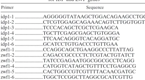
Gene deletions.Gene-specific deletion cassettes containing specific drug re-sistance markers were created using overlap PCR technology (13). The primer pair idp1-1/idp1-4 was used to amplify a 690-bp genomic region corresponding to
on September 8, 2020 by guest
http://ec.asm.org/
the 5⬘region ofIDP1. Primer pair idp1-2/idp1-5 was used for the 3⬘region of the gene (Table 1). An entireIDP1gene open reading frame (1.65-kb genomic fragment) was deleted. Primer pair zwf1-1/zwf1-6 (Table 1) was used to amplify a 3.48-kb region of the genome encompassing theZFWlocus; the entireZWF1
gene open reading frame was deleted. Drug marker cassettes used were Gene-ticin (zwf1⌬) and hygromycin (idp1⌬). KN99␣was transformed using biolistic techniques (64). Cells were grown in rich medium (yeast extract-peptone-dex-trose [YPD]) to late log phase, concentrated, and plated onto YPD agar for transformation. The cells were bombarded with 0.6-m gold beads (Bio-Rad, Richmond, CA) which were coated with DNA of the target construct according to the manufacturer’s recommendations. Following the transformation, the cells were incubated at 25°C for 4 h on nonselective medium to allow for recovery and then transferred with 0.8 ml sterile phosphate-buffered saline (PBS) to the appropriate selective medium. Transformants were observed in 3 to 5 days.
Analysis of transformants.To isolate stable transformants, all transformants were passaged three times on nonselective YPD agar and then tested for resis-tance to the appropriate selective marker. Only those transformants that grew equally well on the selective medium as on nonselective medium were considered stable transformants. A three-primer PCR screen was used to prove homologous integration on both the 5⬘and 3⬘ends of the deletion construct (46). In this manner, homologous recombinants can be distinguished from the wild type. Genomic DNA for the screen was prepared by a modification of the glass bead DNA extraction protocol described by Fujimura and Sakuma (17). A PCR screen using the primers outside the deletion construct will amplify the entire gene region, demonstrating that a single copy of the transforming DNA had been inserted at the desired locus. Southern blots were performed to screen for single integration in the genome. A single band of characteristic size (afterin silico
analysis) was observed for all the deletion colonies used for the experiments. The colonies in which Southern blot analysis revealed multiple insertion of the drug cassette were excluded from further analysis.
Complementation of theC. neoformans idp1mutation.TheIDP1 gene for complementation was amplified from strain K99 using primers idp1-3/idp1-6 (Table 1). A 1.65-kbIDP1gene flanked by 5⬘and 3⬘regions (700 bp and 600 bp, respectively) was cotransformed intoidp1⌬cells with supercoiled G418-contain-ing plasmid. After biolistic transformation, cells initially were incubated at 30°C for 4 h and later were incubated in the presence of G418 at 37°C. The colonies growing at 37°C were passaged immediately on YPD in the absence of drugs and were used for colony PCR. The colonies in which theIDP1gene was reinserted at its genomic locus replacing the hygromycin cassette were confirmed by diag-nostic PCR and also tested sensitive for hygromycin. The two complemented strains designatedidp1⌬::IDP1-A8 andidp1⌬::IDP1-C4 were used for further analysis.
In vitrostress resistance assays.Cells were plated in 10-fold serial dilutions on solid minimal medium (yeast nitrogen base [YNB], pH 4.0) with or without a stressor (1 mM H2O2for oxidative stress and 0.75 mM NaNO2for nitrosative
stress). Plates were incubated and observed at 2, 3, 5, and 7 days.
NADPH assays.For the measurement of NADP⫹and NADPH, cells were grown overnight at 25°C in YNB, pH 4.0. A stressor (1 mM H2O2or 1 mM
NaNO2) was added to the cell cultures at an optical density at 650 nm (OD650)
of 1.0 and incubated for 1 h. Cells were pelleted by centrifugation and frozen at ⫺80°C prior to measurements. The measurements were made using the Enzy-Chrom NADP⫹/NADPH assay kit from BioAssay Systems (ECNP-100; Bio-Assay Systems, Hayward, CA). Briefly, cells were homogenized by bead beating in either acidic extraction buffer for extraction of NADP⫹or alkaline extraction
buffer for extraction of NADPH. Homogenates were heated at 60°C for 5 min and then neutralized by addition of the opposite extraction buffer. For the measurement, a working reagent mix including glucose-6-phosphate dehydroge-nase was added, and an initial reading at 565 nm was taken immediately. Another reading was taken after 30 min of incubation at room temperature. A standard curve of NADP⫹was used for the calculation of total pyridine nucleotide.
For the measurement of NAD⫹and NADH, cells were prepared as for the NADP⫹measurements, using the EnzyChrom NAD⫹/NADH assay kit from BioAssay Systems (ECND-100; BioAssay Systems). The working reagent for this assay contained alcohol dehydrogenase. The first reading at 565 nm was imme-diately following addition of the working reagent, and the final reading was taken after 15 min of incubation at room temperature. A standard curve of NAD⫹was used for the calculation of total pyridine nucleotide.
GC/MS.6-Phosphogluconate (6-PG) was measured by gas chromatography-mass spectrometry (GC/MS) (61). The standard used for comparison was 6-phos-phogluconate potassium salt. This standard was run at multiple concentrations to confirm the retention time of silylated 6-PG and them/zof the corresponding fragments. The derivatized standard eluted at 30.984 min and gave distinct fragments ofm/z299, 315, 333, 357, and 387. The ion ofm/z387 was followed for quantification. Spiked samples were also run to determine matrix effects. For measurement of 6-PG produced in wild-type andzwf1⌬strains under normal conditions, oxidative stress, and nitrosative stress, 50-ml cultures of each strain were incubated in YNB (pH 4.0) in a 30°C shaking incubator overnight. One millimolar H2O2(oxidative stress), 1 mM NaNO2(nitrosative stress), or 1 ml
PBS as a control for no stress was added to the cultures, and the cultures were incubated for an additional 2 h at 30°C. Following treatment, cells were pelleted by centrifugation at 3,220⫻gfor 8 min. The cells were washed three times in sterile PBS. The washed cells were disrupted by vortexing with 0.5 mM glass beads (BioSpec Products, Inc.) for 15 min in sterile, deionized water. The disrupted cells and beads were spun at 16,000⫻gfor 10 min and the superna-tants collected and passed through a 45-m filter.
A specified volume of filtered lysate was transferred to a Reactivial containing the following 11 internal standards: 500 nmold3-creatine; 10 nmold3
-methyl-malonic acid; and 100 nmol each of [13C
3]lactate, [13C3]pyruvate,d3-serine, d5-phenylalanine, [
15
N2]orotate,d4-sebacic acid, [ 13
C6]glucose,d6-inositol, and d5-tryptophan. Forty microliters of triethylammonium trifluoroacetate was added
to each vial, and acetonitrile was added to the tops of the vials. The samples were then placed under a nitrogen stream and held at 70°C in a heating block. Acetonitrile was added as the volume was reduced until a precipitate appeared. Methylene chloride was added, allowed to blow off, and then added again until the sample was anhydrous and reduced to constant volume. Up to 500l
N-methyl-N-trimethylsilyltrifluoroacetamide (MSTFA) was added, depending upon the appearance of the original sample, and the suspension was capped and heated at 70°C for 1 to 2 h. A splitless glass insert was used with the valve program, which turns off the purge for 1 min. The injection temperature was 200°C. A 25-m DB5 capillary column (film thickness, 0.5m; inner diameter, 0.32 mm) was used with a temperature program of 80 to 130°C at 4°C/min, 130 to 200°C at 6 C/min, and 200 to 285°C at 12°C/min, with holds of 1 min at 80°C and 10 min at 285°C for a total run time of 42.5 min. The mass spectrometer, in EI⫹mode, scanned from 50 to 650 amu every 2.46 s after a 3.5-min delay.
Fluorescence microscopy.For staining with fluorescent dyes, 1 mM H2O2or 1
mM NaNO2was added to 50-ml cultures of KN99␣and thezwf1⌬,psi11⌬, and idp1⌬mutants that had been incubated in YNB (pH 4.0) at 25°C overnight. The cultures were incubated for an additional 2 h with the stressors, and then the cells were collected by centrifugation at 1,800 rpm for 8 min. The cells were washed in fresh YNB and then resuspended in 30 ml of YNB prewarmed to 37°C. To 1 ml of each culture, 10g rhodamine 123 (1-mg/ml stock solution, diluted to 10 g/ml), 100g MitoTracker Green FM (lyophilized MitoTracker resuspended in fresh dimethyl sulfoxide [DMSO] to 1 mM, diluted to 100g/ml), or both were added and incubated with shaking at 37°C for 30 min. The cells were collected by centrifugation and washed in fresh YNB three times or until the supernatant was clear. The stained cells were resuspended in 1 ml YNB, and 10 l of the suspension was placed on a microscope slide. The cells were visualized by con-focal microscopy at a magnification of⫻100.
RESULTS
Identification and deletion ofZWF1andIDP1inC.
neofor-mans. The Zwf1 enzyme in C. neoformans (CNAG_03245.1) has been previously purified and characterized as a glucose-6-phosphate dehydrogenase by Niehaus and Mallett (49). We
TABLE 1. Primers used for generating deletion constructs forIDPandZWFgenes
Primer Sequence
idp1-1 ...AGGGGGTATAAGCTGGACAGAAGCCTGC idp1-2 ...CTCGTGGAGCAGAAACAGTCTTGGTGGT idp1-3 ...TCCCACAGCTCGCTCGAAGCA
idp1-4 ...TGCTTCGAGCGAGCTGTGGGA idp1-5 ...TTCAACAGGGTCACAGGATGC idp1-6 ...GCATCCTGTGACCCTGTTGAA zwf1-1 ...CCAGGCAGCTGAAGGCCCTTATTAG zwf1-2 ...CAGACCGCCCCTCTCGTACTGTATCT zwf1-3 ...TATCCGAGAATGGCGGCGCCTCAGG zwf1-4 ...CATGGTCATAGCTGTTTCCTGAGGCG zwf1-5 ...CACTGGCCGTCGTTTTACAACGATGC zwf1-6 ...TGGCTCCGGCTTAGGCGCATCGTTG
on September 8, 2020 by guest
http://ec.asm.org/
compared the sequence of this gene to the currentC. neofor-mansgenome (Broad Assembly 1, May 2006; www.broad.mit .edu/annotation/genome/cryptococcus_neoformans/) and also to the currentS. cerevisiaegenome database (SGD). There are two genes inS. cerevisiae(51), and one inC. neoformans. This gene was disrupted by overlap PCR, and deletion of the gene was confirmed by PCR and Southern blotting.
While there are threeIDPgenes inS. cerevisiae, BLAST homology searches resulted in only one homologue in theC.
neoformans genomic sequence. This gene, IDP1 (CNAG_
03920.1), was homologous to all three of theS. cerevisiae IDP
genes, with highest homology to the mitochondrial IDP1. Mitoprot, a mitochondrial targeting sequence prediction server (http://ihg2.helmholtz-muenchen.de/ihg/mitoprot.html), was used to identify a putative mitochondrial signal sequence
inC. neoformans IDP1. A full-length cDNA ofIDP1was
iso-lated, and the sequencing results confirmed the presence of putative mitochondrial signal sequence.IDP1is only 17% sim-ilar to the C. neoformansNAD⫹-dependent isocitrate dehy-drogenase geneIDH.
Loss ofIDP1influences growth at 37°C, a major virulence
factor.Deletion of theIDP1andZWF1genes and
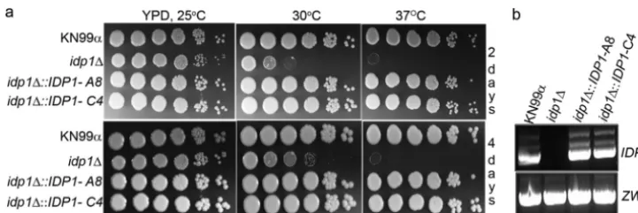
confirma-tion of the deleconfirma-tions were carried out as described in Materials and Methods. A total of 12idp1deletion mutants, generated by four independent biolistic transformations, were selected for phenotypic analysis. Individual colonies grown in YPD were plated in 10-fold serial dilutions on solid YPD plates. Plates were incubated at 25, 30, and 37°C; a representative result is shown in Fig. 1a. Nine of the individual deletion mutants exhibited a consistent temperature-sensitive growth pheno-type. Theidp1⌬cells exhibited growth similar to that of the wild type after 2 and 4 days of incubation at 25°C. However, they showed a significant impairment of growth at 30°C even after 4 days of growth. Loss of Idp1 completely abolished the growth at 37°C. The temperature-sensitive growth phenotype of idp1⌬ was completely restored in the two independent
idp1⌬::IDP1-A8 and idp1⌬::IDP1-C4 complemented strains. Figure 1b confirms the presence of theIDPgene-specific tran-script in the complemented strains. Temperature-sensitive phenotypes were not observed in thezwf1⌬ strain (data not shown).
NADPH levels in wild-type, zwf1⌬, andidp1⌬ cells under
normal growth conditions, oxidative stress, and nitrosative
stress.We measured absolute values of NADP⫹and NADPH
produced inzwf1⌬, idp1⌬, and wild-type cells during normal growth (YNB, pH 4.0, 25°C) and growth during oxidative and nitrosative stress (YNB, pH 4.0, with 1 mM H2O2 or 1 mM
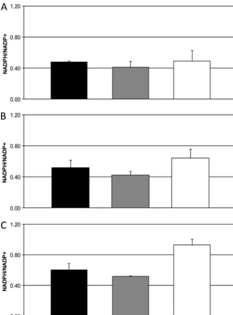
NaNO2, 25°C). NADP⫹ and NADPH were extracted from whole-cell lysate and measured by absorbance at 565 nm. Amounts were quantified by comparison to a standard curve of NADP⫹, and the data were processed as a ratio of NADPH to NADP⫹. The ratio of NADPH to NADP⫹is indicative of the demand for NADPH by the cell. If there was no demand for NADPH, then the ratio of the reduced pyrimidine to the oxi-dized form would be 1. The requirement for NADPH, whether for stress resistance or for biosynthesis, decreases the ratio, as NADPH is used in cellular processes (15, 48, 58). Each mea-surement was taken in triplicate from six biological replicates. In wild-type cells, the same NADPH/NADP⫹ ratio was maintained regardless of growth condition, whether without a stressor or with an oxidative or nitrosative stressor (Fig. 2A, B, and C). We expected the ratio to decrease in this strain during oxidative stress, due to decreased production of NADPH by the oxidative phase of the pentose phosphate pathway in the absence of the rate-limiting enzyme. However, the same ratio found in the wild type was maintained in the zwf1⌬ strain under all growth conditions (Fig. 2A, B, and C). In contrast, the ratio was significantly increased in theidp1⌬strain during oxidative and nitrosative stress compared to in both the wild-type andzwf1⌬strains (Fig. 2B and C). The increase was most dramatic during nitrosative stress (Fig. 2C).
The idp⌬strain, but not the zwf1⌬ strain, is sensitive to
nitrosative stress. Despite the increase in the NADPH/
NADP⫹ratio inidp1⌬cells during nitrosative stress, theidp1⌬
strain was sensitive to nitrosative stress in an in vitro stress assay. Serial dilutions were plated onto solid YNB (pH 4.0) with 1 mM NaNO2or 1 mM H2O2or without a stressor. Plates were incubated at 25°C and observed at 3, 5, and 7 days. Compared to the wild type, theidp1⌬strain exhibited a stress phenotype on nitrosative stress plates. The idp1⌬ strain was not sensitive to oxidative stressin vitro(Fig. 3).
To test whether Zwf1 is important for resistance to oxidative
FIG. 1.C. neoformans IDP1is essential for growth at higher temperature. (a) The temperature-sensitive phenotype of anidp1⌬mutant is completely restored in complemented strains. Tenfold serial dilutions of cell suspensions of wild-typeC. neoformans(KN99), theidp1⌬mutant, and two independentidp1mutants reconstituted with the wild-typeIDP1gene (A8 and C4) were inoculated on YPD and incubated at the indicated temperatures. Pictures were taken at 2 and 4 days. (b) Real-time PCR confirmation of the presence of theIDPgene-specific transcript in the complemented strains.
on September 8, 2020 by guest
http://ec.asm.org/
or nitrosative stress, serial dilutions of zwf1⌬ and wild-type cells were plated on solid YNB (pH 4.0) with 1 mM H2O2or 0.75 mM NaNO2or without a stressor. Plates were incubated at 30°C and observed at 3, 5, and 7 days. UnlikeS. cerevisiae,
the C. neoformans zwf1⌬ strain did not exhibit sensitivity to
either nitrosative or oxidative stress (Fig. 4). Previous reports
onS. cerevisiaehave indicated that thezwf1⌬strain is a
me-thionine auxotroph (62), but theC. neoformans zwf1⌬strain was able to grow in the absence of methionine (data not shown). Thezwf1⌬ and idp1⌬ strains were both tested with lower (0.5 mM) and higher (2.0 mM) concentrations of H2O2, and there was no difference in the phenotypes seen (data not shown).
Production of 6-phosphogluconate in the wild-type and
zwf1⌬strains.Deletion ofZWF1did not cause a
stress-sensi-tive phenotype, as seen in other systems such asS. cerevisiae, or a change in NADPH during stress compared to the wild type. Thus, we investigated whether the second reaction of the oxi-dative phase of the pentose phosphate pathway could be a source of NADPH in the absence of Zwf1.
The two reactions of the oxidative phase of the pentose phos-phate pathway (see Fig. 8) produce NADPH. The first reaction is catalyzed by Zwf1 and converts glucose-6-phosphate to 6-phos-phogluconate. 6-PG is then converted to ribulose-5-phosphate by 6-phosphogluconate dehydrogenase (Gnd), with the con-current reduction of NADP⫹to NADPH. Gnd is highly spe-cific for 6-PG; so far there is no reported evidence that Gnd utilizes any other phosphosugarin vivo(8, 39). Based on cur-rent evidence, this reaction should proceed, thereby producing NADPH, only if 6-PG is provided. In eukaryotes, with the exception ofSchizosaccharomyces pombe(65, 66), the source of 6-PG for the second reaction of the pentose phosphate path-way is the Zwf-catalyzed conversion of glucose-6-phosphate to 6-PG. Therefore, we reasoned that deletion of Zwf1 would
FIG. 2. NADPH inidp1⌬cells increases during nitrosative stress but stays consistent in wild-type andzwf1⌬cells during stress. Absolute amounts of NADPH and NADP⫹were plotted as a ratio. Measure-ments were taken from 1⫻107wild-type (black bars),zwf1⌬(gray
bars), andidp1⌬(white bars) cells. (A) NADPH/NADP⫹ratio during normal growth. (B) NADPH/NADP⫹ratio during oxidative stress (1 mM H2O2). (C) NADPH/NADP⫹ ratio during nitrosative stress (1
mM NaNO2). Values are means⫾standard deviations (n⫽12).ⴱ,P⬍
0.05 compared with control (two-tailed Studentttest).
FIG. 3. Theidp1⌬mutant displays greater sensitivity to nitrosative stress than to H2O2stress. The stress phenotype is completely rescued in
the two independent complemented strains. Serial 10-fold dilutions of the wild type (KN99), theidp1mutant, and two reconstituted strains were spotted on YNB (pH 4.0) and YNB (pH 4.0) supplemented with H2O2(1 mM) or NaNO2(0.75 mM). Plates were incubated at 25°C, and pictures
were taken at days 2 and 4. Theidp1⌬::IDP1-A8 andidp1⌬::IDP-C4 strains contain the wild-typeIDP1gene reinserted at its endogenous locus. FIG. 4. Thezwf1⌬strain is not sensitive to oxidative or nitrosative stress. Three independent null isolates (zwf1⌬-1, zwf1⌬-2, and
zwf1⌬-3) and the wild type (KN99␣) were plated on minimal medium with or without a oxidative (H2O2) or nitrosative (NaNO2) stressor and
incubated at 30°C. Compared to the wild type, the null strains did not have a sensitive phenotype with either stressor. The picture was taken after 7 days of incubation.
on September 8, 2020 by guest
http://ec.asm.org/
eliminate production of 6-PG and thus NADPH by the 6-phos-phogluconate reaction.
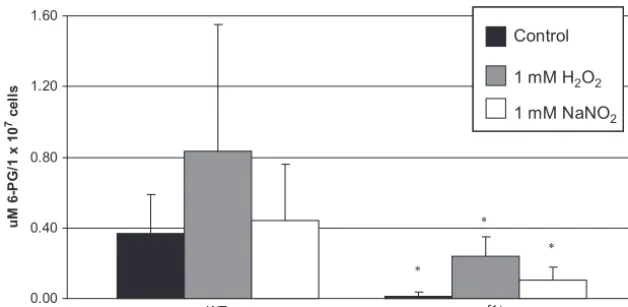
To test this hypothesis, trimethylsilyl derivatives of 6-PG were measured by gas chromatography-mass spectrometry of wild-type andzwf1⌬strains grown with or without oxidative or nitrosative stressors. The results are shown in Fig. 5. The amounts of 6-PG in wild-type cells under normal growth con-ditions (no stressor) and during nitrosative stress were the same. There was a statistically significant increase in 6-PG produced by wild-type cells during oxidative stress, indicating that the pressure of oxidative stress does increase flux through the first reaction of the oxidative phase of the pentose phos-phate pathway in wild-type cells. In contrast to the case for wild-type cells, there was no 6-PG found in the zwf1⌬ cells under normal growth conditions. However, some 6-PG was found in zwf1⌬ cells during oxidative and nitrosative stress. This amount was 4-fold less than that in the wild type under the same conditions. Thus, in C. neoformans, deletion of
ZWF1 attenuates production of 6-PG but does not com-pletely abolish it.
Deletion ofIDP1does not affect NADH production.IDP1is
distinct fromIDH, the NAD⫹-dependent isoform of isocitrate dehydrogenase that functions in the tricarboxylic acid (TCA) cycle. We predict that Idp1 does not participate in this energy-producing cycle; however, we needed to confirm that the slow-growth phenotype of theidp1⌬strain was not due to disruption of the TCA cycle. NADH is a product of the cycle, and so to confirm that deletion ofIDP1does not interfere with the cycle, we measured NADH in wild-type andidp1⌬cells during nor-mal growth and nitrosative stress. We found no difference in the amounts of NAD⫹and NADH in theidp1⌬strain com-pared to the wild type, indicating that deletion ofIDP1does not interfere with normal production of NADH (data not shown).
Integrity of the mitochondrial membrane is compromised
during nitrosative stress.If Idp1 indeed is localized to
mito-chondria, it may have a critical role in supplying mitochondrial antioxidants with NADPH during nitrosative stress. The sen-sitive phenotype despite the increase in NADPH in theidp1⌬
strain during nitrosative stress could be caused by a lack of
sufficient reducing equivalents within the mitochondria due to the absence of a direct source and insufficient membrane trans-port. Thus, NADPH would pool within the cytosol and yet not be accessible to the enzymes essential for nitrosative stress resistance. We decided to use the mitochondrial stains rhoda-mine 123 (Sigma) and MitoTracker Green FM (Invitrogen) to test loss of mitochondrial membrane integrity in idp1⌬ cells during nitrosative stress. Rhodamine 123 passively diffuses across mitochondrial membranes and will be sequestered within the organelles if the membrane is potentiated, as it is in the healthy state. However, if the membrane has lost potential, this dye will efflux out of the organelle (34). Conversely, MitoTracker mitochondrion-selective probes passively diffuse across mitochondrial membranes and will remain within mito-chondria regardless of membrane potential (Invitrogen). As a result, MitoTracker can be used to identify mitochondria by fluorescence microscopy, and rhodamine 123 can be used to determine whether mitochondria have intact, potentiated membranes.
For this experiment, we used the wild-type,idp1⌬, andzwf1⌬
strains. For a control, we used the psi11⌬ strain, which is sensitive to nitrosative stress but in which this sensitivity is likely because of poor cell wall integrity and/or cell signaling (K. J. Gerik and J. K. Lodge, unpublished data) and not pri-marily mitochondrial damage. We looked at the retention of both MitoTracker and rhodamine 123 in strains exposed to oxidative (1 mM H2O2), and nitrosative (0.75 mM NaNO2) stressors for 1 h compared to that in cells not exposed to stress (Fig. 6). To rule out the nonspecific effect of incubating cells at 37°C (employed in this experiment for labeling), we investi-gated the effect of brief exposure of cells (90 min) to 37°C on their viability. By counting the CFU after the exposure of either the wild-type or mutant cells to a temperature of 37°C for up to 90 min, we observed no significant difference in cell viability (data not shown). We found that 100% of wild-type cells not exposed to a stressor that stained with MitoTracker also retained rhodamine 123, indicating healthy mitochondria. Conversely, after exposure to nitrosative stress, only 30% of wild-type cells that stained with MitoTracker also retained rhodamine 123, indicating that nitrosative stress does induce
FIG. 5. Production of 6-PG is attenuated in thezwf1⌬strain compared to the wild type. Trimethylsilyl derivatives of 6-PG were measured by gas chromatography-mass spectrometry from lysates of 1⫻108wild-type (KN99␣) andzwf1⌬cells. Triplicate measurements were made from six
biological replicates of each strain grown in minimal medium without a stressor or after 2 h of exposure to oxidative stress (1 mM H2O2) or
nitrosative stress (1 mM NaNO2). Values are means⫾standard deviations.ⴱ,P⬍0.05 compared to control, using Student’sttest.
on September 8, 2020 by guest
http://ec.asm.org/
mitochondrial damage (Fig. 7). This is in contrast to oxidative stress, which did not result in a decrease in the number of cells that retained both dyes. Similar results were seen with the
zwf1⌬andpsi11⌬strains. However, while 100% of theidp1⌬
cells that took up MitoTracker also retained rhodamine 123 after exposure to no stress or oxidative stress, only 9.0% of cells that retained MitoTracker retained rhodamine 123 as well (Fig. 7). This indicates that theidp1⌬strain is more susceptible to mitochondrial damage during nitrosative stress than any of the other strains tested, supporting the hypothesis that Idp1 is localized to the mitochondria and that its presence there is essential for production of the NADPH used by the antioxi-dant proteins located in the mitochondria.
DISCUSSION
The ability of the fungal pathogenC. neoformansto survive within macrophages of the mammalian host is presumably due to the ability of the fungal cell to protect itself from toxic reactive oxygen and nitrogen species that can be produced by macrophages following phagocytosis. It is well established that several antioxidant proteins are essential for stress resistance
FIG. 7. Percentage of cells that were rhodamine 123 positive after exposure to nitrosative stress. The percentage of cells that retained MitoTracker and also retained rhodamine 123 after 1 h of exposure to nitrosative stress (0.75 mM NaNO2) is shown. Cells that retained both
dyes are considered to have mitochondria with intact, potentiated membranes, while cells that did not retain rhodamine 123 do not have mitochondria with fully potentiated membranes. Three hundred cells of each strain that retained MitoTracker were counted, and the per-centage of these 300 that also retained rhodamine 123 is represented graphically.
FIG. 6. Nitrosative stress induces mitochondrial damage that is pronounced in theidp1⌬strain compared to the wild type. Images were taken by confocal microscopy at a magnification of⫻100. During exposure to nitrosative stress (0.75 mM NaNO2), theidp1⌬cells retain MitoTracker
(MT) (green images) but poorly retain rhodamine 123 (Rh) (red images) compared to the wild type (KN99␣) under the same conditions. During normal conditions (control), both wild-type andidp1⌬cells retain MT and Rh. BF, bright field.
on September 8, 2020 by guest
http://ec.asm.org/
and virulence. Regardless of mechanism or localization, a com-mon requirement of many essential protective enzymes is the reductive cofactor NADPH. Since NADPH crosses membrane bilayers only by facilitated transport, it is important that NADPH be produced within the various cellular compart-ments so that there is a pool of reducing equivalents readily available for stress resistance. Mitochondria particularly are susceptible to damage from NO䡠, as several of the components of the electron transport chain contain metal centers that can be sequestered by NO䡠, leading to disruption of the membrane, impaired function, and ultimately cell death.
Two potential sources of NADPH for stress resistance are the cytosolic pentose phosphate pathway and NADP⫹ -depen-dent isocitrate dehydrogenase. In many other systems, disrup-tion of the pentose phosphate pathway by deledisrup-tion of the first and rate-limiting enzyme, Zwf1, leads to poor growth and insufficient resistance to oxidative stress, presumably due to a decrease in NADPH. Likewise, inS. cerevisiaethe three Idp proteins are important for protection from ROS under various conditions, including peroxisomal fatty acid oxidation (Idp3). Given the need for antioxidant function and the number of NADPH-dependent enzymes essential for nitrosative stress resistance and virulence inC. neoformans, we investigated the roles of Zwf1 and Idp1 as sources of NADPH for resistance to nitrosative stress as well as oxidative stress.
We first investigated the role of Zwf1 in production of NADPH and resistance to stress by creating azwf1⌬ strain, measuring NADPH in this strain, and testing in vitro stress sensitivity. Our results showed that the role of Zwf1 in pro-duction of NADPH and oxidative stress resistance inC.
neo-formansis unlike that in other fungi. InS. cerevisiae, deletion of
Zwf1 results in a slow-growth phenotype and methionine aux-otrophy (35, 47, 62). In contrast, our C. neoformans zwf1⌬
strain had no growth phenotype on rich, minimal, or methi-onine-deficient media, nor was it sensitive to changes in tem-perature. InS. cerevisiae,ZWF1has been shown to be upregu-lated during oxidative stress at both the transcriptional and translational levels (33, 60), and in bothS. cerevisiae and C.
albicans, it is essential for adaptation to oxidative stress (37,
38). In contrast, theC. neoformans zwf1⌬strain was not sen-sitive to oxidative stress, and further, we found no changes in mRNA expression during oxidative stress (data not shown). We also measured the amount of NADPH produced by the
zwf1⌬ strain under normal conditions (no stress), oxidative stress, and nitrosative stress. Finding no difference compared to results for the wild type under any condition, we considered the possibility that NADPH is still made by the pentose phos-phate pathway even in the absence of Zwf1.
While the first two decarboxylation reactions of the pentose phosphate pathway can produce NADPH, the second reaction, driven by Gnd1, is dependent on 6-PG, the substrate produced by the first reaction, and therefore would proceed only if pro-vided this substrate. Thus, without an alternative source of 6-PG, Gnd1 would not be able to reduce NADP⫹in thezwf1⌬
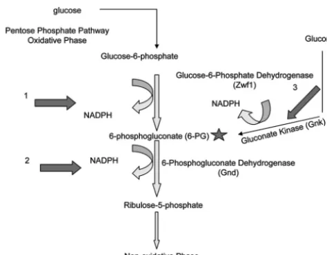
strain. We found that deletion ofZWF1inC. neoformansdid not completely abolish production of 6-PG, and therefore we propose that there is an alternative source of this phospho-sugar inC. neoformans. To date, the only reported source of 6-PG other than the decarboxylation of glucose-6-phosphate by Zwf1 is gluconate kinase (Fig. 8, arrow 3) in the “gluconate
route,” a pathway that, while described mainly for prokaryotes, has been reported forSchizosaccharomyces pombe. While the bacterial gluconate kinase is NAD⫹dependent, theS. pombe
ortholog (CAA93562.1) is NADP⫹ dependent. Despite the authors’ suggestion that the active gluconate route inS. pombe
is a distinguishing factor between fission and budding yeasts (65, 66), bioinformatic searching of theC. neoformansgenome (www .broad.mit.edu/annotation/genome/cryptococcus_neoformans/) re-sulted in identification of a possible homologue (CNAG_03048; E value⫽9e⫺27) to theS. pombegluconate kinase, annotated only as “cytoplasm protein.” We suggest that this enzyme may an alternative source of 6-PG in the absence of Zwf1 as well as another source of cytosolic NADPH (Fig. 8) and that while the amount of 6-PG produced in the absence of Zwf1 is still 4-fold less than that in the wild type, this is enough to support anti-oxidant proteins and biosynthetic pathways. This hypothesis is supported by the lack of an oxidative stress phenotype in the
zwf1⌬strain.
Second, we looked at the role of Idp1 in stress resistance. Ouridp1⌬strain was not susceptible to oxidative stress but was sensitive to both high temperature and nitrosative stress. De-spite the phenotype, the idp1⌬ cells accumulated NADPH during nitrosative stress, as indicated by the increase in NADPH/NADP⫹. In an effort to explain the phenotype, we predicted that the sensitivity to nitrosative stress is due to an inability to reduce mitochondrion-damaging RNS in the ab-sence of Idp1. Containing a mitochondrial signal sequence,C.
neoformans’ Idp1 is likely to be localized to the mitochondria,
and as such it could produce intracompartmental NADPH for use by mitochondrial antioxidant and denitrosylating proteins. We found that theidp1⌬strain accumulated significantly more inviable mitochondria during exposure to nitrosative stress
FIG. 8. Possible cytosolic sources of NADPH inC. neoformans. NADPH is produced during the oxidative phase of the pentose phos-phate pathway after glucose is converted to glucose-6-phosphos-phate, which is then converted to 6-phosphogluconate (6-PG) by glucose-6-phosphate dehydrogenase (Zwf1). This reaction produces NADPH (arrow 1). 6-PG (star) is then converted to ribulose-5-phosphate by 6-phosphogluconae dehydrogenase (Gnd) in a reaction that also pro-duces NADPH (arrow 2). The Gnd reaction requires 6-PG (star) to proceed. InC. neoformans, an alternative source of 6-PG may be gluconate kinase (Gnk), an enzyme that converts gluconate to 6-PG in a reaction that produces NADPH (arrow 3).
on September 8, 2020 by guest
http://ec.asm.org/
than either the wild-type orzwf1⌬strain. Thus, we propose the following working model. Wild-type cells, with both intracom-partmental and cytosolic sources of NADPH, have normal growth and stress resistance. In the absence of Zwf1,C.
neo-formansmaintains normal stress resistance through cytosolic
NADPH produced by an alternative protein or pathway and maintaining intracompartmental levels with Idp1. However, in the absence of Idp1, the cells sustain considerable mitochon-drial damage during nitrosative stress, leading to cell death, while NADPH produced by cytosolic sources pools in the cy-tosol. The presence of antioxidant enzymes in the cytosol pre-vents the idp1⌬ strain from being completely inviable when exposed to nitrosative stress. Of note, we were not able to obtain azwf1⌬ idp1⌬double deletion strain, suggesting that these enzymes have some redundant, essential role.
Our study shows that despite overwhelming evidence for a role of Zwf1 in oxidative stress resistance in many, various systems, its role inC. neoformansis exceptional. One indica-tion from the results is that recycling of oxidized antioxidants, which is purported to be a significant use of NADPH, is not the main source of active enzymes inC. neoformansand that in-stead, increased protein turnover may be the prominent source of reduced enzymes in this species. This hypothesis is sup-ported by proteomic data showing that there are very few changes in steady-state levels of proteins in C. neoformans
during oxidative stress (S. M. Brown and J. K. Lodge, unpub-lished data). We also provide evidence of a functional gluco-nate kinase inC. neoformans. Finally, our results suggest that compartmentalization of NADPH production is important for stress resistance inC. neoformans and reveal a key role for Idp1 in nitrosative stress.
ACKNOWLEDGMENTS
We acknowledge Jack Kennel for helpful discussions and guidance with the mitochondrial staining experiments, John Corbett for many helpful conversations regarding nitrosative stress, Wandy Beatty for microscope images, and Anthony Thomas for excellent technical sup-port.
This work was supported by NIH grant HL088905 to J.K.L.
REFERENCES
1.Alvarez, M., and A. Casadevall.2006. Phagosome extrusion and host-cell survival afterCryptococcus neoformansphagocytosis by macrophages. Curr. Biol.16:2161–2165.
2.Alvarez, M., and A. Casadevall.2007. Cell-to-cell spread and massive vacu-ole formation afterCryptococcus neoformansinfection of murine macro-phages. BMC Immunol.8:16.
3.Ayene, I. S., T. D. Stamato, S. K. Mauldin, J. E. Biaglow, S. W. Tuttle, S. F. Jenkins, and C. J. Koch.2002. Mutation in the glucose-6-phosphate dehy-drogenase gene leads to inactivation of Ku DNA end binding during oxida-tive stress. J. Biol. Chem.277:9929–9935.
4.Boveris, A., N. Oshino, and B. Chance.1972. The cellular production of hydrogen peroxide. Biochem. J.128:617–630.
5.Boveris, A., and E. Cadenas.1982. Production of superoxide radicals and hydrogen peroxide in mitochondria, p. 15–30.InL. W. Oberley (ed.), Su-peroxide dismutase, vol. III. CRC Press, Boca Raton, FL.
6.Brown, G. C., and V. Borutaite.2004. Inhibition of mitochondrial respiratory complex I by nitric oxide, peroxynitrite, and S-nitrosothiols. Biochim. Bio-phys. Acta1658:44–49.
7.Cassanova, N., K. M. O’Brien, B. T. Stahl, T. McClure, and R. O. Poyton.
2005. Yeast flavohemoglobin, a nitric oxide oxidoreductase, is located in both the cytosol and the mitochondrial matrix. J. Biol. Chem.280:7645–7653. 8.Cervellati, C., L. Li, B. Andi, A. Guariento, F. Dallocchio, and P. F. Cook.
2008. Proper orientation of the nicotinamide ring of NADP is important for the precatalytic conformational change in the 6-phosphogluconate dehydro-genase reaction. Biochemistry47:1862–1870.
9.Chaturvedi, V., B. Wong, and S. L. Newman.1996. Oxidative killing of
Cryptococcus neoformansby human neutrophils. J. Immunol.156:3836–3840.
10.Chow, E. D., O. W. Liu, S.O’Brien, and H. D. Madhani.2007. Exploration of whole genome responses of the human AIDS-associated yeast pathogen
Cryptococcus neoformansvar.grubii: NO stress and body temperature. Curr. Genet.52:137–148.
11.Contreras-Shannon, V., and L. McAlister-Henn.2004. Influence of compart-mental localization on the function of yeast NADP⫹-specific isocitrate de-hydrogenase. Arch. Biochem. Biophys.423:235–246.
12.Corpas, F. J., J. B. Barroso, L. M. Sandalio, J. M. Palma, J. A. Lupianez, and L. A. del Rio.1999. Peroxisomal NADP-dependent isocitrate dehydrogenase. Characterization and activity regulation during natural senescence. Plant Physiol.121:921–928.
13.Davidson, R. C., J. R. Blankenship, P. R. Kraus, M. de Jesus Berrios, C. M. Hull, C. D’Souza, P. Wang, and J. Heitman.2002. A PCR-based strategy to generate integrative targeting alleles with large regions of homology. Micro-biology148:2607–2615.
14.De Jesus-Berrios, A., L. Liu, J. C. Nussbaum, G. M. Cox, J. S. Stamler, and J. Heitman.2003. Enzymes that counteract nitrosative stress promote fungal virulence. Curr. Biol.13:1963–1968.
15.Diaz-Flores, M., M. A. Hernandez, R. E. Galvan, M. Gutierrez, G. Duran-Reyes, R. Medina-Navarro, D. Pascoe-Lira, C. Ortega-Camarilla, C. Vilar-Rojas, M. Cruz, and L. A. Baiza-Gutman.2006. Glucose-6-phosphate dehy-drogenase activity and NADPH/NADP⫹ratio in liver and pancreas are dependent on the severity of hyperglycemia in rat. Life Sci.78:2601–2607. 16.Frederiks, W. M., I. Kummerlin, K. Bosch, H. Vreeling-Sindelarova, A.
Jonker, and C. Van Noorden.2007. NADPH production by the pentose phosphate pathway in the zona fasiculata of rat adrenal gland. J. Histochem. Cytochem.55:975–980.
17.Fujimura, H., and Y. Sakuma.1993. Simplified isolation of chromosomal and plasmid DNA from yeasts. Biotechniques14:538–540.
18.Gaetani, G. F., H. N. Kirkman, R. Mangerini, and A. M. Ferraris.1994. Importance of catalase in the disposal of hydrogen peroxide within human erythrocytes. Blood84:325–330.
19.Galvez, S. O., Roche, E. Bismuth, S. Brown, P. Gaelal, and M. Hodges.1998. Mitochondrial localization of a NADPH-dependent isocitrate dehydroge-nase isoenzyme by using the green fluorescent protein as a marker. Proc. Natl. Acad. Sci. U. S. A.95:7813–7818.
20.Gao, Z., and W. H. Loescher.2000. NADPH supply and mannitol biosyn-thesis. Characterization of the non-reversible glyceraldehyde-3-phosphate dehydrogenase in celery leaves. Plant Physiol.124:321–330.
21.Gardner, P. R., A. M. Gardner, L. Martin, Y. Dou, T. Li, J. S. Olson, H. Zhu, and A. F. Riggs.2000. Nitric-oxide dioxygenase activity and function of flavohemoglobins. J. Biol. Chem.275:31581–31587.
22.Geisbrecht, B. V., and Gould, S. J.1999. The human PICD gene encodes a cytoplasmic and peroxisomal NADP(⫹)-dependent isocitrate dehydroge-nase. J. Biol. Chem.274:30527–30533.
23.Gerik, K. J., S. R. Bhimireddy, J. S. Ryerse, C. A. Specht, and J. K. Lodge.
2008. PKC1 is essential for protection against both oxidative and nitrosative stressors, cell integrity, and normal manifestation of virulence factors in the pathogenic fungusCryptococcus neoformans. Eukaryot. Cell7:1685–1698. 24.Giles, S., I. Batinic-Haberle, J. Perfect, and G. Cox.2005.Cryptococcus
neoformansmitochondrial superoxide dismutase: an essential link between antioxidant function and high-temperature growth. Eukaryot. Cell4:46–54. 25.Giles, S., J. Perfect, and G. Cox.2005. Cytochrome c peroxidase contributes to the antioxidant defense ofCryptococcus neoformans. Fungal Genet. Biol.
42:20–29.
26.Giles, S., J. Stajich, C. Nichols, Q. Gerrald, J. A. Alspaugh, F. Dietrich, and J. Perfect.2006. TheCryptococcus neoformanscatalase gene family and its role in antioxidant function. Eukaryot. Cell5:1447–1459.
27.Greub, G., and D. Raoult. 2004. Microorganisms resistant to free-living amoebae. Clin. Microbiol. Rev.17:413–433.
28.Hankinson, O.1974. Mutants of the pentose phosphate pathway in Aspergil-lus nidulans. J. Bacteriol.117:1121–1130.
29.Haselbeck, R. J., and L. McAlister-Henn.1993. Function and expression of yeast mitochondrial NAD- and NADP-specific isocitrate dehydrogenases. J. Biol. Chem.268:12116–12122.
30.He, W., Y. Wang, W. Liu, and C. Zhou.2007. Crystal structure of Saccha-romyces cerevisiae6-phosphogluconate dehydrogenase Gnd1. BMC Struct. Biol.7:38.
31.Holdsworth, J. E., M. Veenhuis, and C. Ratledge.1988. Enzyme activities in oleaginous yeasts accumulating and utilizing exogenous or endogenous lip-ids. J. Gen. Microbiol.134:2907–2915.
32.Idnurm, A., J. L. Reedy, J. C. Nussbaum, and J. Heitman.2004.Cryptococcus neoformansvirulence gene discovery through insertional mutagenesis. Eu-karyot. Cell3:420–429.
33.Izawa, S., K. Maeda, T. Miki, J. Mano, Y. Inoue, and A. Kimura.1998. Importance of glucose-6-phosphate dehydrogenase in the adaptive response to hydrogen peroxide inSaccharomyces cerevisiae. Biochem. J.330:811–817. 34.Johnson, L. V., M. L. Walsh, and L. B. Chen.1980. Localization of mito-chondria in living cells with rhodamine 123. Proc. Natl. Acad. Sci. U. S. A.
77:990–994.
35.Juhnke, H., B. Krems, P. Kotter, and K. D. Entian.1996. Mutants that show increased sensitivity to hydrogen peroxide reveal an important role for the
on September 8, 2020 by guest
http://ec.asm.org/
pentose phosphate pathway in protection of yeast against oxidative stress. Mol. Gen. Genet.252:456–464.
36.Kirkman, H. N., and G. Gaetani.1984. Catalase: a tetrameric enzyme with four tightly bound molecules of NADPH. Proc. Natl. Acad. Sci. U. S. A.
81:4343–4347.
37.Kusch, H., S. Engelman, P. Albrecht, J. Morschhauser, and M. Hecker.
2007. Proteomic analysis of the oxidative stress response inCandida albicans. Proteomics7:686–697.
38.Kusch, H., S. Engelmann, R. Bode, D. Albrecht, J. Morschhauser, and M. Hecker.2008. A proteomic view ofCandida albicansyeast cell metabolism in exponential and stationary growth phases. Int. J. Med. Microbiol.298:291– 318.
39.Li, L., and P. F. Cook.2006. The 2⬘phosphate of NADP is responsible for proper orientation of the nicotinamide ring in the oxidative decarboxylation reaction catalyzed by sheep liver 6-phosphogluconate dehydrogenase. JBC.
281:36803–36810.
40.Ma, H., J. E. Croudace, D. A. Lammas, and R. C. May.2006. Expulsion of live pathogenic yeast by macrophages. Curr. Biol.16:2156–2160. 41.Minard, K. I., and L. McAlister-Henn.1999. Dependence of peroxisomal
-oxidation on cytosolic sources NADPH. J. Biol. Chem.274:3402–3406. 42.Missall, T., J. Cherry-Harris, and J. K. Lodge.2005. Two glutathione
per-oxidases in the fungal pathogenCryptococcus neoformansare expressed in the presence of specific substrates. Microbiology151:2573–2581. 43.Missall, T., and J. Lodge.2005. Function of the thioredoxin proteins in
Cryptococcus neoformansduring stress or virulence and regulation by puta-tive transcriptional modulators. Mol. Microbiol.57:847–858.
44.Missall, T., M. Pusateri, M. Donlin, K. Chambers, J. Corbett, and J. K. Lodge.2006. Posttranslational, translational, and transcriptional responses to nitric oxide stress inCryptococcus neoformans: implications for virulence. Eukaryot. Cell5:518–529.
45.Missall, T., M. Pusateri, and J. Lodge.2004. Thiol peroxidase is critical for virulence and resistance to nitric oxide and peroxide in the fungal pathogen,
Cryptococcus neoformans. Mol. Microbiol.51:1447–1458.
46.Nelson, R. T., J. Hua, B. Pryor, and J. K. Lodge.2001. Identification of virulence mutants of the fungal pathogenCryptococcus neoformansusing signature tagged mutagenesis. Genetics157:935–947.
47.Ng, C., S. Tan, G. Perrone, G. Thorpe, V. Higgins, and I. W. Dawes.2008. Adaptation to hydrogen peroxide inSaccharomyces cerevisiae: the roles of NADPH-generating systems and the SKN7 transcription factor. Free Rad. Biol. Med.44:1131–1145.
48.Nguyen, K., D. Lee, P. J. Anderson, and D. Epstein.1986. Glucose 6-phos-phate dehydrogenase of calf trabecular meshwork. Invest. Opthalmol. Vis. Sci.27:992–997.
49.Niehaus, W., and T. Mallett.1994. Purification and characterization of glu-cose-6-phosphate dehydrogenase fromCryptococcus neoformans: identifica-tion as “nothing dehydrogenase.” Arch. Biochem. Biophys.313:304–309. 50.Nielsen, K., G. M. Cox, P. Wang, D. L. Toffaletti, J. R. Perfect, and J.
Heit-man.2003. Sexual cycle ofCryptococcus neoformansvar.grubiiand virulence of congenic a and alpha isolates. Infect. Immun.71:4831–4841.
51.Nogae, I., and M. Johnston.1990. Isolation and characterization of the ZWF1 gene ofSaccharomyces cerevisiaeencoding glucose-6-phosphate de-hydrogenase. Gene99:161–169.
52.Outten, C. E., and V. C. Culotta.2003. A novel kinase is the mitochondrial source of NADPH inSaccharomyces cerevisiae. EMBO J.22:2015–2024. 53.Paulnock, D. M.1994. The molecular biology of macrophage activation.
Immunol. Ser.60:47–62.
54.Pearce, L. L., A. J. Kanai, M. W. Epperly, and J. Peterson.2005. Nitrosative stress results in irreversible inhibition of purified mitochondrial complexes I and III without modification of cofactors. Nitric Oxide13:254–263. 55.Przybyla-Zawislak, B., D. M. Gadde, K. Ducharme, and M. T. McCammon.
1999. Genetic and biochemical interactions involving tricarboxylic acid cycle (TCA) function using a collection of mutants defective in all TCA cycle genes. Genetics152:153–166.
56.Puskas, F., P. Gergely, K. Banki, and A. Perl.2000. Stimulation of the pentose phosphate pathway and glutathione levels by dehydroascorbate, the oxidized form of vitamin C. FASEB J.14:1352–1361.
57.Rabilloud, T., M. Heller, F. Gasnier, S. Luche, C. Rey, R. Abersold, M. Benahmed, P. Louisot, and J. Lunardi.2002. Proteomic analysis of cellular response to oxidative stress. J. Biol. Chem.277:19396–19401.
58.Rodriguez-Segade, S., M. Freire, and A. Carrion.1978. Regulation of the oxidative phase of the pentose phosphate cycle in mussels. Biochem. J.
170:577–582.
59.Sharma, K. D., L. A. Andersson, T. A. Loehr, J. Terner, and H. Goff.1989. Comparative spectral analysis of mammalian, fungal, and bacterial catalases. J. Biol. Chem.264:12772–12779.
60.Shenton, D., J. Smirnova, J. Selley, K. Carroll, S. Hubbard, G. Pavitt, M. Ashe, and C. Grant.2006. Global translational responses to oxidative stress impact multiple levels of protein synthesis. J. Biol. Chem.281:29011–29021. 61.Shoemaker, J. D., and W. H. Elliott.1991. Automated screening of urine samples for carbohydrates, organic and amino acids after treatment with urease. J. Chromatogr.562:125–138.
62.Slekar, K., D. Kosman, and V. Culotta.1996. The yeast copper/zinc super-oxide dismutase and the pentose phosphate pathway play overlapping roles in oxidative stress protection. J. Biol. Chem.271:28831–28836.
63.Song, B. C., T. K. Kim, Y. M. Jung, and Y. H. Lee.2006. Modulation of talA gene in pentose phosphate pathway for overproduction of poly-beta-hy-droxybutyrate in transformedEscherichia coliharboring phbCAB operon. J. Biosci. Bioeng.102:237–240.
64.Toffaletti, D. L., T. H. Rude, S. A. Johnston, D. T. Durack, and J. R. Perfect.
1993. Gene transfer inCryptococcus neoformansby use of biolistic delivery of DNA. J. Bacteriol.175:1405–1411.
65.Tsai, C. S., J. L. Shi, and H. G. Ye.1995. Kinetic studies of the gluconate pathway enzymes fromSchizosaccharomyces pombe. Arch. Biochem. Bio-phys.316:163–168.
66.Tsai, C. S., H. G. Ye, and J. L. Shi.1995. Carbon 13 NMR studies and purification of gluconate pathway enzymes from Schizosaccharomyces pombe. Arch. Biochem. Biophys.316:155–162.
67.Tsutsui, E. A., and P. A. Marks.1962. A study of the mechanism by which triphosphopyridine nucleotide affects human erythrocyte glucose-6-phos-phate dehydrogenase. Biochem. Biophys. Res. Commun.8:338–341. 68.Vives Corrons, J. L., D. Colomer, A. Pujades, A. Rovira, M. Aymerich, A.
Merino, and J. L. Aguilar i Bascompte.1996. Congenital 6-phosphoglu-conate dehydrogenase (6PGD) deficiency associated with chronic hemolytic anemia in a Spanish family. Am. J. Hemotol.53:221–227.
69.Welter, R., L. Yu, and C. A. Yu.1996. The effects of nitric oxide on electron transport complexes. Arch. Biochem. Biophys.331:9–14.
70.Yoshihara, T., T. Hamamoto, R. Munakata, R. Tajiri, N. Ohsumi, and S. Yokota.2001. Localization of cytosolic NADP-dependent isocitrate dehy-drogenase in the peroxisomes of rat liver cells: biochemical and immunocy-tochemical studies. J. Histochem Cytochem.49:1123–1131.