Cryptococcus
Species
Aubrey E. Frazzitta, Haily Vora, Michael S. Price, Jennifer L. Tenor, Marisol Betancourt-Quiroz, Dena L. Toffaletti, Nan Cheng, John R. Perfect
Department of Medicine, Duke University Medical Center, Durham, North Carolina, USA
Cryptococcus neoformansandC. gattiicause meningoencephalitis and are an increasing human health threat. These pathogenic
Cryptococcusspecies are neurotropic and persist in the cerebrospinal fluid (CSF) of the mammalian host during infection. In order to survive in the host, pathogenic fungi must procure nutrients, such as carbon and nitrogen, from the CSF. To enhance our understanding of nutrient acquisition during central nervous system infection byCryptococcusspecies, we examined the utilization of nitrogen sources available in CSF. We screened for the growth and capsule production of 817 global environmental and clinical isolates on various sources of nitrogen. Both environmental and clinical strains grew robustly on uric acid, Casa-mino Acids, creatinine, and asparagine as sole nitrogen sources. Urea induced the greatest magnitude of capsule induction. This induction was greater inCryptococcus gattiithan inC. neoformans. We confirmed the ability of nonpreferred nitrogen sources to increase capsule production in pathogenic species ofCryptococcus. Since urea is metabolized to ammonia and CO2(a known
signal for capsule induction), we examined urea metabolism mutants for their transcriptional response to urea regarding cap-sule production. The transcriptional profile ofC. neoformansunder urea-supplemented conditions revealed both similar and unique responses to other capsule-inducing conditions, including both intra- and extracellular urea utilization. As one of the most abundant nitrogen sources in the CSF, the ability ofCryptococcusto import urea and induce capsule production may sub-stantially aid this yeast’s survival and propagation in the host.
C
ryptococcus neoformansis a pathogenic, basidiomycete yeast responsible for causing meningoencephalitis in immunocom-promised individuals. Distributed worldwide, it is the fourth larg-est infectious disease killer in sub-Saharan Africa as a complica-tion of the AIDS epidemic (1). Because of the mortality and increasing incidence of cryptococcosis, it is imperative that we understand all aspects of this pathogen-host interaction and the methods ofC. neoformanssurvival in an attempt to disrupt them. As with carbon assimilation (2), nitrogen metabolism is fun-damentally critical for the survival of fungal pathogens within their plant or animal hosts. For instance, Lau and Hamer reported the influence of nitrogen regulatory elements on the expression of theMagnaporthe griseapathogenicity gene MPG1(3). In their study, novel nitrogen regulatory elementsNPR1andNPR2were shown to be required for expression ofMPG1and thenpr1and npr2mutant strains were avirulent on barley. Lau and Hamer also demonstrated that the global nitrogen regulatory transcription factorNUT1, a homolog of theNeurospora crassa NIT2and Asper-gillus nidulans areAtranscription factor genes, was also partly re-sponsible forMPG1expression under nitrogen starvation. How-ever, the complexity of the regulation of pathogenicity through nitrogen pathways in fungi is apparent, as strains with deletion of NUT1showed no loss of pathogenicity (4), but deletion of the relevantNIT2/areAhomolog resulted in attenuated virulence (5). The role of nitrogen metabolism in fungal pathogenicity of animals has also been investigated. Deletion of the global nitrogen regulatory transcription factorareAresults in attenuation of vir-ulence in the murine model of invasive pulmonary aspergillosis (IPA) (6). While the various components of the nitrate assimila-tion pathway have yet to be tested for roles in pathogenicity inA. fumigatus, another regulatory protein, RhbA, was shown to facil-itate growth on nitrate, histidine, and proline and to promote virulence (7). The link between nitrogen utilization andpatho-genicity has also been observed in human-pathogenic yeasts, such asCandida albicans. Limjindaporn and colleagues showed that theareAhomologGAT1regulates nitrogen utilization in C. albicansand is required for pathogenicity in a murine model of candidiasis, even in the absence of defects in dimorphism or growth in serum (8).
Nitrogen metabolism has begun to be understood inC. neofor-mansin the context of pathobiology. Early studies on nitrogen utilization inC. neoformansfocused on the metabolism of nitrog-enous compounds influencing melanin production during growth in areas of ecological or medical importance. Recently, Lee and colleagues demonstrated the necessity of the Gat1 global tran-scription factor for utilization of uric acid, creatinine, and urea (9). Indeed, the researchers noted the ability of various nitrogen sources to greatly enhance capsule production, with the greatest increase shown with creatinine in minimal medium. Underscor-ing the differences between pathogenic Cryptococcus species, Ngamskulrungroj and colleagues showed thatGAT1regulation of amino acid utilization differs betweenC. neoformansandC. gattii (10). Regulation of nitrogen metabolism and its impact on viru-lence in cryptococcosis have begun to be appreciated. Results
ob-Received12 July 2013 Accepted19 August 2013 Published ahead of print23 August 2013
Address correspondence to John R. Perfect, [email protected].
A.E.F., H.V., and M.S.P. contributed equally to this article.
Supplemental material for this article may be found athttp://dx.doi.org/10.1128 /EC.00169-13.
Copyright © 2013, American Society for Microbiology. All Rights Reserved.
doi:10.1128/EC.00169-13
on September 8, 2020 by guest
http://ec.asm.org/
tained after deletion of theC. neoformans GAT1gene, which is homologous toareAinAspergillusspp., the deletion of which re-sulted in decreased virulence (6), are mixed; one study showed increased thermotolerance and slightly increased virulence in a murine model of cryptococcosis (9), whereas another study showed no effect of this mutation on virulence (11). Mechanisti-cally, Wilson and colleagues showed inMagnaporthethat control of nitrogen metabolism and pathogenicity was integrated with trehalose metabolism; Tps1 is required for proper regulation of the nitrogen repressorNMR1(12). The latter finding is intriguing, as recent data regarding theNMR1homologTAR1inC. neofor-mansdemonstrate the relationship of nitrogen regulation to mel-anin production, which is a major virulence factor forC. neofor-mans(13), and the trehalose pathway has signal control on the cryptococcal virulence composite (14,15).
Urea is a product of metabolism of proteins and exists in copi-ous amounts in cerebrospinal fluid (CSF) (16). Extracellular ure-ase (URE1) production byC. neoformanshas been shown to be a virulence factor in cryptococcosis (17). Cox and colleagues showed that a urease-deficient mutant (ure1⌬) was impaired for virulence in murine models of both inhalation and intravenous disease, yet the mutant strain was completely virulent in the rabbit CSF model of cryptococcosis (17). The explanation for the differ-ence in viruldiffer-ence of theure1⌬mutant between animal models of disease was tentatively explained by a study showing that Ure1 is important for sequesteringCryptococcusyeasts in the microvascu-lature of the brain and promoting the blood-brain barrier crossing of the yeast into the central nervous system (CNS) (18). This re-quirement for urease was bypassed in the rabbit CSF model, where the CSF was inoculated directly. Though rare, urease-deficient strains ofC. neoformans have been isolated from patients with invasive cryptococcosis (19,20).
Like melanin production (21), capsule production inC. neo-formanshas been a topic of great interest in understanding cryp-tococcal pathobiology, and certain aspects of capsule synthesis are known. Increased CO2concentrations, iron deprivation, and se-rum have all been shown to induce capsule production by this yeast (22–24). A carbon substrate, the availability of vitamins, and amino acid availability have also been described to promote cap-sule growth (25).In vivoinfections produce differently sized cap-sules, depending on the organ site of infection; the lung and the brain show greater induction than other organs (26). This is im-portant, as increased capsule size has been linked to decreased phagocytosis (27).
Nitrogen has also been shown to influence the production of capsule to various degrees, and this induction is subject to nitro-gen catabolite repression (9). Furthermore, differences betweenC. neoformansandC. gattiiin the utilization of nitrogen sources, includingD-proline, phenylalanine, tryptophan, andD-alanine, exist (10,28), but the extension of these differences to capsule production has not been studied. In order to enhance our under-standing of the relationship between nitrogen acquisition and capsule production during infection byC. neoformans, we evalu-ated capsule production during growth on five different nitrogen sources: asparagine-Casamino Acids, creatinine, ammonium, uric acid, and urea. In our study, the nitrogen source was shown to profoundly affect capsule size, with urea being the most dramatic inducer of capsule production in this pathogenic yeast.
MATERIALS AND METHODS
Strains and media.Yeast extract-peptone-dextrose (YPD; 1% yeast ex-tract, 2% peptone, 2% dextrose) medium was used for maintaining the strains. Cultures were grown with continuous shaking (220 rpm) at 30°C on agar plates unless otherwise noted. Five strains representing four sero-types ofCryptococcuspathogenic species, serotype A strain H99, serotype B strain R265, serotype B strain WM276, serotype C strain DUMC106.97, and serotype D strain JEC21, were selected for study in assays, unless stated otherwise. H99-derived deletion mutants ure1⌬ and amt1/2⌬
(JR5), obtained as a gift from Joe Heitman, were used for urea metabolism pathway experiments (17,29).
Cryptococcussp. strain library. Cryptococcussp. isolates were ob-tained from our laboratory collection or as a generous gift from Joe Heit-man. The isolates in the combined collection of 817Cryptococcusisolates were genotyped using multilocus sequence typing (MLST) strategies, and the isolates are described in Table S1 in the supplemental material. The strain collection was propagated from⫺80°C freezer stocks in 1-ml cul-tures of YPD plus Hogness freezing medium (30) in 96-well growth blocks at 37°C for 48 h. The strains were then plated onto 6 different nitrogen source plates (ammonium sulfate, sodium nitrate, urea, uric acid, aspar-agine, and Casamino Acids). Agar plates were composed of yeast carbon base (YCB) minimal medium and 5 g/liter of nitrogen source. The plates were incubated for 72 h at 37°C and subsequently analyzed by inspection for growth and mucoid and filamentous appearance. The plates were also observed following 120 h growth.
Capsule induction by nitrogen source.The four media described above, YCB plus asparagine, YCB plus creatinine, YCB plus ammonium, and YCB plus urea, were induced with each of the 5 strains, H99, R265, WM276, DUMC 106.97, and JEC21, in a 32-well plate. Nitrogen source concentrations were 5 g/liter. The cultures were grown for 48 h, and im-ages were taken using a Zeiss Axioskop microscope. Capsule measure-ments were calculated as previously described (22). Briefly, 2l of India ink (Becton, Dickinson) and 3l of culture were placed on glass slides. Images were captured with a⫻63 differential interference contrast oil lens using AxioVision AC imaging software. Capsule-to-cell-diameter ratios were measured forⱖ50 cells per strain culture using the Ruler tool in Adobe Photoshop CS4 (Adobe Systems Inc., San Jose, CA).
Growth curve.Growth curves were assessed forCryptococcusyeast strains H99, R265, and WM276 in YCB plus ammonium, YCB plus urea, and YCB plus uric acid broth. The nitrogen concentration in the medium in each culture was 0.5 g/liter. Fifty milliliters of overnight cultures of each strain in YPD was diluted to 1⫻106CFU and used to inoculate the 3 types
of YCB broth. The optical density at 600 nm was determined in triplicate three times per day for 70 h and plotted on a log10scale.
Benomyl assay.Strains R265 and H99 were grown for 24 h in 40 ml of YCB plus ammonium medium (5 g/liter) at 37°C. After 24 h, the cultures were divided into 10-ml aliquots and washed with sterile water. Cells were then resuspended in 50 ml of either YCB plus ammonium or YCB plus urea at 5 g/liter nitrogen source. Two microliters of 50 mM benomyl was added to one of each of the media and strain types. Cells were grown for 24 more hours and were imaged using the method described above for Zeiss Axioskop capsule measurement.
Urea titration assay.Strains R265 and H99 were grown at 37°C for 24 h in 40 ml YCB plus uric acid (0.5 g/liter) medium. After 24 h, the cultures were washed with sterile water and placed in new medium with incremen-tally increasing amounts of urea added to YCB plus uric acid medium. The concentrations of urea tested were 0 g/liter, 0.05 g/liter, 0.5 g/liter, and 5 g/liter. After growing for 24 h, the cells were imaged utilizing the method described above for capsule measurement using a Zeiss Axioskop micro-scope.
Urea mutant capsule induction assay.Mutants of the urea metabo-lism pathway were grown in YCB plus proline (0.25 g/liter) liquid me-dium for 18 h at 30°C. Cultures were then spun down and washed three times with deionized water and resuspended in YCB plus proline (0.25 g/liter) and urea (5 g/liter) liquid medium. The capsules of the H99
on September 8, 2020 by guest
http://ec.asm.org/
amt1/2⌬andure1⌬mutants and wild-type (WT) strain H99 were mea-sured as described above. Additionally, theamt1/2⌬,ure1⌬, and H99 strains were grown in YPD, washed, and then transferred to Dulbecco’s modified Eagle’s medium (DMEM) or DMEM plus urea (0.25 g/liter) in 12-well plates and grown for 48 h at 37°C in a CO2incubator (data not
shown). Capsule images were made as described above.
Microarrays.YCB plus proline (0.25 g/liter) medium was inoculated with the wild-type H99 and theure1⌬andamt1/2⌬mutant strains and grown overnight at 30°C. Cells were washed in sterile water and resus-pended in 25 ml of either YCB plus proline (0.25 g/liter) or YCB plus proline (0.25 g/liter) and urea (0.25 g/liter). All media were pH 5⫾0.1. H99 was also resuspended in YCB plus 6 mM ammonium and 10 mM urea at the concentrations outlined by Lee et al. (9). The cultures were grown for 1 h at 30°C, pelleted, and frozen at⫺80°C. RNA for all samples was prepared for microarray analysis as previously described (2). Follow-ing RNA extraction, data acquisition and microarray hybridizations were executed at the Duke Microarray Facility (http://www.genome.duke.edu /cores/microarray/) according to established protocols for custom-spot-ted arrays. Data were normalized using the analysis of variance process in JMP Genomics (v6.0) software (31), and calculations of fold change were performed using the Adaptive Holm multiple-testing correction to set statistical significance at aPvalue of⬍0.000024434.
Gene transcriptional response comparisons.Lists of genes with sta-tistically significant responses generated from the microarrays were com-piled into Venn diagram comparisons using the JMP Genomics (v6.0) Venn diagram function. Gene lists were generated from a comparison of WT H99 growth under YCB plus proline or YCB plus proline and urea-supplemented conditions and a comparison of the growth of the WT,
ure1⌬, andamt1/2⌬strains in YCB plus proline and urea medium. The 2 lists and the gene list in Table S2 in the work of Haynes (32) were com-pared for determination of identical locus identifiers.
GO enrichment.Gene ontology (GO) enrichment analysis was car-ried out by utilizing the R package TopGo. Gene ontology information for
C. neoformans H99 was downloaded from http://genome.jgi-psf.org /Cryne_H99_1/Cryne_H99_1.download.ftp.html. P values were cor-rected for multiple-hypothesis testing, and significance was determined using a cutoff value of 0.01.
Statistical analysis.All data analysis was executed in the Microsoft Excel program (Microsoft Inc., Redmond, WA), unless otherwise noted. Data were expressed as the mean⫾standard deviation of at least three repeat experiments. Each experiment was performed in triplicate. Differ-ences in capsule sizes were assessed using Student’sttest or repeated-measures analysis of variance to determine the statistical significance be-tween the parental strain control and mutant treatment groups. APvalue of⬍0.05 was considered statistically significant.
Microarray data accession number.The accession number assigned to the microarray data isGSE46829.
RESULTS
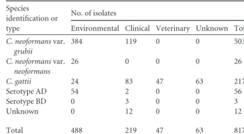
Nitrogen assimilation varies by serotype, as revealed in a survey of North American and AfricanCryptococcusisolates.Using a collection of 817Cryptococcusstrains from numerous locations in Africa and North America (Table 1), we evaluated the ability of the different pathogenic serotypes to assimilate ecological and CNS-relevant nitrogen sources and assess their impact on gross mor-phology and virulence trait expression. A qualitative assessment of growth using ammonium, nitrate, urea, uric acid, asparagine, or Casamino Acids revealed some interesting observations (Table 2); whileC. neoformansandC. gattiistrains share aspects of nitrogen utilization, stark differences also exist. Uric acid was utilized as a nitrogen source by at least 96% of theC. neoformansandC. gattii strains. Interestingly, moreC. gattiistrains (76.96%) than either C. neoformans(11.54%) or hybrid serotype (39.30 and 33.33%) isolates grew robustly on urea. Casamino Acids and ammonium
supported the growth of 95.90% of all the non-hybrid serotype isolates.
The nitrogen source also affected the colony morphology dif-ferently among the strains (Fig. 1). First, isolates in this collection exhibited a wrinkled colony appearance and filamentous behavior (see Table S1 in the supplemental material), and these were pro-nounced along serotype lines. These phenotypes were primarily seen when strains were grown on uric acid and were equally ob-served in both clinical and environmentalC. gattiistrains. Fila-mentous growth could indicate hyphal formation or haploid fruiting, and these observations could be further explored, but evidence of spores was not examined, since these cultures were not protected from light. Second, the different nitrogen sources in-duced a mucoid colony phenotype at various frequencies among the different serotypes. This phenotype was most pronounced in C. gattiiandC. neoformansvar.grubiistrains, where 66.80% and 57.10%, respectively, exhibited a mucoid appearance. Although no serotype BD isolates exhibited mucoid colonies on any of the nitrogen sources selected, this observation may be due to the small sample size of BD isolates included in this study. Overwhelmingly, urea as a nitrogen source produced the most mucoid colonies in all strains (54.5%), followed by asparagine at 23.1%.
The mucoid colony phenotype has been associated with in-creased virulence in strains (33). We found that this mucoid phe-notype was correlated with increased capsule size, a known viru-lence trait, when a selection of these strains was mixed with India ink and viewed microscopically (data not shown). This observa-tion prompted us to focus on the impact of the nitrogen source on capsule production.
Nitrogen sources present in CSF affect capsule production differently between pathogenicCryptococcusspecies.The global screen of Cryptococcus isolates on various nitrogen sources showed that urea induces a mucoid colony appearance and the resulting increased capsule size in a wide breadth of strains. Urea exists throughout the body at various concentrations. It is the most prevalent nitrogen source in the CSF, the critical site of cryp-tococcal infection (16). Previously, our laboratory defined the dif-ferences inC. neoformanscarbon metabolism during growth at different body sites, such as the CSF, during infection (2). In the interest of better defining the nitrogen metabolites’ importance for growth and survival within this host site, we assessed urea and 3 other readily assimilated nitrogen sources available in CSF (cre-TABLE 1Environmental and clinical isolate librarya
Species identification or type
No. of isolates
Environmental Clinical Veterinary Unknown Total
C. neoformansvar.
grubii
384 119 0 0 503
C. neoformansvar.
neoformans
26 0 0 0 26
C. gattii 24 83 47 63 217
Serotype AD 54 2 0 0 56
Serotype BD 0 3 0 0 3
Unknown 0 12 0 0 12
Total 488 219 47 63 817
a
Tabulation of strains screened by species, variety, serotype, molecular type, and origin. Strains came from 7 U.S. states and 8 countries. Strains were isolated between 2002 and 2007.
on September 8, 2020 by guest
http://ec.asm.org/
atinine, ammonium, and asparagine) for their impact on growth and virulence factor expression inC. neoformansandC. gattii(34). Furthermore, we aimed to determine whether differences in cap-sule size for the most CNS-relevant nitrogen sources correlated with a mucoid colony appearance in the isolate screen. As shown
inFig. 2, nitrogen source influences capsule production
differ-ently in isolates representing the 4 major serotypes. Overall, urea was the most potent inducer of capsule across the strains. Inter-estingly, theC. gattiistrains, including Vancouver outbreak strain R265, produced the most robust capsules in urea-containing me-dium. Indeed, the capsules produced by theseC. gattiistrains in urea were⬎33% larger than the capsule produced by serotype A strain H99 (C. neoformans) in creatinine, the nitrogen source shown to induce its largest capsule, confirming published results (9). While we found that urea induced the greatest capsule
induc-tion response overall, there was some variability among the strains.
We noticed in our screens that the strains did not grow as rapidly on urea as on other nitrogen sources at the levels used in this study. Therefore, growth rates on urea compared to those on ammonium were measured for serotype A and B strains, and the results precisely confirmed this observation (Fig. 3). Poor growth conditions have been shown to induce a larger capsule size. There-fore, we wanted to separate the impact of inhibited or slow cell growth on capsule production from that of the potential direct capsule signaling produced by the urea molecule. To confirm that the observed impact of urea on the capsule was not solely due to reduced growth, we compared capsule production for both H99 and R265 on ammonium and urea in the presence and absence of benomyl. Benomyl, a benzimidazole fungicide, blocks mitosis by TABLE 2Effect of nitrogen source on growth and colony appearancea
Species identification or type
% of isolates with growth ability (% of growing isolates that were mucoid)
% mucoidb
NH4 Nitrate Urea Uric acid Asparagine Casamino Acids
C. neoformansvar.grubii 99.60 (1.59) 27.63 (2.78) 73.36 (57.10) 99.80 (4.20) 100.00 (7.20) 99.60 (3.00) 59.64
C. neoformansvar.neoformans 100.00 (0.00) 26.92 (7.69) 11.54 (0.00) 100.00 (0.00) 96.15 (0.00) 100.00 (0.00) 7.69
C. gattii 96.31 (5.07) 81.11 (44.20) 76.96 (66.80) 96.31 (57.00) 95.85 (67.00) 92.17 (49.00) 84.79
Serotype AD 100.00 (0.00) 14.29 (0.00) 39.29 (16.10) 100.00 (0.00) 100.00 (0.00) 100.00 (0.00) 16.07
Serotype BD 66.67 (0.00) 33.33 (0.00) 33.33 (0.00) 33.33 (0.00) 33.33 (0.00) 33.33 (0.00) 0.00
Unknown 91.67 (16.70) 75.00 (33.33) 66.67 (33.33) 83.33 (42.00) 91.67 (58.00) 91.67 (50.00) 58.33
aStrains were grown on YCB plus NH
4, NO3, urea, uric acid, asparagine, and Casamino Acids. Overall viability, filamentous growth, and mucoid colony appearance were assessed
on a⫹⫹,⫹, and⫺scale.
bOverall percentage of isolates within each species identification or type that displayed a mucoid colony appearance.
D
E
F
C
A
B
FIG 1Environmental and clinical isolate screen findings. (A)C. gattiiserotype C strains on uric acid; (B) aC. gattiistrain on uric acid, capsule production; (C)
C. neoformansserotype A strains on urea, mucoid appearance; (D)C. gattiion uric acid, filamentous growth; (E)C. neoformansserotype A strains on urea, growth differences on a white light box; (F) an African clinical isolate on urea, capsule production.
on September 8, 2020 by guest
http://ec.asm.org/
disrupting microtubule formation. It was therefore useful to as-sess the impact of impeded growth on capsule production inde-pendently of any effect caused by urea as a nonpreferred nitrogen source. Capsule production was consistently higher in the pres-ence of urea, regardless of the prespres-ence or abspres-ence of benomyl, indicating that urea itself directly increases or signals capsule pro-duction in pathogenicCryptococcusspecies. Nutrient starvation is an unlikely cause of the observed large capsule phenotype in the urea-containing medium.
Urea acts as a signal for increased capsule production in
Cryptococcusspecies.Since urea was shown to prominently in-crease capsule production, we sought to describe how the urea molecule actually signaled the observed increase in capsule
pro-duction. This is important to understand, as urea exists at concen-trations comparable to those for glucose in human CSF (16) and may be a factor contributing to the formation of the protective large capsules observed in patients with cryptococcosis. Therefore, we increased urea concentrations in the presence of a constant con-centration of an alternate nonpreferred nitrogen source (uric acid). Indeed, increasing urea concentrations in culture yielded increased capsule sizes in both the H99 and R265 strains (Fig. 4), indicating that capsule induction by urea is dose responsive.
Since urea can be broken down to ammonia and CO2, we ex-amined previously described mutants for their response to urea regarding capsule production (Fig. 5). The urease geneURE1 en-codes the major urease inC. neoformansand has previously been 0
0.5 1 1.5 2 2.5 3
Ammonium Asparagine Creatinine
Urea
Ammonium Asparagine Creatinine
Urea
Ammonium Asparagine Creatinine
Urea
Ammonium Asparagine Creatinine
Urea
Ammonium Asparagine Creatinine
Urea
H99 JEC21 R265 WM276 DUMC106.97
Capsule thickness (total dia./cell dia.)
FIG 2Capsule thickness in serotypes A, B, C, and D in YCB medium plus a nitrogen source. Serotypes A, B, C, and D were grown in YCB medium with ammonium, asparagine, creatinine, or urea as the nitrogen source. The ratios of the cell diameter (dia.) to the total diameter including the capsule forⱖ50 cells per group are shown. Bars represent standard errors.
0.0001 0.001 0.01 0.1 1 10
0 10 20 30 40 50 60 70
log
10
OD600
Time (hrs)
H99 NH4 H99 Urea R265 NH4 R265 Urea WM276 NH4 WM276 Urea
FIG 3Growth on different nitrogen sources. Strains were grown overnight and then used to inoculate either YCB plus ammonium, YCB plus urea, or YCB plus uric acid medium (not shown). The optical density at 600 nm (OD600) was recorded three times per day until the cultures entered stationary phase.
on September 8, 2020 by guest
http://ec.asm.org/
shown by us to yield no impact on CNS virulence in a rabbit CNS model of cryptococcosis (17). Likewise, a strain lacking both am-monia transporters (Amt1 and Amt2) was shown to have no vir-ulence defects in mice, lacks the ability to take up ammonia from the extracellular milieu (29), and thus helps control urea break-down product effects. However,CAN2, encoding the final step in the conversion of CO2to bicarbonate, was not evaluated due to the strict requirement of the mutant strain for elevated CO2levels for growth that would confound the effect of urea on capsule induction. Theure1⌬andamt1⌬amt2⌬mutants were compared to wild-type strain H99 for capsule formation in the presence of urea as the sole nitrogen source. Theure1⌬mutant strain showed elevated capsule production when grown in defined medium with urea and a low level of CO2(that found in ambient air) (Fig. 6) relative to that of both the WT and theamt1/2⌬mutant. This finding supports the hypothesis that the urea molecule itself acts as an external or internal signal for capsule production in Cryptococ-cusspecies.
A variety of conditions presentin vivo, including the CO2 con-centration and the presence of serum, can induce capsule
produc-tion. In order to determine whether urea can further enhance capsule production when cells are already under capsule-inducing conditions, we grew the urea pathway mutants and WT at 5% CO2 in either DMEM or DMEM plus urea (0.25 g/liter). No discernible capsule size difference was detected between strains grown with or without urea at the concentrations used.
Transcriptional responses to urea exposure.To examine the direct effects of urea onC. neoformansgene expression, we carried out DNA microarray analysis on WT strain H99 grown with either proline alone or urea-supplemented proline medium. Identifying significant modifications to the cryptococcal transcriptome may better inform us of the mechanism by which urea directly induces capsule. Proline was selected as the control nitrogen source be-cause it appears to be a poor inducer of capsule on its own (9). Following growth in proline medium,C. neoformanswas resus-pended in either proline medium or biologically significant con-centrations of urea plus proline for 1 h. Following this 1-h expo-sure, 626 genes in strain H99 showed significantly different expression in urea-supplemented medium than in proline me-dium lacking urea (P⬍0.000024434; see Table S2 in the supple-mental material) (Table 3).
GO enrichment analysis indicated that oxidoreductase activity, various nitrogen transmembrane activities, and flavin mononu-cleotide binding were molecular functions all significantly en-riched in the response to the presence of urea. Induced oxi-doreductase activity indicates nonpreferred nitrogen assimilation, as nonpreferred nitrogen sources are generally reduced to ammo-nia (35). Biological processes enriched by the presence of urea included defense and organismal responses, ribosome-related biogenesis, and transport of carboxylic and amino acids.
Gene transcription altered by the presence of urea covered a variety of functional categories, though many genes were specifi-FIG 4Capsule production increases with increasing urea concentration.
Strains H99 and R265 were grown for 24 h in YCB plus uric acid medium and then shifted to YCB plus uric acid medium with various amounts of urea. After the strains were grown for an additional 24 h, cell capsule size was imaged and measured forⱖ50 cells per group displayed. *,P⬍0.05.
H2N NH2 O
CO2 + 2 NH3
Ure1
H2O
NH3 CO2
Amt1/2
Can2
HCO3
FIG 5Schematic diagram of urease and urea metabolism. To pinpoint the product of urea metabolism that induces capsule production, H99-derived deletion mutants were compared to the wild type. The urease geneURE1
encodes the major urease inC. neoformans, and the genesAMT1andAMT2
encode ammonia transporters. Likewise, the geneCAN2encodes a-class carbonic anhydrase.
0 0.2 0.4 0.6 0.8 1 1.2 1.4 1.6 1.8
H99 amt1/2 ure1
Capsule thickness (total dia./cell dia.)
FIG 6Theure1⌬mutant strain shows elevated capsule production in the presence of urea. Mutants of the urea metabolism pathway were grown in YCB plus proline for 18 h. Cultures were then shifted to YCB plus proline and urea. Only theure1⌬mutant showed elevated capsule production.
on September 8, 2020 by guest
http://ec.asm.org/
cally related to nitrogen catabolism and the stress response (see Table S2 in the supplemental material). In general, genes highly upregulated in urea-supplemented medium were uncharacter-ized, whereas downregulated genes were enriched for those related to nitrogen transport (see Table S2 in the supplemental material). Several genes either directly or indirectly related to capsule regulation were upregulated in the presence of urea.
For example, the CNAG_03395, CNAG_00815, and
CNAG_02083 genes, which are involved in iron homeostasis, were induced 1.9-, 3.2-, and 6.3-fold, respectively. Various sugar transporters (CNAG_03140, CNAG_04210, and CNAG_06932) were also induced up to 11-fold. Rim101 (CNAG_05431), known to regulate the attachment of the capsule to the cell surface (31), was also induced 2.3-fold in urea. Therefore, the upregulation of genes for many cellular processes known to result in increased capsule forma-tion support the observaforma-tion that urea induces capsule formaforma-tion.
Theure1⌬urea pathway mutant affects theC. neoformans
transcriptome.In order to gain further insight into how urea affects capsule induction on the transcriptional level, we evaluated the gene expression differences of urea catabolism pathway mu-tants under urea-supplemented conditions. Since theure1⌬ mu-tant showed a further increased capsule size when grown in de-fined medium with urea and a low CO2concentration compared to the size for both the WT and theamt1/2⌬mutant, we reasoned that theure1⌬mutant could provide an even more specific subset of genes involved in capsule production in response to urea. The inability of theure1⌬mutant to degrade urea to ammonia and carbon dioxide should enhance urea levels in the direct extracel-lular environment relative to those for the WT and amt1/2⌬
strains. Among the 10,601 gene probes on the array, those for 1,623 genes showed significantly different expression. After ex-cluding genes differentially expressed in the WT versus the amt1/2⌬mutant, 504 genes inC. neoformansdisplayed signifi-cantly different expression in theure1⌬mutant than in the two other strains (Fig. 7).
In comparing the transcriptional responses of the 3 strains, the response to urea was more similar in the WT and theamt1/2⌬ mutant compared with that in theure1⌬mutant. As expected, there were more differentially expressed genes in theure1⌬ mu-tant strain (which showed the highest level of capsule induction) than in the WT and theamt1/2⌬mutant (which showed lower levels of capsule induction). Intuitively, the strains that displayed the least capsule induction in urea-supplemented medium, the WT and theamt1/2⌬mutant, exhibited the most similar tran-scriptional responses and displayed the smallest number of differ-entially expressed genes (Fig. 7).
GO enrichment analysis more specifically revealed differences in trends among strains in response to urea medium (Fig. 8). While WT genes differentially expressed fromure1⌬genes were enriched for genes for defense-related responses and ion binding, genes of theamt1/2⌬mutant differentially expressed from genes of theure1⌬mutant were related to lipid glycosylation and amino acid transporter activity. An increased extracellular urea environ-ment appears to alter the transcription of cellular processes known to result in increased capsule and further supports our observa-tion that urea leads to inducobserva-tion of capsule.
Theure1⌬ urea pathway mutant and urea-supplemented
conditions provide a capsule-specific gene list.Both our urea-supplemented microarray andure1⌬urea pathway mutant mi-croarray experiments provided lists of potential genes that might explain the level of transcription of the large capsule phenotype that we observed whenCryptococcusspecies were exposed to urea. In order to obtain a subset of genes linking capsule with urea-specific gene expression (rather than urea metabolism or other ure1⌬mutant functions), we compared these two lists and looked for common genes.
TABLE 3Urea-dependent gene expression in WT strain H99a
Gene
identification Functional annotation
Fold
changeb Pvalue
CNAG_02777 Phosphate transporter 0.08 1.04E⫺07
CNAG_06932 Sugar transporter 0.09 1.05E⫺09
CNAG_00539 Membrane transport protein 0.10 9.10E⫺11
CNAG_05160 Predicted protein 0.11 8.43E⫺09
CNAG_06346 Conserved hypothetical protein 0.16 1.03E⫺08 CNAG_02083 Siderochrome-iron transporter 0.16 4.01E⫺08 CNAG_05201 Conserved hypothetical protein 0.18 4.39E⫺11 CNAG_04325 Conserved hypothetical protein 0.24 3.05E⫺07 CNAG_06242 Conserved hypothetical protein 0.24 1.16E⫺09 CNAG_04675 Conserved hypothetical protein 0.25 4.68E⫺06
CND02430 Amino acid transporter 9.21 2.22E⫺13
CNAG_02851 Threonine aldolase 9.21 5.01E⫺14
CNAG_06329 Hypothetical protein 9.44 1.89E⫺07
CNAG_07448 Urea transporter 10.85 3.34E⫺22
CNAG_07902 Amino acid transporter 11.15 5.55E⫺16
CNAG_06374 Malate dehydrogenase 15.32 5.32E⫺15
CNAG_07448.2 Urea transporter 16.30 5.64E⫺13
CNAG_04758 Ammonium transporter 16.93 1.47E⫺13
CNAG_02049 Proline dehydrogenase 24.09 3.10E⫺13
CNAG_01118 Amino acid transporter 27.06 1.63E⫺19
a
Genes with the greatest fold change for WT strain H99 in urea-supplemented proline medium. The gene identification and functional annotation were obtained from the Broad Institute database (http://www.broadinstitute.org/annotation/genome /cryptococcus_neoformans/) or the NCBI database (http://www.ncbi.nlm.nih.gov/) with hand editing.
bFold change represents the level of expression on proline minus the level of expression
on proline plus urea.
9 177
5
104 10 223
5
1090
WT vs amt1/2Δ WT vs ure1Δ
amt1/2Δ vs ure1Δ
FIG 7Genes differentially expressed in urea metabolism mutants. Cultures were grown for 24 h in proline medium and then shifted to either YCB plus proline (0.25 g/liter) or YCB plus proline (0.25 g/liter) and urea (0.25 g/liter) for 1 h. Transcription profiles were generated for WT strain H99, theamt1/2⌬
mutant, and theure1⌬mutant in proline or proline-urea. The Venn diagram compares the genes differentially expressed by the strains in proline plus urea.
on September 8, 2020 by guest
http://ec.asm.org/
The comparisons of the transcription profiles from the two microarray experiments did show similarities. For example, genes for several sugar transporters were significantly upregulated when strains were grown in the presence of urea (see Table S2 in the supplemental material). The genes for these sugar transporters were also significantly upregulated in theure1⌬strain compared to the levels of expression in both the WT and theamt1/2⌬mutant in the presence of urea. This is consistent with our observations of increased capsule under urea-supplemented conditions (Fig. 2) and in theure1⌬strain (Fig. 6). Additionally, genes involved in vesicle transport were also upregulated in theure1⌬background and WT in the presence of urea. Lastly, the gluconeogenesis
gate-way genePCK1, encoding phosphoenolpyruvate carboxykinase, exhibited significant induction in theure1⌬mutant background as well as in the WT under urea-supplemented conditions. Pck1 presumably supports increased pentose and hexose anabolism from 2- and 3-carbon precursors, which would be important for making the building blocks of capsule. As these genes were differ-entially expressed in both microarray experiments, they provide a specific subset of genes linking urea exposure to an increased cap-sule phenotype.
Urea induces unique genes, in addition to known capsule-associated genes, compared to those induced under other cap-sule-inducing conditions.In a comprehensive study, Haynes and
Broad ID Function
FC Pro, Pro+Urea
FC WT,
ure1
FC
amt1/2 ,
ure1
Haynes
Signature GO ID GO Term Name
CNAG_04053 expressed protein 0.35 Not Sig 0.31 N None None
CNAG_02190 hypothetical protein 0.38 Not Sig 0.40 N None None
CNAG_03140 sugar transporter 0.41 2.21 Not Sig P
GO:0055085 GO:0016021 GO:0022857
Transmembrane transport integral to membrane transmembrane transporter activity
CNAG_05769 cytoplasm protein 0.43 2.34 Not Sig N None None
CNAG_03992 GTP-binding protein 1 1.73 Not Sig 1.90 N GO:0003924GO: 0005525 GTPase activity GTP binding CNAG_04379 conserved hypothetical protein 1.89 0.57 Not Sig N None None
CNAG_04314 NAD+ kinase 1.91 1.89 2.54 P GO:0008152GO:0003951 metabolic process NAD+ kinase activity CNAG_05336 glucose transporter 1.92 1.85 2.76 P
GO:0055085 GO:0016020 GO:0016021 GO:0022857 GO:0022891
transmembrane transport membrane integral to membrane transmembrane transporter activity
substrate-specific transmembrane transporter activity
CNAG_04196 actin binding protein 1.95 Not Sig 1.87 P None None
CNAG_03187 protoporphyrinogen oxidase 1.98 Not Sig 2.17 P
GO:0006779 GO:0055114 GO:0004729
porphyrin-containing compound biosynthetic process oxidation-reduction process
oxygen-dependent protoporphyrinogen oxidase activity
CNAG_05388 DNA glycosylase 2.07 Not Sig 2.01 P
GO:0006284 GO:0006289 GO:0003676 GO:0003684 GO:0003906 GO:0008270
base-excision repair nucleotide-excision repair nucleic acid binding damaged DNA binding
DNA-(apurinic or apyrimidinic site) lyase activity zinc ion binding
CNAG_01387 conserved hypothetical protein 2.08 Not Sig 2.25 P None None
CNAG_02996 flavoprotein oxygenase 2.11 Not Sig 2.29 P GO:0010181GO:0042602 FMN binding flavin reductase activity CNAG_07319 hypothetical protein 2.19 Not Sig 2.89 P None None
CNAG_05246 conserved hypothetical protein 2.38 Not Sig 1.92 N GO:0005622GO:0003779 intracellular actin binding CNAG_01751 expressed protein 2.55 2.8 4.39 P None None
CNAG_04475 conserved hypothetical protein 2.59 3.72 5.34 N None None
CNAG_04275 metalloendopeptidase 2.61 Not Sig 2.87 P
GO:0006508 GO:0016020 GO:0004222
proteolysis membrane
metalloendopeptidase activity
CNAG_06067 conserved hypothetical protein 2.69 0.25 0.33 N GO:0006807 nitrogen compound metabolic process
CNAG_03531 3-beta hydroxysteroid
dehydrogenase/isomerase 2.9 0.4 Not Sig N
GO:0044237 GO:0003824 GO:0050662
cellular metabolic process catalytic activity coenzyme binding
CNAG_07775 conserved hypothetical protein 2.97 Not Sig 5.27 N None None
CNAG_03048 cytoplasmic protein 3.07 0.44 Not Sig P GO:0004765
GO:0005524
shikimate kinase activity ATP binding
CNAG_03400 oxidoreductase 3.26 Not Sig 3.82 N GO:0006694
GO:0003854
steroid biosynthetic process
3-beta-hydroxy-delta5-steroid dehydrogenase activity
CNAG_01417 hypothetical protein 3.52 Not Sig 2.54 P None None
CNAG_02966 carboxypeptidase D 4.03 Not Sig 8.38 P GO:0006508GO:0004185 proteolysis serine-type carboxypeptidase activity CNAG_02444 UPF0103 protein 4.55 Not Sig 3.72 P None None
FIG 8Genes differentially expressed in urea, urea pathway mutant, and capsule signature microarray experiments. The genes listed were identified by differential expression analysis and cross comparisons, as shown inFig. 9. Dark gray shading, fold change (FC) values of⬍1 or genes whose expression shows a negative correlation with capsule size; white shading, fold change values of⬎1 or genes whose expression shows a positive correlation with capsule size according to the signature of Haynes et al. (32); Not Sig, in gray, fold change values that did not show a statistically significant change. Broad, Broad Institute; ID, identifier; N, negative; P, positive.
on September 8, 2020 by guest
http://ec.asm.org/
colleagues made a step toward defining the transcriptional signa-ture of capsule induction using microarray data fromC. neofor-mansexposed to various capsule-inducing conditions (32). This composite included 826 genes in which gene transcript levels were significantly higher. These genes can be further defined as either positively or negatively correlating with capsule size, and these data for patterns of transcriptional regulators are consistent with the previously demonstrated roles of the genes in capsule regula-tion. We compared our differentially expressed gene lists from our two microarray studies to this transcriptional signature.
Our microarray experiment examining wild-type strain H99 under urea-supplemented conditions provided us with a general list of genes differentially expressed in the presence of urea. In order to gain insight into genes specifically involved in capsule induction, rather than urea metabolism, we compared our gene list to the list of capsule transcriptional signature genes of Haynes et al. (32). A total of 78 unique genes appeared on both lists and were differentially expressed under both urea-supplemented con-ditions and the concon-ditions of Haynes et al. (32). Twenty-eight of the genes appearing on both lists were upregulated under urea-supplemented conditions (see Table S3 in the supplemental ma-terial). Notably, 4 genes upregulated under urea-supplemented conditions were also positively correlated with capsule size, ac-cording to Haynes et al. (32). Two of these genes are conserved hypothetical proteins (CNAG_05057, CNAG_02041) and may be important in inducing capsule in the presence of urea in novel ways.
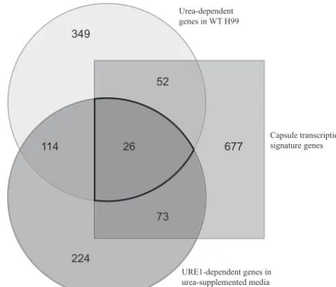
In addition, we compared these two lists (i.e., the capsule tran-scriptional signature genes and genes differentially expressed in urea-supplemented medium) to data from our second microarray experiment where we further examined urea exposure. We noted genes differentially expressed in theure1⌬mutant strain com-pared to either the WT or theamt1/2⌬mutant within the context of urea-supplemented medium and compiled them together (Fig. 9). There were 99 genes displaying differential expression in the ure1⌬mutant strain under urea-supplemented conditions that also appeared on the capsule transcriptional signature list. From these 99 genes, we compiled a list of 26 genes, shown inFig. 8, differentially expressed at significant levels linking our studies of urea exposure in the medium, studies of theure1⌬mutant strain with further urea exposure, and an investigation of consensus cap-sule-inducing conditions.Figure 8 presents the genes most ro-bustly urea associated and most likely to link the influence of urea to capsule production. The GO terms associated with this gene list spanned from GTPase activity and redox processes to steroid bio-synthesis, but many of the genes are putatively involved in trans-membrane transport and nitrogen metabolism. Most notably, one particular gene exhibited increased expression in urea and was positively correlated with capsule size in the study of Haynes et al. (32): the sugar transporter CNAG_03140 (Fig. 8). Sugar transport is known to be involved in capsule production (36).
DISCUSSION
As a cosmopolitan, pathogenic fungus found in a wide variety of environments, nutrient acquisition and nitrogen assimilation are vital forCryptococcus. One recent study illustrated the differences in utilization of 23 amino acids, such as phenylalanine, trypto-phan,D-alanine, andD-proline, between 67 strains ofC. neofor-mansandC. gattii(10). In a more expansive survey of 42 nitrogen sources, 16 strains ofCryptococcusspecies were revealed to utilize
large macromolecules, including amino acids and purines (9). For our study, we assessed an ecologically and biologically relevant subset of these nitrogen sources in a larger pool of 817 strains collected from clinical and environmental locations throughout North America and Africa. These clinical, veterinary, and envi-ronmental strains represent all 8 currently recognized molecular types. Confirming the previous studies, Casamino Acids were al-most universally assimilated as the sole nitrogen source, indicat-ing the fundamental utility of amino acids for nitrogen assimila-tion; the same was also true for uric acid. Since uric acid is present in high quantities in pigeon guano, the environmental reservoir most typically associated withC. neoformansvar.neoformansand C. neoformansvar.grubiistrains (37), the ubiquitous assimilation of uric acid byC. gattiistrains is more surprising.C. gattiiis typ-ically associated with a variety of trees, especially those in tropical to subtropical climates and, more recently, in temperate and high mountain climates (38–42). It is tempting to speculate thatC. gattiiis associated with the guano of bird species associated with these trees, as no link between C. gattiiand pigeons currently exists.
Our global survey of nitrogen utilization amongCryptococcus strains also confirmed the paradigm-shifting observation previ-ously made in the characterization ofTAR1(13): a significant number of isolates, particularlyC. gattiistrains, appear to utilize nitrate as the sole nitrogen source. This ability to use nitrate is less frequent in strains of theC. neoformansspecies. While these re-sults conflict with the prevailing understanding of nitrate nonuti-lization by all pathogenicCryptococcusspecies, they do confirm the published results of Jiang and colleagues showing a latent abil-ity of pathogenic cryptococci to utilize nitrate as the sole nitrogen source (13). Furthermore, we confirmed the accepted paradigm that nonpathogenic species, such asCryptococcus albidus, display robust growth on nitrate (data not shown). Our results suggest
677
224 349
114 26 52
73
FIG 9Gene expression under capsule formation-implicated conditions. Lists of genes from 3 microarray experiments were compared for overlapping genes. Differential expression of WT strain H99 in proline plus urea medium, differ-ential expression ofure1⌬genes in proline plus urea medium, and the capsule transcriptional signature from Haynes et al. (32) were examined for overlap-ping genes.
on September 8, 2020 by guest
http://ec.asm.org/
that the loss of nitrate utilization may have correlated with the pathogenic phenotype in C. neoformansandC. gattiiand that possible selection is occurring in the environment to reestablish this phenotype in some of these strains. Further work needs to be done to establish the genetic basis for this reversal in phenotype.
Nutrient acquisition within the central nervous system (CNS) is essential for the fungus at this site and has significant clinical implications. We have clearly described how critical carbon me-tabolism is in this sequestered sanctuary (2), and in this study, we have begun to identify the nitrogen products’ impact on Crypto-coccus physiology. Uric acid is present in cerebrospinal fluid (CSF), but only in small amounts. Urea, however, comprises the most abundant nitrogen metabolite in the CSF, surpassing even glucose in concentration. Impressively, the sum of all average amino acid concentrations does not equal a quarter of the concen-tration of urea (16). While we demonstrated with our capsule induction assay that uric acid can increase capsule size, particu-larly inC. gattiistrains, and we confirmed the observation of Lee et al. (9) that creatinine as the sole nitrogen source induces the larg-est capsule in certain serotype A strains, we found that urea pro-duced the largest capsule sizes in all other serotypes, especially the Vancouver outbreak VGIIa strain R265. We have identified this abundant nitrogenous product as a signaling molecule for induc-tion of cryptococcal capsule, and this mechanism is generalizable across all species and genotypes.
Furthermore, we observed that this induction of capsule by urea is dose dependent. At the concentrations selected, capsule size increased with increasing urea concentration; this effect was observed more prominently inC. gattiistrains. As urea is highly soluble in water, practically nontoxic, and slightly alkaline in aqueous solutions, this induction effect could result in a substan-tial, high upper limit. While these values may exceed the estab-lished biological concentrations in CSF, urea is frequently used at very high concentrations as a soil fertilizer, and this exposure may have selected for prominent capsule induction in response to ele-vated urea in certain strains. As fertilizer lacks typical reagents known to induce capsule production, including high CO2and serum, nitrogen sources such as urea may help this fungus survive environmental predatory scavengers, such as amoebae or worms that phagocytose the fungus, and killing by inducing the same large protective capsule that we observed during persistence in human CSF.
Our phenotypic observations that the presence of urea in-creases capsule size were further reinforced by our microarray results. Analysis of the WT under urea-supplemented conditions revealed enrichment for a variety of molecular functions related to capsule production. In the presence of various extracellular urea concentrations, the urea pathway mutants yielded further differ-ences in their transcriptional responses to the presence of urea; in the presence of higher local urea concentrations, theure1⌬strain was indeed enriched for various molecular functions related to capsule production. Numerous sugar transporters and vesicle transport-related genes were upregulated in theure1⌬mutant. Taken together, the increased transcription of these genes indi-cates cellular conditions favorable for increased capsule produc-tion in response to intact urea and corroborates our phenotypic observation of increased capsule production in theure1⌬strain in the presence of urea compared to that for the WT. The genes differentially expressed in both of these microarray experiments
with increased urea concentrations provide a specific group of genes linking capsule production to urea exposure.
From our microarray studies, we also found that the mecha-nism of urea catabolism may be concentration dependent inC. neoformans. InArabidopsisspecies, urea degradation to CO2and NH3by urease occurs extracellularly at high concentrations. At low concentrations, though, urea is imported via Arabidopsis thalianaDUR3, a high-affinity urea/H⫹symporter (43). Saccha-romyces cerevisiaealso follows a dual transport mechanism for urea transport. The first mode of uptake involves active transport, is subject to nitrogen catabolite repression, and makes use of the two-step urea carboxylase/allophanate hydrolase mechanism, while the second mode occurs via passive or facilitated diffusion at concentrations greater than 0.5 mM (44). The results from our microarray experiments support a similar regulated mechanism inC. neoformans. The urea transporter is upregulated under low-urea conditions or conditions without low-urea relative to the regula-tion under urea-supplemented condiregula-tions in all strains tested (⬃16-fold increase). This finding suggests the importance of urea exposure for Cryptococcus species and that urea catabolism is probably controlled in a concentration-dependent manner, as ob-served in other model organisms.
The work by Haynes and colleagues established a capsule tran-scriptional signature and assembled a comprehensive list of genes that may be directly vital to capsule induction (32). Their selection of 8 media for performing microarray analysis included low-iron medium (LIM) with and without the chelating agent EDTA, phos-phate-buffered saline (PBS) with and without fetal bovine serum (FBS), Dulbecco’s modified Eagle’s medium (DMEM) in room air (RA) or in 5% CO2, and Littman’s medium (LIT) with two con-centrations of thiamine (LO-THI and HI-THI). These conditions are well established as being variably capsule inducing. However, several other nutrient sources, including urea, have also been shown to increase capsule size and, in fact, may not do so by the same mechanisms that these environments exploit (45). Compar-ison of their transcriptional signature genes with our lists of dif-ferentially expressed genes determined from the use of urea-sup-plemented conditions and urea pathway mutants did reveal a number of genes differing between the two lists. As the phenotypes were comparable among the various conditions, this could indi-cate that urea may induce a unique pathway(s) for capsule pro-duction inCryptococcusspecies.
On the other hand, there were some transcriptional similarities between the lists of differentially expressed genes determined from the use of urea-supplemented conditions and capsule-in-ducing conditions that included 26 genes. This finding likely iden-tifies common responses among the transcriptional controls of capsule induction pathways. These comparisons may help us not only determine which genes are uniquely required for urea-spe-cific capsule induction but also identify common genetic control over capsule induction. The consensus 26 differentially expressed genes that appeared at significant levels under all urea-supple-mented conditions that we studied and that were on the signature list of Haynes et al. (32) may provide the best opportunity for further direct investigation into the ability of urea to induce cap-sule production inC. neoformans. While many of these genes are hypothetical proteins (CNAG_01751, CNAG_04475) and may present novel pathways linking urea with capsule induction, other genes on this list have already been clearly implicated in capsule production. For instance, the sugar transporter CNAG_03140
on September 8, 2020 by guest
http://ec.asm.org/
may transport vital raw sugar materials for synthesizing capsule in the presence of urea, and a nitrogen compound metabolic process gene (CNAG_06067) may function in regulation of the large cap-sule phenotype observed in the presence of urea. We have now identified a list of genes with which to dissect networks and the linkage of genes to phenotypes.
The pathobiological consequences of urea at the site of CNS infection are less apparent. As noted in our studies with the phys-iological CO2concentrations, further induction of capsule with urea is not detected in theure1⌬mutant, and in the model of acute rabbit meningitis, this mutant appears to survive as well as the wild-type strain in the CNS (17). However, the impact on long-term yeast survival in the leptomeninges and in CNS cryptococ-comas has not been addressed, and cryptococcal urease functions and urea exposure in the lung have been shown to have influences on host immunity (46). Furthermore, there are many known pos-itive and negative effects of the capsular polysaccharide on local immune functions that could be impacted by its induction. As we have shown, there is a dose effect of urea concentrations on cap-sular polysaccharide production, and increased urea in the host is dynamic and can occur in patients with renal dysfunction from an underlying disease and/or can be caused by polyene therapy, which could directly affect capsule production in the host to the benefit of the yeast at the CNS site. On another note, the urea effect on reducing expression of the gene for uracil phosphoribosyl-transferase (CNAG_02337) might even have a deleterious effect on the fungicidal activity of flucytosine treatment at the site of infection. This observation will need further examination. Clearly, urea in the mammalian host can be co-opted by this encapsulated yeast as a signal to help it survive the hostility of the host environ-ment and, potentially, its treatenviron-ment.
In summary, we have demonstrated that urea, as a signal, is a significant inducer of capsule at biologically relevant concentra-tions in the model fungal pathogenC. neoformansorC. gattii. Our results indicate that this increase in capsule size is not due to growth deficiency in the presence of urea but is due to urea signal-ing. Furthermore, these results may explain why pathogenic Cryp-tococcusspecies have such a pronounced capsule size following persistence within the CSF. As one of the most abundant nitrogen sources in the CSF, urea may prove significant in inducing the prominent capsular phenotype within the CNS.
ACKNOWLEDGMENTS
We thank the members of the J. A. Alspaugh lab for their helpful conver-sations on this work. We thank Joseph Heitman and Ana Litvintseva for providing isolates for characterization and Yuan Chen for advice on GO enrichment.
This work was supported by NIH PHS grant AI73896 (to J.R.P.). A.E.F. was supported by a Bill & Melinda Gates Foundation University Scholars Program Scholarship and the Duke University Undergraduate Research Scholars Program.
REFERENCES
1.Park BJ, Wannemuehler KA, Marston BJ, Govender N, Pappas PG, Chiller TM.2009. Estimation of the current global burden of cryptococcal meningitis among persons living with HIV/AIDS. AIDS23:525–530. 2.Price MS, Betancourt-Quiroz M, Price JL, Toffaletti DL, Vora H, Hu G,
Kronstad JW, Perfect JR.2011.Cryptococcus neoformansrequires a func-tional glycolytic pathway for disease but not persistence in the host. mBio 2(3):e00103– 00111. doi:10.1128/mBio.00103-11.
3.Lau G, Hamer JE.1996. Regulatory genes controllingMPG1expression and pathogenicity in the rice blast fungusMagnaporthe grisea. Plant Cell 8:771–781.
4.Froeliger EH, Carpenter BE.1996.NUT1, a major nitrogen regulatory gene inMagnaporthe grisea, is dispensable for pathogenicity. Mol. Gen. Genet.251:647– 656.
5.Bolton M, Thomma B.2008. The complexity of nitrogen metabolism and nitrogen-regulated gene expression in plant pathogenic fungi. Physiol. Mol. Plant Pathol.72:104 –110.
6.Hensel M, Arst HN, Aufauvre-Brown A, Holden DW.1998. The role of theAspergillus fumigatus areAgene in invasive pulmonary aspergillosis. Mol. Gen. Genet.258:553–557.
7.Panepinto JC, Oliver BG, Fortwendel JR, Smith DL, Askew DS, Rhodes JC.2003. Deletion of theAspergillus fumigatusgene encoding the Ras-related protein RhbA reduces virulence in a model of invasive pulmonary aspergillosis. Infect. Immun.71:2819 –2826.
8.Limjindaporn T, Khalaf RA, Fonzi WA.2003. Nitrogen metabolism and virulence ofCandida albicansrequire the GATA-type transcriptional ac-tivator encoded byGAT1. Mol. Microbiol.50:993–1004.
9.Lee IR, Chow EWL, Morrow CA, Djordjevic JT, Fraser JA. 2011. Nitrogen metabolite repression of metabolism and virulence in the hu-man fungal pathogen Cryptococcus neoforhu-mans. Genetics188:309 –323. 10. Ngamskulrungroj P, Chang Y, Roh J, Kwon-Chung KJ.2012. Differ-ences in nitrogen metabolism betweenCryptococcus neoformansandC. gattii, the two etiologic agents of cryptococcosis. PLoS One7:e34258. doi: 10.1371/journal.pone.0034258.
11. Kmetzsch L, Staats CC, Simon E, Fonseca FL, Oliveira DL, Joffe LS, Rodrigues J, Lourenço RF, Gomes SL, Nimrichter L, Rodrigues ML, Schrank A, Vainstein MH.2011. The GATA-type transcriptional activa-tor Gat1 regulates nitrogen uptake and metabolism in the human patho-gen Cryptococcus neoformans. Fungal Genet. Biol.48:192–199. 12. Wilson RA, Jenkinson JM, Gibson RP, Littlechild JA, Wang Z-Y, Talbot
NJ.2007. Tps1 regulates the pentose phosphate pathway, nitrogen metab-olism and fungal virulence. EMBO J.26:3673–3685.
13. Jiang N, Xiao D, Zhang D, Sun N, Yan B, Zhu X.2009. Negative roles of a novel nitrogen metabolite repression-related gene,TAR1, in laccase production and nitrate utilization in the basidiomyceteCryptococcus neo-formans. Appl. Environ. Microbiol.75:6777– 6782.
14. Petzold EW, Himmelreich U, Mylonakis E, Rude T, Toffaletti D, Cox GM, Miller JL, Perfect JR.2006. Characterization and regulation of the trehalose synthesis pathway and its importance in the pathogenicity of
Cryptococcus neoformans. Infect. Immun.74:5877–5887.
15. Ngamskulrungroj P, Himmelreich U, Breger JA, Wilson C, Chayakul-keeree M, Krockenberger MB, Malik R, Daniel HM, Toffaletti D, Djordjevic JT, Mylonakis E, Meyer W, Perfect JR.2009. The trehalose synthesis pathway is an integral part for the virulence composite for Cryp-tococcus gattii. Infect. Immun.77:4584 – 4596.
16. Mandal RGA, Chaudhary KK, Liu P, Yallou FS, Dong E, Aziat F, Wishart DS.2012. Multi-platform characterization of the human cere-brospinal fluid metabolome: a comprehensive and quantitative update. Genome Med.4:38. doi:10.1186/gm337.
17. Cox GM, Mukherjee J, Cole GT, Casadevall A, Perfect JR.2000. Urease as a virulence factor in experimental cryptococcosis. Infect. Immun.68: 443– 448.
18. Olszewski MA, Noverr MC, Chen GH, Toews GB, Cox GM, Perfect JR, Huffnagle GB.2004. Urease expression byCryptococcus neoformans pro-motes microvascular sequestration, thereby enhancing central nervous system invasion. Am. J. Pathol.164:1761–1771.
19. Bava AJ, Negroni R, Bianchi M.1993. Cryptococcosis produced by a urease negative strain ofCryptococcus neoformans. J. Med. Vet. Mycol. 31:87– 89.
20. Ruane PJ, Walker LJ, George WL.1988. Disseminated infection caused by urease-negativeCryptococcus neoformans. J. Clin. Microbiol.26:2224 – 2225.
21. Chaskes S, Tyndall RL.1975. Pigment production byCryptococcus neo-formansfrom para- and ortho-diphenols: effect of the nitrogen source. J. Clin. Microbiol.1:509 –514.
22. Granger DL, Perfect JR, Durack DT.1985. Virulence ofCryptococcus neoformans.Regulation of capsule synthesis by carbon dioxide. J. Clin. Invest.76:508 –516.
23. Vartivarian SE, Anaissie EJ, Cowart RE, Sprigg HA, Tingler MJ, Jacob-son ES.1993. Regulation of cryptococcal capsular polysaccharide by iron. J. Infect. Dis.167:186 –190.
24. Zaragoza O, Fries BC, Casadevall A.2003. Induction of capsule growth inCryptococcus neoformansby mammalian serum and CO(2). Infect. Im-mun.71:6155– 6164.
on September 8, 2020 by guest
http://ec.asm.org/
25. Littman ML.1958. Capsule synthesis byCryptococcus neoformans. Trans. N. Y. Acad. Sci.20:623– 648.
26. Rivera J, Feldmesser M, Cammer M, Casadevall A. 1998. Organ-dependent variation of capsule thickness in Cryptococcus neoformans during experimental murine infection. Infect. Immun.66:5027–5030. 27. Kozel TR, Pfrommer GS, Guerlain AS, Highison BA, Highison GJ.
1988. Strain variation in phagocytosis ofCryptococcus neoformans: disso-ciation of susceptibility to phagocytosis from activation and binding of opsonic fragments of C3. Infect. Immun.56:2794 –2800.
28. Dufait R, Velho R, De Vroey C.1987. Rapid identification of the two varieties of Cryptococcus neoformans byD-proline assimilation. Mykosen 30:483.
29. Rutherford JC, Lin X, Nielsen K, Heitman J.2008. Amt2 permease is required to induce ammonium-responsive invasive growth and mating in
Cryptococcus neoformans. Eukaryot. Cell7:237–246.
30. Werner E, Holder AA, Hoheisel JD.1997. Growth and storage of YAC clones in Hogness freezing medium. Nucleic Acids Res.25:1467–1468. 31. O’Meara TR, Norton D, Price MS, Hay C, Clements MF, Nichols CB,
Alspaugh JA.2010. Interaction of Cryptococcus neoformans Rim101 and protein kinase A regulates capsule. PLoS Pathog.6:e1000776. doi:10.1371 /journal.ppat.1000776.
32. Haynes BC, Skowyra ML, Spencer SJ, Gish SR, Williams M, Held EP, Brent MR, Doering TL.2011. Toward an integrated model of capsule regulation in Cryptococcus neoformans. PLoS Pathog.7:e1002411. doi:10 .1371/journal.ppat.1002411.
33. Jain N, Guerrero A, Fries BC.2006. Phenotypic switching and its impli-cations for the pathogenesis of Cryptococcus neoformans. FEMS Yeast Res.6:480 – 488.
34. Wishart DS, Lewis MJ, Morrissey JA, Flegel MD, Jeroncic K, Xiong Y, Cheng D, Eisner R, Gautam B, Tzur D, Sawhney S, Bamforth F, Greiner R, Li L. 2008. The human cerebrospinal fluid metabolome. J. Chro-matogr. B Analyt. Technol. Biomed. Life Sci.871:164 –173.
35. Marzluf GA. 1997. Genetic regulation of nitrogen metabolism in the fungi. Microbiol. Mol. Biol. Rev.61:17–32.
36. Cottrell TR, Griffith CL, Liu H, Nenninger AA, Doering TL.2007. The
pathogenic fungus Cryptococcus neoformans expresses two functional GDP-mannose transporters with distinct expression patterns and roles in capsule synthesis. Eukaryot. Cell6:776 –785.
37. Levitz SM, Tabuni A.1991. Binding ofCryptococcus neoformansby hu-man cultured macrophages. Requirements for multiple complement re-ceptors and actin. J. Clin. Invest.87:528 –535.
38. Byrnes EJ, III, Li W, Lewit Y, Ma H, Voelz K, Ren P, Carter DA, Chaturvedi V, Bildfell RJ, May RC, Heitman J.2010. Emergence and pathogenicity of highly virulent Cryptococcus gattii genotypes in the northwest United States. PLoS Pathog.6:e1000850. doi:10.1371/journal .ppat.1000850.
39. MacDougall L, Kidd SE, Galanis E, Mak S, Leslie MJ, Cieslak PR, Kronstad JW, Morshed MG, Bartlett KH.2007. Spread of Cryptococcus gattii in British Columbia, Canada, and detection in the Pacific Northwest, USA. Emerg. Infect. Dis.13:42–50.
40. Ellis DH, Pfeiffer TJ.1990. Ecology, life cycle, and infectious propagule of
Cryptococcus neoformans. Lancet336:923–925.
41. Kwon-Chung J, Bennett J.1984. High prevalence of Cryptococcus neo-formans var. gattii in tropical and subtropical regions. Zentralbl. Bakte-riol. Mikrobiol. Hyg. A257:213–218.
42. Granados DP, Castañeda E.2006. Influence of climatic conditions on the isolation of members of the Cryptococcus neoformans species complex from trees in Colombia from 1992-2004. FEMS Yeast Res.6:636 – 644. 43. Liu L-H, Ludewig U, Frommer WB, von Wirén N. 2003. AtDUR3
encodes a new type of high-affinity urea/H⫹symporter in Arabidopsis. Plant Cell15:790 – 800.
44. Cooper TG, Sumrada R.1975. Urea transport in Saccharomyces cerevi-siae. J. Bacteriol.121:571–576.
45. Zaragoza O, Casadevall A.2004. Experimental modulation of capsule size in Cryptococcus neoformans. Biol. Proced. Online6:10 –15. 46. Osterholzer JJ, Surana R, Milam JE, Montano GT, Chen G-H, Sonstein
J, Curtis JL, Huffnagle GB, Toews GB, Olszewski MA.2009. Crypto-coccal urease promotes the accumulation of immature dendritic cells and a non-protective T2 immune response within the lung. Am. J. Pathol. 174:932–943.