The PROFILE Feasibility Study: Targeted Screening of Men With a Family
History of Prostate Cancer
ELENACASTRO,a,bCHRISTOSMIKROPOULOS,a,bELIZABETHK. BANCROFT,a,bTOKHIRDADAEV,aCHEEGOH,a,bNATALIETAYLOR,a,b EDWARDSAUNDERS,aNIGELBORLEY,bDIANAKEATING,aELIZABETHC. PAGE,aSIBELSAYA,aSTEPHENHAZELL,cNAOMILIVNI,c
NANDITA DESOUZA,dDAVIDNEAL,e,fFREDDIEC. HAMDY,gPARDEEPKUMAR,bANTONISC. ANTONIOU,hZSOFIAKOTE-JARAI,aTHEPROFILE STUDYSTEERINGCOMMITTEE, ROSALINDA. EELESa,b
a
Oncogenetics Team, The Institute of Cancer Research, London, United Kingdom;bAcademic Urology Unit andcHistopathology
Department, The Royal Marsden National Health Service Foundation Trust, London, United Kingdom;dDivision of Radiotherapy and
Imaging, The Institute of Cancer Research, Sutton, United Kingdom; Departments ofeOncology andfSurgery, Cancer Research UK
Cambridge Institute, Cambridge, United Kingdom;gNuffield Department of Surgical Sciences, University of Oxford, Oxford, United
Kingdom;hCentre for Cancer Genetic Epidemiology, Department of Public Health and Primary Care, University of Cambridge, Cambridge,
United Kingdom
Disclosures of potential conflicts of interest may be found at the end of this article.
Key Words. Prostate cancer x Family history x Single nucleotide polymorphisms x Prostate-specific antigen
ABSTRACT
Background.A better assessment of individualized prostate cancer (PrCa) risk is needed to improve screening. The use of the prostate-specific antigen (PSA) level for screening in the general population has limitations and is not currently advocated. Approximately 100 common single nucleotide polymorphisms (SNPs) have been identified that are associated with the risk of developing PrCa. The PROFILE pilot study explored the feasibility of using SNP profiling in men with a family history (FH) of PrCa to investigate the probability of detecting PrCa at prostate biopsy (PB). The primary aim of this pilot study was to determine the safety and feasibility of PrCa screening using transrectal ultrasound-guided PB with or without diffusion-weighted magnetic resonance imaging (DW-MRI) in men with a FH. A secondary aim was to evaluate the potential use of SNP profiling as a screening tool in this population.
Patients and Methods.A total of 100 men aged 40–69 years with a FH of PrCa underwent PB, regardless of their baseline
PSA level. Polygenic risk scores (PRSs) were calculated for each participant using 71 common PrCa susceptibility alleles. We treated the disease outcome at PB as the outcome variable and evaluated its associations with the PRS, PSA level, and DW-MRI findings using univariate logistic regression.
Results.Of the 100 men, 25 were diagnosed with PrCa, of whom 12 (48%) had clinically significant disease. Four adverse events occurred and no deaths. The PSA level and age at study entry were associated with PrCa at PB (p5.00037 andp5 .00004, respectively).
Conclusion.The results of the present pilot study have demon-strated that PB is a feasible and safe method of PrCa screening in men with a FH, with a high proportion of PrCa identified requiring radical treatment. It is feasible to collect data on PrCa-risk SNPs to evaluate their combined effect as a potential screening tool. A larger prospective study powered to detect statistical associa-tions is in progress.The Oncologist2016;21:716–722
Implications for Practice:Prostate biopsy is a feasible and safe approach to prostate cancer screening in men with a family history and detects a high proportion of prostate cancer that needs radical treatment. Calculating a polygenic risk score using prostate cancer risk single nucleotide polymorphisms could be a potential future screening tool for prostate cancer.
INTRODUCTION
Prostate cancer (PrCa) is the most common cancer in men in Europe and constitutes a significant health burden. Screening for PrCa in the general population using the prostate-specific antigen (PSA) level is controversial and not currently advocated owing to its inability to distinguish between clinically significant and indolent disease and conflicting evidence on the effect on
mortality [1–3]. Overdiagnosis and overtreatment are signifi-cant limitations of PSA screening. However, targeted screening aimed at higher risk groups might have a greater impact [4]. Excluding age and African-American ancestry, the strongest risk factor for PrCa is a family history (FH) [5]. First-degree relatives (FDRs) of men with PrCa have approximately twice the risk of the
Correspondence: Rosalind A. Eeles, F.Med.Sci., Ph.D., The Institute of Cancer Research and Royal Marsden National Health Service Foundation Trust, 15 Cotswold Road, Sutton SM2 5NG, United Kingdom.Telephone: 44-208-722-4094; E-Mail: ros.eeles@icr.ac.uk Received August 17, 2015; accepted for publication February 9, 2016; published Online First on May 5, 2016. ©AlphaMed Press 1083-7159/2016/$20.00/0 http://dx.doi. org/10.1634/theoncologist.2015-0336
CM
E
by guest on October 26, 2016
http://theoncologist.alphamedpress.org/
general population, which increases to more than fourfold if PrCa was diagnosed in a relative when he was younger than 60 years [6]. Several germline single nucleotide polymorphisms (SNPs) have been associated with PrCa risk [5]. Although the effect of each of these variants is small, they act multiplicatively and together explain∼30% of the genetic variance of PrCa [5]. Several studies have reported that polygenic risk scores (PRSs), based on the combined effects of these SNPs, could be used to predict a man’s future PrCa risk [6, 7] and could be useful for targeted screening. However, it is unclear how they relate to the probability of detecting existing PrCa in asymptomatic men.
In the present cross-sectional pilot study, the primary aim was to determine the feasibility of using prostate biopsy (PB) as a screening tool (irrespective of the PSA level) in men with a FH of PrCa. The secondary aims were to evaluate the feasibility of collecting data on SNP profiles, PSA levels, and diffusion-weighted magnetic resonance imaging (DW-MRI) and assess whether they could be used as screening tools in this population.
PATIENTS ANDMETHODS
Patients
Eligible men were identified as follows: (a) through cancer genetics and uro-oncology clinics at a National Health Service cancer hospital in London; (b) men taking part in the U.K. Genetic Prostate Cancer study (UKGPCS; http://www.icr.ac. uk/ukgpcs); and (c) advertisements and newsletters. For the first two methods, men with PrCa were approached either in clinic or by letter and invited to pass on an information sheet to eligible relatives. The eligibility criteria included (a) age 40–69 years; (b) a FH of PrCa; (c) if previous PB had been performed, it must have been performed$1 year earlier; and (d) provision of informed consent. Men with a previous diagnosis of PrCa, currently undergoing evaluations for symptoms suggestive of PrCa, or diagnosed with cancer with a terminal prognosis of,5 years were excluded. A FH was defined as having (a) one FDR with PrCa diagnosed at,70 years; (b) two relatives (FDRs or second-degree relatives) on the same side of the family with at least one diagnosed at age,70 years; or (c) three relatives on the same side of the family diagnosed at any age.
Study Design
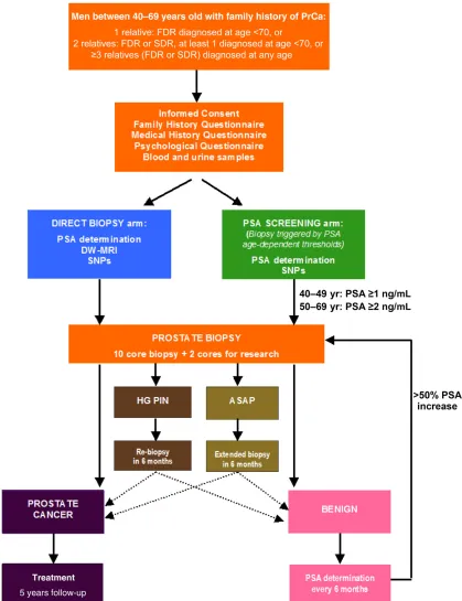
After providing written informed consent, the participants completed a family and medical history questionnaire and provided blood samples for PSA measurement and DNA extraction. All men underwent a 10-core (standard practice at the time the protocol was written) transrectal ultrasound-guided PB, regardless of the baseline PSA level, within 8 weeks of study entry. Standard antibiotic prophylaxis was used, and the biopsies were performed to a standard template by two urologists (N.B., P.K.). An expert pathologist reviewed all of the biopsy specimens (S.H.). The participants who rejected an initial PB were followed up with 6-month PSA measurements and were excluded from the present analyses. The study algorithm is detailed in Figure 1.
The first 50 patients enrolled in the direct biopsy arm were also offered DW-MRI before biopsy [8]. The images were assessed by an experienced observer using a combination of T-weighted and DW images and scored as positive or negative
for tumor. This was not used to guide the biopsy, because this component of the study was to gauge the acceptability of this technique to inform the future main study design. The results of this component of the study have been reported separately [8].
For those patients diagnosed with PrCa at PB, management was offered either at the cancer center or at their local hospital according to the standard U.K. national guidelines [9]. The outcomes of the different treatments for these patients will be followed up for 5 years. Those patients who presented with atypical small acinar proliferation (ASAP) or high-grade prostatic intraepithelial neoplasia (HG-PIN) underwent repeat biopsy after 6 months. Men with negative biopsy findings were monitored with PSA testing every 6 months, with the biopsy repeated if the PSA level increased by.50% (data not presented).
Genetic Profiling
The participants’ DNA samples were genotyped using a custom Illumina iSelect genotyping array [10, 11]. We used the data for 71 known PrCa susceptibility SNPs; 61 were di-rectly genotyped and, for 10 loci, the data for a proxy SNP with pairwise correlation of r2 .0.75 were used (supplemental online Table 1).
Statistical Analysis
For each patient, the PRS was calculated as the weighted sum of the number of risk alleles at the 71 loci, for which the weights were the estimated log-odds ratios (ORs) associated with each allele, obtained from published studies [5]. In an exploratory analysis, we used univariate logistic regression to evaluate the associations of age at study entry, PSA, and PRS with a diagnosis of PrCa at PB. R, version 3.1.2 (October 31, 2014), was used for statistical analysis [12].
RESULTS
From December 2010, 897 men aged 40–69 years with a FH of PrCa were invited into the study, and 285 (32%) replied. The enrollment was closed in January 2013 when 115 men had entered the study and 100 had agreed to undergo PB (Table 1). Six men had previously undergone PB at a mean of 45.3 months (range 24–96). Fifteen men declined PB and were followed up with PSA measurements only and were excluded from the present analyses.
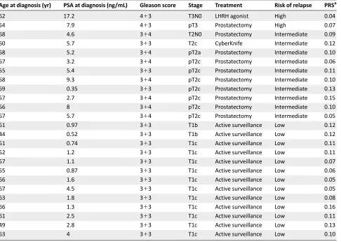
The median age of the participants was 53 years (range 40–69), with a median PSA level of 1.3 ng/mL (range 0.2–9.8; Table 2). Ten-core PB identified 7 cases of HG-PIN, 5 cases of ASAP, and 25 cases of PrCa. The median age of PrCa diagnosis was 61 years (range 44–69), with median PSA level of 2.7 ng/mL (range 0.35–9.3). The median age of men diagnosed with PrCa was older than that of those with negative biopsy findings (61 vs. 50 years;p5.00004). No differences were found in the rates of cancer diagnosis between the men according to their family history (Table 3). Thirteen tumors were low-risk (52%), 10 were intermediate-risk (40%), and 2 (8%) were high-risk as classified by the National Institute for Health and Care Excellence (NICE) criteria [9] (Table 4).
Complications occurred in 4 of the 100 participants (4%), with 3 postbiopsy infections (3%) reported and treated with a course of oral (n52) or intravenous (n51) antibiotics. One patient (1%) was kept under observation because he had fainted after the biopsy.
CM
E
by guest on October 26, 2016
http://theoncologist.alphamedpress.org/
Univariate association analyses showed that the PSA level was associated with the PrCa diagnosis (OR 1.73, 95% CI 1.28–2.33; p 5 .00037). Age at study entry was also significant (OR 1.16, 95% CI 1.08–1.25; p 5 .00004); however, we found no significant association between the PRS and PB outcome (p5.25802; Fig. 2). When stratified by the Gleason score of the tumor (7 or greater vs. less than 7),
no association was observed between the PRS and tumors with a Gleason score of 7 or greater (p5.21057). The PSA level was associated with a high Gleason score (OR 1.85, 95% CI 1.13–3.03;p5.01456).
We also evaluated the association between the PRS for the different levels of the presenting PSA. The PRS did not differ between men found to have prostate cancer and those without
Figure 1. PROFILE study algorithm.
Abbreviations: ASAP, atypical small acinar proliferation; DW-MRI, diffusion-weighted magnetic resonance imaging; FDR, first-degree relative; HG PIN, high-grade prostatic intraepithelial neoplasia; PrCa, prostate cancer; PSA, prostate-specific antigen; SDR, second-degree relative; SNPs, single nucleotide polymorphisms.
T
CM
E
by guest on October 26, 2016
http://theoncologist.alphamedpress.org/
the disease after biopsy among men with a PSA level,3 ng/mL (p 5 .15) or PSA level ,1 ng/mL (p 5 .08). This group constituted 38 men with 5 PrCas diagnosed; the median age at PrCa diagnosis was 57 years (range 44–69).
All patients diagnosed with low-risk PrCa opted for active surveillance. For those choosing radical treatment, radical prostatectomy was chosen by 10 men (40%). One patient underwent radiotherapy with CyberKnife (Accuray, Sunnyvale, CA, http://www.cyberknife.com), and one patient with metastatic disease received hormonal treatment alone.
DISCUSSION
The present study was undertaken to assess the feasibility of using primary PB in a known higher risk group without the
confounder of the PSA level used to trigger biopsy. The reason for undertaking the present study was to be able to inform a larger PROFILE study, which will be powered to be able to associate the PRS outcome with the PB outcome. A FH group was chosen because this increased the power approximately fourfold. These results provide evidence that it is feasible to screen for PrCa using upfront PB in men with a FH of the disease, and approximately one third of men will agree to such screening. Of the men undergoing PB, 25% were diagnosed with PrCa, of whom 48% had disease that was intermediate- or high-risk and warranting treatment using the NICE guidelines.
Interestingly, of the 25 study participants diagnosed with PrCa, 13 (52%) had a PSA level,3 ng/mL, and these patients would most likely have not undergone PB within the traditional PSA-based screening schedules. This is a finding that raises questions about the reliability of PSA determination, especially for men with a significant FH. In contrast, the Prostate Cancer Prevention Trial (PCPT) reported an overall PrCa detection rate of 15.2% in 2,950 men with a PSA level ,4.0 ng/mL and unsuspicious digital rectal examination findings. They found that only 19.6% of tumors had high-grade disease (defined as a Gleason score.7), and the men were older, with an age range of 62–91 years [13]. Although the two studies are not directly comparable because of the different classifications of disease risk and different PSA levels used, a remarkable difference was found in the proportion of clinically significant tumors. Larger studies are required to confirm the differences observed. Little is known about the effect of FH on PrCa progression and the benefits of early diagnosis in this group [14]. All but one man diagnosed with low-risk PrCa accepted active surveillance, highlighting the growing understanding of the natural history of PrCa and the increasing acceptance of a structured moni-toring strategy.
Another major concern with using PB for PrCa screening is safety. Although hematuria and hematospermia are common, severe postprocedural infections have been reported to occur in approximately 1% of cases, with major complications rare [15]. A similar incidence was observed in our series, with a 4% infection rate, 1% requiring hospitalization, and no major complications reported. Therefore, the potential negative consequences of PrCa screening, including biopsy complica-tions and cancer diagnosis, were regarded as acceptable for proceeding to the main study, which will be powered to detect the effect of the PRS in the screening algorithm. Men in the PROFILE study might be more predisposed to accept more screening-related side effects than the general population owing to their previous experience of relatives developing PrCa. However, a recent study evaluating the psychological effect of prostate biopsy in 1,147 men who had participated in the Prostate Testing for Cancer and Treatment (ProtecT) trial reported that postbiopsy symptoms such as discomfort, pain, or bleeding were experienced relatively commonly and for most patients were tolerated as a minor problem or no problem [16].
In the present pilot study, we performed exploratory analyses to assess the performance of age, PSA level, and PrCa risk SNPs in predicting the biopsy outcome in this setting. It is important to note that the SNP risk scores and PSA level were considered in our study as tools for predicting the PB outcome
Table 1. Responses of men invited into the study
Description n(%)
Total men invited 897 (100.0)
Did not reply 612 (68.2)
Replied 285 (31.8)
Declined 85 (9.5)
Accepted 115 (12.8)
Accepted but ineligible 31 (3.5)
Expressed an interest 47 (5.2)
Would take part if available locally 7 (0.8)
Table 2. Age at study entry, PSA level, and polygenic risk score
Variable
Prostate
cancer Mean Median Range (SD)
Age at study entry
No 51.2 50.2 40.3–67.3 (7.6)
Yes 59.9 60.9 44.6–68.5 (6.8)
Baseline PSA No 1.4 1.1 0.19–9.8 (1.35)
Yes 3.4 2.7 0.52–9.3 (2.54)
Polygenic risk score
No 0.1 0.1 0.023–0.222 (0.039)
Yes 0.1 0.1 0.041–0.156 (0.033)
Abbreviation: PSA, prostate-specific antigen.
Table 3. Family history of prostate cancer
Variable %
Total cohort,n5100
One FDR with PrCa diagnosed at age,70 yr 36
Two relatives (FDR or SDR) on same side of family;
one diagnosed at age,70 yr
37
Three relatives on same side of the family diagnosed at any age
27
PrCa cases,n525
One FDR with PrCa diagnosed at age,70 yr 36
Two relatives (FDR or SDR) on same side of the family;
one diagnosed at age,70 yr
40
Three relatives on same side of the family diagnosed at any age
24
Abbreviations: FDR, first-degree relative; PrCa, prostate cancer; SDR, second-degree relative.
CM
E
by guest on October 26, 2016
http://theoncologist.alphamedpress.org/
rather than predicting the future risk of developing PrCa. The latter could not be evaluated in the present study design; however, as prospective data are accumulated, it will be assessed in the future.
In the present small pilot study using univariate analyses, the SNP profile was not associated with prostate cancer at PB, although the estimated OR was greater than 1. However, much larger studies are required to address this question reliably.
Table 4. Characteristics of prostate cancers diagnosed in the study
Age at diagnosis (yr) PSA at diagnosis (ng/mL) Gleason score Stage Treatment Risk of relapse PRSa
62 17.2 413 T3N0 LHRH agonist High 0.04
54 7.9 413 pT3 Prostatectomy High 0.07
68 4.6 314 T2N0 Prostatectomy Intermediate 0.09
60 5.7 313 T2c CyberKnife Intermediate 0.12
68 5.2 314 pT2a Prostatectomy Intermediate 0.10
67 3.2 314 pT2c Prostatectomy Intermediate 0.06
55 5.4 313 pT2c Prostatectomy Intermediate 0.11
68 9.3 314 pT2c Prostatectomy Intermediate 0.10
69 0.35 313 pT2c Prostatectomy Intermediate 0.13
57 2.7 314 pT2c Prostatectomy Intermediate 0.15
56 8 314 pT2c Prostatectomy Intermediate 0.10
67 5.7 314 pT2c Prostatectomy Intermediate 0.05
61 0.97 313 T1b Active surveillance Low 0.12
44 0.52 313 T1b Active surveillance Low 0.12
51 0.74 313 T1c Active surveillance Low 0.11
52 1.2 313 T1c Active surveillance Low 0.11
57 1.1 313 T1c Active surveillance Low 0.07
55 0.87 313 T1c Active surveillance Low 0.06
66 1.6 313 T1c Active surveillance Low 0.05
67 4.5 313 T1c Active surveillance Low 0.05
63 1.8 313 T1c Active surveillance Low 0.08
66 1.3 313 T1c Active surveillance Low 0.16
61 2.5 313 T1c Active surveillance Low 0.11
49 2.8 313 T1c Active surveillance Low 0.13
63 4 313 T1c Active surveillance Low 0.10
a
Risk of developing prostate cancer by 85 years old.
Abbreviations: LHRH, luteinizing hormone-releasing hormone; PRS, polygenic risk score; PSA, prostate-specific antigen.
Figure 2. The distribution of PRSs in the cohort. No significant association was found between the PRS and the prostate biopsy outcome
(p5.25802).
Abbreviations: PrCa, prostate cancer; PRS, polygenic risk score.
T
CM
E
by guest on October 26, 2016
http://theoncologist.alphamedpress.org/
Furthermore, this might be improved as further common variants are identified, which will increase the discrimination of the risk strata in populations. A possible confounder that might have caused this is that the men in the present study had a strong FH and their genetic risk was high, resulting in poor discrimination. Analyses of larger series of participants with adjustment for all relevant confounders are therefore warranted. Others have previously reported similar results, with areas under the curve that ranged from 0.57 to 0.67, although their prediction models included fewer genetic variants than did ours [7, 17–20].
The predictive value of SNP profiling in men presenting with a PSA level of 1–3 ng/mL was assessed by Nordstr¨om et al. [21], who found that a risk score based on 49 SNPs was a significant predictor of positive biopsy findings (p5.028). Based on current clinical practice, if these men were mon-itored using a PSA screening protocol, they would not undergo PB. In the PROFILE study, we analyzed the predictive value of the genetic score for men with a FH and a very low presenting PSA level of#1 ng/mL. These men would have normally been reassured by the PSA result, and we found no significant association between the PRS and PrCa diagnosis. However, the number of PrCas diagnosed in asymptomatic young men with a very low PSA level was sizeable (13% had PrCa at PB).
SNP profiling is an important tool in PrCa risk prediction algorithms. Risk prediction models serve to identify those men who could potentially benefit from screening, through early diagnosis and treatment [22]. Nam et al. reported a prediction model using FH, PSA level, and four of the published genome-wide association study (GWAS) risk SNPs, which improved the positive predictive value of the PSA level [18]. Zheng et al. included 11 SNPs in their model and showed that the prediction of the combination of genetic variants and FH was similar to that of the PSA level [23]. Pashayan et al. reported the use of a polygenic risk model to personalize screening and compared this with a theoretical model in which only age was used to determine whether to screen a population [24]. They showed that the use of SNPs reduced the risk of an overdiagnosis [24]. Macinnis et al. developed a model for predicting the probability of developing PrCa in the future using 26 SNPs and FH [25]. Kader et al. developed a multivariable predictive model for patients who had previously had negative PB findings [7].The predictive performance of the model, which included age, FH, PSA, prostate volume, and number of cores at PB, was improved after the addition of the genetic score based on 33 PrCa risk-associated SNPs. The proposed PROFILE study offers a unique opportunity to prospectively determine the benefit of the model, as the participants will be followed up for 5 years.
We acknowledge that the present pilot study had several weaknesses. The PROFILE study was designed as a feasibility study and, therefore, had a small sample size that did not allow definitive conclusions. Therefore, we did not test the ability of the genetic score to distinguish between the risk of indolent versus aggressive disease. Debate has ensued whether the SNPs identified to date are able to distinguish between these two forms of PrCa [26]. Second, most SNPs were selected by their association with a lifetime risk of developing prostate cancer, rather than predicting cancer at any given time.
Therefore, a prospective study with long-term follow-up is necessary to provide the data required to evaluate their use in a predictive risk model. Third, the present analysis only included men of European ancestry, because several GWASs included only men of this ethnicity, and it is not clear to what extent these results are applicable to other ethnic groups. Furthermore, all men had a FH of PrCa and, therefore, an increased risk of PrCa compared with the general population, which might have made them more receptive to undergoing PB and might also explain the high incidence of PrCa and pre-cancerous findings in the present series. Moreover, by definition, all the participants in the present study were expected to have higher PRSs compared with the general population, which would limit the ability of the PRS to discriminate between those with PrCa at PB and those without PrCa.
The main PROFILE study started recruiting in 2015, with the aim of recruiting 350 men of European ancestry with a FH and 350 men of African-Caribbean ancestry. In addition to DW-MRI, other predictive biomarkers will be incorporated, such as urinary PCA3 and TMPRSS-ERG translocation status.We aim to use the most up-to-date SNP set available to calculate the risk score by ethnic group.
CONCLUSION
PROFILE is the first study conducted of men with a FH of PrCa to incorporate biopsy, genetic profiling, imaging, and biomarkers in an innovative screening model.The feasibility study we have reported showed that one quarter of the men who underwent a PB were found to have PrCa, and 48% of these had disease that required radical treatment using the NICE guidelines. Our results indicate that direct PB is feasible and safe as a method of PrCa screening in men with an FH of PrCa. A larger study is underway for the development of a prediction model com-bining clinical variables and PrCa risk-associated SNPs that would help to determine which men at high risk of PrCa owing to their FH of PrCa should undergo PB.
ACKNOWLEDGMENTS
We thank all the participants and families who took part in this research. This work was supported by The Ronald and Rita McAulay Foundation and Cancer Research UK Grant C5047/ A13232. E.C. was supported by a European Society for Medical Oncology Clinical Research Fellowship, and the American Society of Clinical Oncology (ASCO) Cancer Foundation awarded her a 2013 ASCO Annual Meeting Merit Award. C.G. was supported by the Genetic Associations and Mecha-nisms in Oncology (GAME-ON) Initiative (NIH ELLIPSE Grant U19CA148537). A.C.A. is a Cancer Research U.K. Senior Cancer Research Fellow (C12292/A11174). The investigators at The Institute of Cancer Research and The Royal Marsden National Health Service (NHS) Foundation Trust are supported by an NIH research grant to the Biomedical Research Centre at The Institute of Cancer Research and The Royal Marsden NHS Foundation Trust. Funding for the iCOGS infrastructure came from the European Community’s Seventh Framework Pro-gramme under Grant 223175 (HEALTH-F2-2009-223175) (COGS), Cancer Research U.K. (Grants C1287/A10118, C1287/A 10710, C12292/A11174, C1281/A12014, C5047/ A8384, C5047/A15007, C5047/A10692, C8197/A16565), the NIH (Grant CA128978), and the Post-Cancer Genome-Wide
CM
E
by guest on October 26, 2016
http://theoncologist.alphamedpress.org/
Association Study Initiative (Grants 1U19 CA148537, 1U19 CA148065, and 1U19 CA148112—the GAME-ON initiative), the Department of Defense (Grant W81XWH-10-1-0341), the Canadian Institutes of Health Research (CIHR) for the CIHR Team in Familial Risks of Breast Cancer, Komen Foundation for the Cure, the Breast Cancer Research Foundation, and the Ovarian Cancer Research Fund. The PROFILE Study Steering Committee: A. Ardern-Jones, Royal Marsden NHS Foundation Trust; P. Ardern-Jones, Patient representative; N. van As, Royal Marsden NHS Foundation Trust; D. Dearnaley, Royal Marsden NHS Foundation Trust and The Institute of Cancer Research; C. Foster, HCA Laboratories; V. Khoo, Royal Marsden NHS Foundation Trust; S. Lewis, University of Bristol; H. Lilja, Memorial Sloan Kettering Cancer Center, Lund University, and the University of Oxford; J. Melia, University of Cambridge; C. Moynihan, The Institute of Cancer Research, P. Pharoah, University of Cambridge; and A. Sohaib, Royal Marsden NHS Foundation Trust. E.C. is currently affiliated with the Spanish National Cancer Research Centre, Madrid, Spain.
AUTHORCONTRIBUTIONS
Conception/Design:Elena Castro, Elizabeth K. Bancroft, Nandita deSouza, David Neal, Freddie C. Hamdy, Antonis C. Antoniou, Zsofia Kote-Jarai, PROFILE Study Steering Committee, Rosalind A. Eeles
Provision of study material or patients:Elena Castro, Natalie Taylor, Nigel Borley, Stephen Hazell, Pardeep Kumar, Rosalind A. Eeles
Collection and/or assembly of data: Elena Castro, Christos Mikropoulos, Elizabeth K. Bancroft, Chee Goh, Natalie Taylor, Edward Saunders, Nigel Borley, Diana Keating, Elizabeth C. Page, Sibel Saya, Stephen Hazell, Naomi Livni, Nandita deSouza
Data analysis and interpretation: Elena Castro, Christos Mikropoulos, Elizabeth K. Bancroft, Tokhir Dadaev, Chee Goh, Edward Saunders, Elizabeth C. Page, Sibel Saya, Nandita deSouza, Freddie C. Hamdy, Pardeep Kumar, Antonis C. Antoniou, Zsofia Kote-Jarai, Rosalind A. Eeles
Manuscript writing:Elena Castro, Christos Mikropoulos, Elizabeth K. Bancroft, Tokhir Dadaev, Chee Goh, Edward Saunders, Sibel Saya, Nandita deSouza, David Neal, Freddie C. Hamdy, Pardeep Kumar, Antonis C. Antoniou, Zsofia Kote-Jarai, PROFILE Study Steering Committee, Rosalind A. Eeles
Final approval of manuscript:Elena Castro, Antonis C. Antoniou, Rosalind A. Eeles
DISCLOSURES
The authors indicated no financial relationships.
REFERENCES
1.Carlsson S, Vickers AJ, Roobol M et al. Prostate cancer screening: Facts, statistics, and interpreta-tion in response to the US Preventive Services Task Force Review. J Clin Oncol 2012;30:2581–2584.
2.Schr¨oder FH. Landmarks in prostate cancer screening. BJU Int 2012;110(suppl 1):3–7.
3.Andriole GL Jr. PSA screening and prostate cancer risk reduction. Urol Oncol 2012;30:936–937.
4.Basch E, Oliver TK, Vickers A et al. Screening for prostate cancer with prostate-specific antigen test-ing: American Society of Clinical Oncology Provisional Clinical Opinion. J Clin Oncol 2012;30:3020–3025.
5.Eeles R, Goh C, Castro E et al. The genetic epidemiology of prostate cancer and its clinical implications. Nat Rev Urol 2014;11:18–31.
6.Xu J, Sun J, Kader AK et al. Estimation of absolute risk for prostate cancer using genetic markers and family history. Prostate 2009;69:1565–1572.
7.Kader AK, Sun J, Reck BH et al. Potential impact of adding genetic markers to clinical parameters in predicting prostate biopsy outcomes in men follow-ing an initial negative biopsy: Findfollow-ings from the REDUCE trial. Eur Urol 2012;62:953–961.
8.deSouza NM, Morgan VA, Bancroft E et al. Diffusion-weighted MRI for detecting prostate tumour in men at increased genetic risk. Eur J Radiol Open 2014;1:22–27.
9.National Institute for Health and Care Excel-lence. Prostate Cancer: Diagnosis and Treatment. London, United Kingdom: NICE, 2008.
10.Eeles RA, Olama AA, Benlloch S et al. Identi-fication of 23 new prostate cancer susceptibility loci using the iCOGS custom genotyping array. Nat Genet 2013;45:385–391.
11.Bahcall OG. iCOGS collection provides a collaborative model. Foreword. Nat Genet 2013; 45:343.
12.R Core Team. R: A language and environment for statistical computing. Vienna, Austria: R Foun-dation for Statistical Computing, 2014. Available at http://www.R-project.org/. Accessed August 17, 2015.
13.Thompson I, Pauler DK, Goodman PJ et al. Prevalence of prostate cancer among men with a prostate-specific antigen level,or54.0 ng per milliliter. N Engl J Med 2004;350:2239–2246.
14.Goh CL, Saunders EJ, Leongamornlert DA et al. Clinical implications of family history of prostate cancer and genetic risk single nucleotide poly-morphism (SNP) profiles in an active surveillance cohort. BJU Int 2013;112:666–673.
15.Loeb S, Carter HB, Berndt SI et al. Is repeat prostate biopsy associated with a greater risk of hospitalization? Data from SEER-Medicare. J Urol 2013;189:867–870.
16.Wade J, Rosario DJ, Macefield RC et al. Psychological impact of prostate biopsy: Physical symptoms, anxiety, and depression. J Clin Oncol 2013;31:4235–4241.
17.Salinas CA, Koopmeiners JS, Kwon EM et al. Clinical utility of five genetic variants for predicting prostate cancer risk and mortality. Prostate 2009;69: 363–372.
18.Nam RK, Zhang WW,Trachtenberg J et al. Utility of incorporating genetic variants for the early detection of prostate cancer. Clin Cancer Res 2009; 15:1787–1793.
19.Akamatsu S, Takahashi A, Takata R et al. Reproducibility, performance, and clinical utility of
a genetic risk prediction model for prostate cancer in Japanese. PLoS One 2012;7:e46454.
20.Klein RJ, Hallden C, Gupta A et al. Evaluation of multiple risk-associated single nucleotide polymor-phisms versus prostate-specific antigen at baseline to predict prostate cancer in unscreened men. Eur Urol 2012;61:471–477.
21.Nordstr¨om T, Aly M, Eklund M et al. A genetic score can identify men at high risk for prostate cancer among men with prostate-specific antigen of 1-3 ng/ml. Eur Urol 2014;65:1184–1190.
22.Freedman AN, Seminara D, Gail MH et al. Cancer risk prediction models: A workshop on development, evaluation, and application. J Natl Cancer Inst 2005;97:715–723.
23.Zheng SL, Sun J, Wiklund F et al. Genetic variants and family history predict prostate cancer similar to prostate-specific antigen. Clin Cancer Res 2009;15:1105–1111.
24.Pashayan N, Pharoah P, Tab ´ar L et al.Validation of a modelling approach for estimating the likely effectiveness of cancer screening using cancer data on prevalence screening and incidence. Cancer Epidemiol 2011;35:139–144.
25.Macinnis RJ, Antoniou AC, Eeles RA et al. A risk prediction algorithm based on family history and common genetic variants: Application to prostate cancer with potential clinical impact. Genet Epide-miol 2011;35:549–556.
26.Teerlink CC, Thibodeau SN, McDonnell SK et al. Association analysis of 9,560 prostate cancer cases from the International Consortium of Prostate Cancer Genetics confirms the role of reported prostate cancer associated SNPs for familial disease. Hum Genet 2014;133:347–356.
See http://www.TheOncologist.com for supplemental material available online.
CME This article is available for continuing medical education credit at CME.TheOncologist.com.
T
CM
E
by guest on October 26, 2016
http://theoncologist.alphamedpress.org/