REVI E W
Open Access
Feeding critically ill patients the right
‘
whey
’
:
thinking outside of the box. A personal view
Paul E Marik
Abstract
Atrophy of skeletal muscle mass is an almost universal problem in survivors of critical illness and is associated with significant short- and long-term morbidity. Contrary to common practice, the provision of protein/amino acids as a continuous infusion significantly limits protein synthesis whereas intermittent feeding maximally stimulates skeletal muscle synthesis. Furthermore, whey-based protein (high in leucine) increases muscle synthesis compared to soy or casein-based protein. In addition to its adverse effects on skeletal muscle synthesis, continuous feeding is unphysiological and has adverse effects on glucose and lipid metabolism and gastrointestinal function. I propose that critically ill patients’be fed intermittently with a whey-based formula and that such an approach is likely to be associated with better glycemic control, less hepatic steatosis and greater preservation of muscle mass. This paper provides the scientific basis for my approach to intermittent feeding of critically ill patients.
Keywords:Nutrition; Whey; Muscle mass; Muscle atrophy; Intermittent feeding; Continuous feeding; Metabolic function; Insulin; Incretin; MTOR; Autophagy; Ubiquitin–proteasome complex
Review
Survivors of critical illness suffer from marked muscle wasting which may take years to recover. The loss of muscle mass is associated with muscle weakness, pro-longed mechanical ventilatory support, fatigue and de-layed recovery [1–3]. This disorder is known as critical illness myopathy (CIM) or intensive care unit-acquired weakness (ICUAW) [1–3]. CIM is characterized by a diffuse non-necrotizing myopathy accompanied by fiber atrophy, fatty degeneration of muscle fibers and fibrosis [4]. Multiple factors are likely to play a role in inducing CIM including muscle inactivity, inflammation, cellular energy stress, corticosteroids, hyperglycemia, neuro-muscular blocking agents and inadequate nutritional support [2, 4]. CIM is exceedingly common in ICU survi-vors, being reported in up to 46 % of cases [5]. Herridge et al. followed 109 survivors of ARDS for up to 5 years after discharge from the ICU [6, 7]. All patients reported poor functional status with proximal weakness and fatigue at discharge. At 1 year, the distance walked in 6 minutes was 66 % of predicted which increased to 76 % of pre-dicted at 5 years [7].
Muscle breakdown during acute illness
In health, net muscle synthesis is stimulated in the post-prandial state while net muscle breakdown occurs be-tween meals with muscle mass being maintained through balanced protein synthesis and breakdown [8]. Distinct metabolic pathways are involved in the synthesis and breakdown of muscle. Figure 1 provides an overview of these pathways. Muscle protein synthesis and not break-down is more responsive to anabolic stimuli [9]. In healthy individuals, the anabolic effects of feeding occurs due to an increase in the synthetic rate of muscle protein synthe-sis of approximately 300 % with a concomitant 50 % de-crease in the rate of protein breakdown [8, 10]. In healthy young men following an oral bolus of essential amino acids, there is a lag period of 45–90 min followed by an increase in the muscle protein synthetic response which continues for about 90 min then rapidly returns to base-line [8, 10, 11]. The duration and degree of the muscle protein synthetic response following protein ingestion is influenced by exercise, age and the dose and type of pro-tein ingested and the anabolic/catabolic state of the indi-vidual [8]. It should be noted that other macronutrients have no additive anabolic effects and that the addition of carbohydrate to protein does not enhance muscle protein synthesis or attenuate muscle protein breakdown [8, 12].
Correspondence:[email protected]
Division of Pulmonary and Critical Care Medicine, Eastern Virginia Medical School, 825 Fairfax Av, Suite 410, Norfolk, VA 23507, USA
In critical illness, loss of muscle mass results from an imbalance between muscle proteolysis and protein synthe-sis, with proteolysis overwhelming an inadequate synthetic response [13]. Proteolysis is mediated by several cellular signalling networks, but the predominant proteolytic pathway activated in models of muscle atrophy is the ubiquitin–proteasome system [14–16]. Two muscle-specific E3-ligases belonging to the ubiquitin–proteasome complex, muscle RING-finger 1 (MuRF1) and muscle atrophy F-box (MAFbx), have been identified as key regulators of proteasome-mediated protein breakdown [4, 17–19]. Forkhead box O (FOXO) are a family of transcriptional factors that plays a major role in muscle wasting primarily by increasing expression of MuRF-1 and MAFbx [20, 21]. FOXO is activated (dephosphory-lated) by inflammation and sepsis.
Puthucheary and colleagues demonstrated a 17 % re-duction in the rectus femoris cross-sectional area in crit-ically ill patients after 10 days of mechanical ventilation [13]. Loss of muscle mass was greatest in those with multisystem failure and increased with increasing length of stay. In this study, the pattern of intracellular signalling demonstrated increased muscle breakdown and decreased synthesis. Wollersheim and colleagues investigated the dy-namics of myosin degradation in patients requiring mech-anical ventilation for at least 15 days [4]. These authors demonstrated decreased gene expression of the myosin heavy chain isoforms with significantly increased
expression of MuRF-1, MAFbx and FOXO-1 mRNA. Constantin et al. reported similar findings; in addition, these authors reported widespread dephosphorylation (in-activation) of the proteins regulating translation initiation factor activation and protein synthesis (AKt1, mTOR, 4E-BP1) and increased expression of myostatin [22].
Activation of muscle synthesis and the role of leucine In skeletal muscle, the binding of insulin or insulin growth factor-1 (IGF1) activates the phosphoinositol-3 kinase/pro-tein kinase B (PI3K/AKT) pathway inducing muscle syn-thesis by stimulating translation via mammalian target of rapamycin (mTOR) kinases [23]. In addition, IGF-1 sup-presses MuRF1 transcription in part via the phosphatidyl-inositol 3 kinase/AKT pathway; Akt phosphorylates FOXO which is then sequestered in the cytoplasm preventing transcription of FOXO target genes [24]. mTOR exerts a critical role in mediating signal transduction necessary for mRNA translation initiation [8]. Rapamycin, a specific in-hibitor of mTOR, signalling inhibits muscle protein syn-thesis in humans after an oral load of essential amino acids [25]. Key targets for mTOR activation include the 70-kDA ribosomal protein S6 kinase (p70S6K) and the eukaryotic initiation factor 4e-binding protein (4E-BP1) (see Fig. 1) [26]. Ingestion of protein in the form of free amino acids, milk protein or meat stimulates skeletal muscle protein synthesis at rest which increases further with exer-cise [27]. Postprandial muscle protein synthesis depends
Fig. 1A simplified overview of the anabolic and catabolic pathways in skeletal muscle.AKT= protein kinase b;FOXO-1=forkhead box class O-1; IRS-1=insulin receptor substrate-1; MAFBx=muscle atrophy f-box-1; MURF-1=muscle ring finger protein 1; NFκB= nucelar factorκB; IKK = inhibitor of nuclear factorκB kinase; IκB=inhibitor of nuclear factorκB; 4E-BP1= eukaryotic initiation factor (eIF) 4E binding protein 1; P70S6K= 70-kDa
on the quantity and type of protein ingested. Activation of mTOR pathway is markedly increased following the inges-tion of essential amino acids, particularly leucine. Activa-tion of protein synthesis after acute resistance training is significantly reduced in the absence of essential amino acids [28, 29]. Essential amino acids have been described as ‘priming molecules’ whose phosphorylation of mTOR at Ser2448 is a prerequisite for further phosphorylation by Akt [26, 30]. Both leucine and AKt activate mTOR through phosphorylation of the Ser 2448 site [26, 31]. The activation of translation initiation by essential amino acids is independent of upstream IGF-1 signalling, with mTOR acting as a convergence point for the separate actions of amino acids and resistance exercise [26, 29]. Insulin increases muscle synthesis by multiple mecha-nisms including increased AKT/mTOR signalling and endothelial-dependent vasodilatation with an increase in nutritive flow [32].
Whey protein accounts for about 20 % and casein com-promises about 80 % of total milk protein [33]. Whey pro-tein is a rich source of leucine (14 %) and branched chain amino acids (26 %) [26]. The peak activation of muscle protein synthesis is reported to be proportional to the leu-cine content of the meal [34]. Whereas both casein and whey contain all the amino acids required to effectively stimulate muscle protein synthesis, whey has a consider-ably higher leucine concentration and has been demon-strated to have a greater postprandial muscle protein synthetic response than casein or soy [27, 35, 36]. Whey protein has been demonstrated to preserve muscle mass during intentional weight loss in obese adults [37]. Whey protein ingestion results in greater muscle protein synthe-sis than ingestion of its constituent amino acid content [38]. Bioactive peptides generated from whey protein have been demonstrated to stimulate the release of several gut hormones including cholecystokinin, peptide YY and the incretins glucose-dependent insulinotropic polypeptide 1 (GIP-1) and glucagon-like peptide (GLP-1) that potentiate insulin secretion [33, 39, 40]. In addition, these bioactive peptides inhibit dipeptidyl peptidase-4 (DPP4) preventing incretin degradation [33]. This may partly explain the greater insulin response following whey protein com-pared to casein [35]. In addition to its effects on protein synthesis, whey protein may limit autophagy by increas-ing activation of mTOR [41]. In a lipopolysaccharide (LPS) sepsis model, Tsutsumi et al. demonstrated that mice on a whey-based diet demonstrated improved sur-vival with less mitochondrial autophagy and improved mitochondrial bioenergetics compared to mice on a casein-based diet [42]. Smith and colleagues demon-strated that dietary omega-3 fatty acid supplementation augments the hyperaminoacidemia–hyperinsulinemia-induced increase in the rate of protein synthesis [43, 44]. While the mechanism of this effect is not clear, the
authors of this study demonstrated increased activation of the mTOR-p70s6k signalling pathway without an ef-fect on Akt signalling, suggesting increased activation at the level of mTOR.
The kinetics of muscle protein synthesis
group while these biomarkers were at basal levels in the continuously fed animals.
The muscle synthetic response and the‘muscle full’ ef-fect are influenced by exercise, age, the type of protein ingested and the anabolic/catabolic state of the individual [8]. Decreased postabsorptive muscle protein synthesis termed ‘anabolic resistance’ is common with aging and may partly explain the sarcopenia of the elderly [8, 52]. In-creased doses of protein and high-quality protein (whey) appear to overcome age-related anabolic resistance [53]. A 20-g dose of whey protein is required for the maximal myofibrillar synthetic rate in rested and exercised muscle of resistance-trained, young men [54] while 40 g is require in older adults [55]. Immobility [56–58], sepsis [59–61] and inflammation [62] result in anabolic resistance. Vary demonstrated that high dose leucine increases muscle protein synthesis and overcomes anabolic resistance in a murine sepsis model [63].
It is important to emphasize that the kinetics of muscle protein synthesis, the comparison of intermittent versus continuous supply of protein and the effect of whey- ver-sus casein-based protein formula on protein synthesis have not been studied in critically ill patients. However, while the magnitude of the synthetic response may likely be blunted in critically ill patients as compared to healthy individuals, there is no physiological reason to believe that the stereotypic pattern of muscle synthesis noted in healthy individuals and animal models should not apply to the critically ill patient. In combat troops, protein dosing at a minimum of 20 g of high-quality protein every 4–5 h (during waking hours) has been recom-mended for optimal functional recovery [64]. While the optimal protein dose and dosing strategy in critically ill patients is unknown, I suggest that an approach similar to that of combat troops may limit the loss of muscle in these patients.
Clinical studies fail to demonstrate improved outcome with more protein (provided continuously)
Four randomized controlled trials have been performed comparing permissive underfeeding to full feeding or standard feeding to ‘PepUp’ feeding [65–68]. None of these trials demonstrated an improvement in any clinical outcome by providing more calories and more protein. The largest of these trials, the EDEN study randomized patients (n = 1000) with acute lung injury to receive either trophic feeding at 20 kcal/h (which is about 7 cal/ kg/day) or full feeding at 25–30 kcal/kg/day for the first 6 days (the average protein dose received in each group was not reported!) [65]. After day 6, all patients who were still receiving mechanical ventilation received the full feeding protocol. There was no difference in the num-ber of ventilator-free days (primary outcome), 60-day mortality and other secondary end-points between groups.
Follow-up of these patients showed no difference in phys-ical, psychological and cognitive function as well as quality of life at 12 months [69, 70]. In the study by Arabi et al., hospital mortality was lower in the permissive under-feeding group than in the target under-feeding group (30.0 % vs 42.5 %; RR 0.71; 95 % CI: 0.50, 0.99;P= 0.04) [66]. In all of these studies, patients were fed enterally with a continuous supply of amino acids. The Early Parenteral Nutrition Completing Enteral Nutrition in Adult Critic-ally Ill Patients (EPaNIC) was a prospective RCT that compared early with late initiation of parenteral nutrition in ICU patients unable to tolerate adequate enteral nutri-tion [71]. In a post hoc analysis of this study, Casear et al. demonstrated a strong association between increasing cu-mulative protein intake (given as a continuous infusion) with a lower likelihood of an earlier alive-discharge from the ICU [72]. I postulate that the negative outcomes of these studies are related to the fact that the increased dose of protein/amino acids were given as a continuous infu-sion. It is noteworthy that in the study of Puthucheary and colleagues (referenced above) a higher protein delivery during the first week of critical illness was associated with greater muscle wasting [13].
Intermittent vs continuous feeding
No species eats continuously (day and night) and such an evolutionary design would seem absurd. The alimen-tary tract and metabolic pathways of humans appear de-signed for intermittent ingestion of nutrients a few times a day. Humans have evolved, as intermittent meal eaters are not adapted to a continuous inflow of nutrients; nor-mal physiology appears to be altered when this approach is adopted. However, continuous enteral feeding of crit-ically ill patients appears to be the standard of care around the world [73]; such an approach is clearly un-physiological and likely to be associated with significant complications. Rapid syringe bolus feeding was the norm prior to the introduction of continuous infusion pumps. Rapid bolus feeding was associated with sudden gastric distension and a high incidence of nausea and vomiting. Continuous feeding with an infusion pump was associ-ated with less intolerance and soon become considered the standard of care. However, when the ‘bolus’ of en-teral feed is given over a longer period of time (20–40 min), the incidence of nausea and vomiting is not in-creased [74]; this method of feeding is best referred to as intermittent feeding.
pancreatic function and nutrient absorption [75]. The majority of these hormones are secreted within minutes of nutrient ingestion and rise transiently in the circulation with levels falling back to basal levels after termination of feeding. This entero-hormonal response to nutrient ingestion is almost completely abolished following continuous tube feeding. The incretins, glucose-dependent insulinotropic polypeptide (GIP) and glucagon-like peptide-1 (GLP-1) play an important role in the coordinated response to the incoming carbohydrate load [75]. Both these hormones potentiate insulin secretion from the islet-βcell in a glucose-dependent manner and account for up to 70 % of insulin release [76, 77]. Stoll et al. studied the kinetics of incretin release and gastro-intestinal function in neonatal pigs who received continu-ous or intermittent enteral feeding [78]. In this study, blood GIP and GLP-1 levels as well as insulin receptor phosphorylation in liver and muscle were significantly re-duced in the continuously fed as compared to the inter-mittently fed animals. Furthermore, ileal mass and villus height were significantly less while hepatic steatosis and hepatic inflammation were significantly greater in the con-tinuously fed animals. Similarly, Shulman et al. compared bolus versus continuous tube feedings on small-intestinal growth and development in newborn pigs [79]. In this study, small-intestinal mucosal weight, ileal protein mass and mucosal enzymatic activity were significantly less in the continuously fed animals. In a randomized crossover study, Chowdhury et al. compared bolus with continuous nasogastric feeding in healthy human adults [80]. In this study, bolus feeding led to a significant increase in mesen-teric artery blood flow and an increase in the concentration of insulin and peptide YY; these variables remained virtu-ally flat in the continuously fed group. Furthermore, the
mean blood glucose concentration was significantly lower in the bolus fed group over the 4-h study period (P < 0.0001). It should be noted that GLP-1 results in skeletal muscle microvascular recruitment with in-creased blood flow [81, 82] and nutrient delivery and this may play a role in the coordinated postprandial muscle synthetic response as already discussed. Recent studies have demonstrated that there are GLP-1 recep-tors on many organs and tissues including the kidney, brain and heart and that GLP-1 has neuro-protective, cardio-protective, reno-protective and anti-inflammatory properties [77, 83, 84]. It is possible that the blunted re-lease of GLP-1 may contribute to organ dysfunction in continuously fed critically ill patients. Intermittent oral feeding results in pulsatile cholecystokinin (CCK) release with gall bladder emptying whereas continuous enteral feeding results in a blunted CCK response and an enlarged non-contractile gall bladder [85, 86]. Impaired release of bile may result in impaired lipid absorption and diarrhoea commonly noted in the critically ill. Furthermore, im-paired gall bladder contractility may account for the high incidence of acalculous cholecystitis which occurs in this patient population [87].
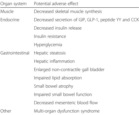
Based on these data, I suggest that it is illogical to feed patients with a continuous infusion of enteral nutrition (a parenteral infusion would be more illogical). A limited number of studies have been performed comparing con-tinuous to intermittent enteral nutrition [74, 88, 89]. While these studies did not evaluate patient centered out-comes such as mortality, ventilator-free days, muscle func-tion or metabolic parameters they demonstrated that this approach is both safe and feasible. MacLeod and col-leagues randomized 164 trauma patients to an intermit-tent feeding regimen (one-sixth of daily needs infused every 4 h) or a continuous feeding regimen [74]. The intermittent feed was delivered via an enteral feeding pump over a 30- to 60-min period of time. These investi-gators reported no difference in the complication rate be-tween groups (diarrhoea and pneumonia); however, the caloric goal was achieved earlier in the intermittently fed patients. We currently have experience with feeding over 300 patients by the intermittent method. This feeding technique has been very well tolerated by our patients with no evidence of an increase in the risk of aspiration or diarrhoea. Compared to historical controls, our data sug-gests that glycemic control improves with intermittent feeding (significantly lower percentage of patients with blood glucose >180 mg/dl). Due to the improved gastro-intestinal tolerance, improved glycemic control, perceived clinical benefits and ease of administration, intermittent bolus feeding is preferred over continuous feeding by our ICU nursing staff and dieticians. It should be noted that the intermittent boluses are given using an enteral feeding pump over a 20- to 40-min period. While the optimal Table 1Potential harm associated with continuous tube feed
Organ system Potential adverse effect
Muscle Decreased skeletal muscle synthesis
Endocrine Decreased secretion of GIP, GLP-1, peptide YY and CCK Decreased insulin release
Insulin resistance Hyperglycemia Gastrointestinal Hepatic steatosis
Hepatic inflammation
Enlarged non-contractile gall bladder Impaired lipid absorption
Small bowel atrophy Impaired small bowel function Decreased mesenteric blood flow Other Multi-organ dysfunction syndrome
GIPglucose-dependent insulinotropic polypeptide,GLP-1glucagon-like
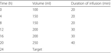
amount of calories and protein that should be given with the intermittent approach is unknown, we target 20–25 cal/kg/day divided into 6 aliquots given every 4 h. We use a whey-based formula (with omega-3 fatty acids) with a caloric density of 1.2 calories/ml with an average target of 1800 calories (250 ml q 4 hr). The protocol for escalation of intermittent feeds is provided in Table 2.
It is my opinion that continuous enteral nutrition is unphysiological, limits preservation of muscle mass and is associated with adverse effects on glucose and lipid me-tabolism and that this approach to nutritional support should be abandoned. Experimental and clinical studies have shown that‘mechanical silencing’of skeletal muscle plays a major role in CIM [56, 90]. Loss of muscle mass and function can be attenuated by early mechanical load-ing [91, 92], supportload-ing early physical therapy in immobi-lized patients [93–95]. It is therefore my belief that optimal nutritional support provided by intermittent feed-ing of a whey-based enteral formula combined with early physical therapy may attenuate CIM.
Conclusions
In conclusion, I believe that critically ill patients should be fed intermittently with a whey-based formula which contains omega-3 fatty acids. Such an approach is likely to limit muscle atrophy and promote metabolic stability. Continuous tube feeding is unphysiological and likely harmful and should be abandoned. Large randomized controlled trials are urgently required to demonstrate the clinical benefits of an intermittent feeding strategy.
Abbreviations
CCK:Cholecystokinin; CIM: Critical illness myopathy; FOXO: Forkhead box O; GIP: Glucose-dependent insulinotropic polypeptide; GLP-1: Glucagon-like peptide-1; GRV: Gastric residual volumes; IGF1: Insulin growth factor-1; ICUAW: Intensive care unit-acquired weakness; LPS: Lipopolysaccharide; MuRF1: Muscle RING-finger 1; MAFbx: Muscle atrophy F-box; mTOR: Mammalian target of rapamycin; TNF-α: Tumour necrosis factor-α; TGF-β: Transforming growth factor-β; PI3K/AKT: Phosphoinositol-3 kinase/protein kinase B (PI3K/AKT).
Competing interests
The author has given educational lectures sponsored by Abbott Nutrition and Nestle Health Sciences. The author has no other financial ties with these companies and no other conflicts or competing interests.
Acknowledgements
I acknowledge the nurses and nurse managers of our GICU who were willing to trial the intermittent feeding approach, who provided valuable feedback allowing us to refine this technique and who are passionate about proving the best bedside care possible.
Disclosure
Figure 1 and Tables 1 and 2 are original and have not been copied from other sources.
Received: 3 March 2015 Accepted: 15 May 2015
References
1. Batt J, dos Santos CC, Herridge MS. Muscle injury during critical illness. JAMA. 2013;310:1569–70.
2. Kress JP, Hall JB. ICU-acquired weakness and recovery from critical illness. N Engl J Med. 2014;370:1626–35.
3. Fan E, Cheek F, Chlan L, Gosselink R, Hart N, Herridge MS, et al. An Official American Thoracic Society Clinical Practice Guideline: the diagnosis of intensive care unit-acquired weakness in adults. Am J Respir Crit Care Med. 2014;190:1437–46.
4. Wollersheim T, Woehlecke J, Krebs M, Hamati J, Lodka D, Luther-Schroeder A, et al. Dynamics of myosin degradation in intensive care unit-acquired weakness during severe critical illness. Intensive Care Med. 2014;40:528–38. 5. Stevens RD, Dowdy DW, Michaels RK, Mendez-Tellez PA, Pronovost PJ,
Needham DM. Neuromuscular dysfunction acquired in critical illness: a systematic review. Intensive Care Med. 2007;33:1876–91.
6. Herridge MS, Cheung AM, Tansey CM, Matte-Martyn A, Diaz-Granados N, Al-Saidi F, et al. One-year outcomes in survivors of the acute respiratory distress syndrome. N Engl J Med. 2003;348:683–93.
7. Herridge MS, Tansey CM, Matte A, Tomlionson G, Diaz-Granados N, Cooper A, et al. Functional disability 5 years after acute respiratory distress syndrome. N Engl J Med. 2011;364:1293–304.
8. Phillips BE, Hill DS, Atherton PJ. Regulation of muscle protein synthesis in humans. Curr Opin Clin Nutr Metab Care. 2012;15:58–63.
9. Rennie MJ, Wackerhage H, Spangenburg EE, Booth FW. Control of the size of the human muscle mass. Annu Rev Physiol. 2004;66:799–828. 10. Atherton PJ, Etheridge T, Watt PW, Wilkinson D, Selby A, Rankin D, et al.
Muscle full effect after oral protein: time-dependent concordance and discordance between human muscle protein synthesis and mTORC1 signaling. Am J Clin Nutr. 2010;92:1080–8.
11. Mitchell WK, Phillips BE, Williams JP, Rankin D, Lund JN, Smith K, et al. A dose- rather than delivery profile-dependent mechanism regulates the "muscle-full" effect in response to oral essential amino acid intake in young men. J Nutr. 2015;145:207–14.
12. Staples AW, Burd NA, West DW, Currie KD, Atherton PJ, Moore DR, et al. Carbohydrate does not augment exercise-induced protein accretion versus protein alone. Med Sci Sports Exerc. 2011;43:1154–61.
13. Puthucheary ZA, Rawal J, McPhail M, Connolly B, Ratnayake G, Chan P. Acute skeletal muscle wasting in critical illness. JAMA. 2013;310:1591–600. 14. Lecker SH, Jagoe RT, Gilbert A, Gomes M, Baracos V, Bailey J, et al. Multiple
types of skeletal muscle atrophy involve a common program of changes in gene expression. FASEB J. 2004;18:39–51.
15. Gomes MD, Lecker SH, Jagoe RT, Navon A, Goldberg AL. Atrogin-1, a muscle-specific F-box protein highly expressed during muscle atrophy. Proc Natl Acad Sci USA. 2001;98:14440–5.
16. Bodine SC, Latres E, Baumhueter S, Lai VK, Nunez L, Clarke BA, et al. Identification of ubiquitin ligases required for skeletal muscle atrophy. Science. 2001;294:1704–8.
17. Klaude M, Mori M, Tjader I, Gustafsson T, Wernerman J, Rooyackers O. Protein metabolism and gene expression in skeletal muscle of critically ill patients with sepsis. Clin Sci. 2012;122:133–42.
18. Lecker SH, Lecker SH. Ubiquitin-protein ligases in muscle wasting: multiple parallel pathways? Curr Opin Clin Nutr Metab Care. 2003;6:271–5. 19. Levine S, Biswas C, Dierov J, Barsotti R, Shrager JB, Nguyen T, et al.
Increased proteolysis, myosin depletion, and atrophic AKT-FOXO signaling in human diaphragm disuse. Am J Respir Crit Care Med. 2011;183:483–90.
20. Smith IJ, Alamdari N, O'Neal P, Gonnella P, Aversa Z, Hasselgren PO. Sepsis increases the expression and activity of the transcription factor Forkhead Table 2Intermittent feeding schedule
Time (h) Volume (ml) Duration of infusion (min)
0 100 20
4 150 20
8 150 20
12 200 30
16 200 30
20 250 40
Box O 1 (FOXO1) in skeletal muscle by a glucocorticoid-dependent mechanism. Int J Biochem Cell Biol. 2010;42:701–11.
21. Sandri M, Sandri C, Gilbert A, Skurk C, Calabria E, Picard A, et al. Foxo transcription factors induce the atrophy-related ubiquitin ligase atrogin-1 and cause skeletal muscle atrophy. Cell. 2004;117:399–412.
22. Constantin D, McCullough J, Mahajan RP, Greenhaff PL, Constantin D, McCullough J, et al. Novel events in the molecular regulation of muscle mass in critically ill patients. J Physiol. 2011;589:3883–95.
23. Latres E, Amini AR, Amini AA, Griffiths J, Martin FJ, Wei Y, et al. Insulin-like growth factor-1 (IGF-1) inversely regulates atrophy-induced genes via the phosphatidylinositol 3-kinase/Akt/mammalian target of rapamycin (PI3K/Akt/mTOR) pathway. J Biol Chem. 2005;280:2737–44.
24. Zhao X, Gan L, Pan H, Kan D, Majeski M, Adam SA, et al. Multiple elements regulate nuclear/cytoplasmic shuttling of FOXO1: characterization of phosphorylation- and 14-3-3-dependent and -independent mechanisms. Biochem J. 2004;378:839–49.
25. Dickinson JM, Fry CS, Drummond MJ, Gundermann DM, Walker DK, Glynn EL, et al. Mammalian target of rapamycin complex 1 activation is required for the stimulation of human skeletal muscle protein synthesis by essential amino acids. J Nutr. 2011;141:856–62.
26. Farnfield MM, Carey KA, Gran P, Trenerry MK, Cameron-Smith D. Whey protein ingestion activates mTOR-dependent signalling after resistance exercise in young men: a double-blinded randomized controlled trial. Nutrients. 2009;1:263–75.
27. Burd NA, Yang Y, Moore DR, Tang JE, Tarnopolsky MA, Phillips SM. Greater stimulation of myofibrillar protein synthesis with ingestion of whey protein isolate v. micellar casein at rest and after resistance exercise in elderly men. Br J Nutr. 2012;108:958–62.
28. Karlsson HK, Nilsson PA, Nilsson J, Chibalin AV, Zierath JR, Blomstrand E. Branched-chain amino acids increase p70S6k phosphorylation in human skeletal muscle after resistance exercise. Am J Physiol Endocrinol Metab. 2004;287:E1–7.
29. Blomstrand E, Eliasson J, Karlsson HK, Kohnke R. Branched-chain amino acids activate key enzymes in protein synthesis after physical exercise. J Nutr. 2006;136:269S–73.
30. Anthony JC, Anthony TG, Kimball SR, Jefferson LS. Signaling pathways involved in translational control of protein synthesis in skeletal muscle by leucine. J Nutr. 2001;131:856S–60.
31. Reynolds TH, Bodine SC, Lawrence Jr JC. Control of Ser2448 phosphorylation in the mammalian target of rapamycin by insulin and skeletal muscle load. J Biol Chem. 2002;277:17657–62.
32. Timmerman KL, Lee JL, Dreyer HC, Dhanani S, Glynn EL, Fry CS, et al. Insulin stimulates human skeletal muscle protein synthesis via an indirect mechanism involving endothelial-dependent vasodilation and mammalian target of rapamycin complex 1 signaling. J Clin Endocrinol Metab. 2010;95:3848–57.
33. Jakubowicz D, Froy O. Biochemical and metabolic mechanisms by which dietary whey protein may combat obesity and Type 2 diabetes. J Nutr Biochem. 2013;24:1–5.
34. Norton LE, Layman DK, Bunpo P, Anthony TG, Brana DV, Garlick PJ. The leucine content of a complete meal directs peak activation but not duration of skeletal muscle protein synthesis and mammalian target of rapamycin signaling in rats. J Nutr. 2009;139:1103–9.
35. Pennings B, Boirie Y, Senden JM, Gijsen AP, Kuipers H, van Loon LJ. Whey protein stimulates postprandial muscle protein accretion more effectively than do casein and casein hydrolysate in older men. Am J Clin Nutr. 2011;93:997–1005.
36. Tang JE, Moore DR, Kujbida GW, Tarnopolsky MA, Phillips SM. Ingestion of whey hydrolysate, casein, or soy protein isolate: effects on mixed muscle protein synthesis at rest and following resistance exercise in young men. J Appl Physiol. 2009;107:987–92.
37. Verreijen AM, Verlaan S, Engberink MF, Swinkels S, deVogel-van den Bosch J, Weijs PJ. A high whey protein-, leucine-, and vitamin D-enriched supplement preserves muscle mass during intentional weight loss in obese older adults: a double=blind randomized controlled trial. Am J Clin Nutr. 2015;101:279–86. 38. Katsanos CS, Chinkes DL, Paddon-Jones D, Zhang XJ, Aarsland A, Wolfe RR.
Whey protein ingestion in elderly persons results in greater muscle protein accrual than ingestion of its constituent essential amino acid content. Nutr Res. 2008;28:651–8.
39. Nilsson M, Stenberg M, Frid AH, Holst JJ, Bjorck IM. Glycemia and insulinemia in healthy subjects after lactose-equivalent meals of milk and
other food proteins: the role of plasma amino acids and incretins. Am J Clin Nutr. 2004;80:1246–53.
40. Nilsson M, Holst JJ, Bjorck IM. Metabolic effects of amino acid mixtures and whey protein in healthy subjects: studies using glucose-equivalent drinks. Am J Clin Nutr. 2007;85:996–1004.
41. Blagosklonny MV. Hypoxia, MTOR and autophagy: converging on senescence or quiescence. Autophagy. 2013;9:260–2.
42. Tsutsumi R, Horikawa Y, Kume K, Tanaka K, Kasai A, Kadota T, Tsutsumi Y. Whey-protein based formulas are protective in LPS mediated sepsis. JPEN. 2014; ePub. 43. Smith GI, Atherton P, Reeds DN, Mohammed BS, Rankin D, Rennie MJ, et al.
Dietary omega-3 fatty acid supplementation increases the rate of muscle protein synthesis in older adults: a randomized controlled trial. Am J Clin Nutr. 2011;93:402–12.
44. Smith GI, Atherton P, Reeds DN, Mohammed BS, Rankin D, Rennie MJ, et al. Omega-3 polyunsaturated fatty acids augment the muscle protein anabolic response to hyperinsulinaemia-hyperaminoacidaemia in healthy young and middle-aged men and women. Clin Sci. 2011;121:267–78.
45. Suryawan A, O'Connor PM, Bush JA, Nguyen HV, Davis TA. Differential regulation of protein synthesis by amino acids and insulin in peripheral and visceral tissues of neonatal pigs. Amino Acids. 2009;37:97–104.
46. O'Connor PM, Bush JA, Suryawan A, Nguyen HV, Davis TA. Insulin and amino acids independently stimulate skeletal muscle protein synthesis in neonatal pigs. Am J Physiol Endocrinol Metab. 2003;284:E110–9. 47. Wilson FA, Suryawan A, Orellana RA, Kimball SR, Gazzaneo MC, Nguyen HV,
et al. Feeding rapidly stimulates protein synthesis in skeletal muscle of neonatal pigs by enhancing translation initiation. J Nutr. 2009;139:1873–80. 48. Bohe J, Low JF, Wolfe RR, Rennie MJ. Latency and duration of stimulation of
human muscle protein synthesis during continuous infusion of amino acids. J Physiol. 2001;532:575–9.
49. Moore DR, Robinson MJ, Fry JL, Tang JE, Glover EI, Wilkinson SB, et al. Ingested protein dose response of muscle and albumin protein synthesis after resistance exercise in young men. Am J Clin Nutr. 2009;89:161–8. 50. West DW, Burd NA, Coffey VG, Baker SK, Burke LM, Hawley JA, et al. Rapid
aminoacidemia enhances myofibrillar protein synthesis and anabolic intramuscular signaling responses after resistance exercise. Am J Clin Nutr. 2011;94:795–803.
51. Gazzaneo MC, Suryawan A, Orellana RA, Torrazza RM, El-Kadi SW, Wilson FA, et al. Intermittent bolus feeding has a greater stimulatory effect on protein synthesis in skeletal muscle than continuous feeding in neonatal pigs. J Nutr. 2011;141:2152–8.
52. Kumar V, Selby A, Rankin D, Patel R, Atherton P, Hildebrandt W, et al. Age-related differences in the dose–response relationship of muscle protein synthesis to resistance exercise in young and old men. J Physiol. 2009;587:211–7.
53. Cuthbertson D, Smith K, Babraj J, Leese G, Waddell T, Atherton P, et al. Anabolic signaling deficits underlie amino acid resistance of wasting, aging muscle. FASEB J. 2005;19:422–4.
54. Witard OC, Jackman SR, Breen L, Smith K, Selby A, Tipton KD. Myofibrillar muscle protein synthesis rates subsequent to a meal in response to increasing doses of whey protein at rest and after resistance exercise. Am J Clin Nutr. 2014;99:86–95. 55. Yang Y, Breen L, Burd NA, Hector AJ, Churchward-Venne TA, Josse AR, et al.
Resistance exercise enhances myofibrillar protein synthesis with graded intakes of whey protein in older men. Br J Nutr. 2012;108:1780–8. 56. Kortebein P, Ferrando A, Lombeida J, Wolfe R, Evans WJ. Effect of 10 days of
bed rest on skeletal muscle in healthy older adults. JAMA. 2007;297:1772–4. 57. Breen L, Stokes KA, Churchward-Venne TA, Moore DR, Baker SK, Smith K,
et al. Two weeks of reduced activity decreases leg lean mass and induces "anabolic resistance" of myofibrillar protein synthesis in healthy elderly. J Clin Endocrinol Metab. 2013;98:2604–12.
58. Glover EI, Phillips SM, Oates BR, Tang JE, Tarnopolsky MA, Selby A, et al. Immobilization induces anabolic resistance in human myofibrillar protein synthesis with low and high dose amino acid infusion. J Physiol. 2008;586:6049–61.
59. Lang CH, Frost RA, Nairn AC, MacLean DA, Vary TC. TNF-alpha impairs heart and skeletal muscle protein synthesis by altering translation initiation. Am J Physiol Endocrinol Metab. 2002;282:E336–47.
60. Lang CH, Frost RA. Sepsis-induced suppression of skeletal muscle translation initiation mediated by tumor necrosis factor alpha. Metabolism. 2007;56:49–57. 61. Lang CH, Frost RA. Endotoxin disrupts the leucine-signaling pathway
62. Balage M, Averous J, Remond D, Bos C, Pujos-Guillot E, Papet I, et al. Presence of low-grade inflammation impaired postprandial stimulation of muscle protein synthesis in old rats. J Nutr Biochem. 2010;21:325–31. 63. Vary TC. Acute oral leucine administration stimulates protein synthesis
during chronic sepsis through enhanced association of eukaryotic initiation factor 4G with eukaryotic initiation factor 4E in rats. J Nutr. 2007;137:2074–9. 64. Atherton PJ. Is there an optimal time for warfighters to supplement with
protein? J Nutr. 2013;143:1848S–51.
65. Initial trophic vs full enteral feeding in patients with acute lung injury. The EDEN randomized trial. JAMA. 2012; 307:795–803.
66. Arabi YM, Tamim HM, Dhar GS, Al-Dawood A, Al-Sultan M, Sakkijha MH, et al. Permissive underfeeding and intensive insulin therapy in critically ill patients: a randomized controlled trial. Am J Clin Nutr. 2011;93:569–77. 67. Heyland DK, Murch L, Cahill N, McCall M, Muscedere J, Stelfox HT, et al.
Enhanced protein-energy provision via the enteral route feeding protocol in critically ill patients: Results of a cluster randomized trial. Crit Care Med. 2013;41:2743–53.
68. Charles EJ, Petroze RT, Metzger R, Hranjec T, Rosenberger LH, Riccio LM, et al. Hypocaloric compared with eucaloric nutritional support and its effect on infection rates in a surgical intensive care unit: a randomized controlled trial. Am J Clin Nutr. 2014;100:1337–43.
69. Needham DM, Dinglas VD, Bienvenu OJ, Colantuoni E, Wozniak AW, Rice TW, et al. One year outcomes in patients with acute lung injury randomised to initial trophic or full enteral feeding: prospective follow-up of EDEN randomised trial. BMJ. 2013;346:f1532.
70. Needham DM, Dinglas VD, Morris PE, Jackson JC, Hough CL, Mendez-Tellez PA, et al. Physical and cognitive performance of acute lung injury patients one year after initial trophic vs full enteral feeding: EDEN trial follow-up. Am J Respir Crit Care Med. 2013;188:567–76.
71. Casaer MP, Mesotten D, Hermans G, Wouters PJ, Schetz M, Meyfroidt G, et al. Early versus late parenteral nutrition in critically ill adults. N Engl J Med. 2011;365:506–17.
72. Casaer MP, Wilmer A, Hermans G, Wouters PJ, Mesotten D, van den Berghe G. Role of disease and macronutrient dose in the randomized controlled EPaNIC trial. A Post Hoc Analysis Am J Respir Crit Care Med. 2013;187:247–55. 73. McClave SA, Martindale RG, Vanek VW, McCarthy M, Roberts P, Taylor B,
et al. Guidelines for the provision and assessment of nutrition support therapy in the adult critically ill patient: Society of Critical Care Medicine (SCCM) and American Society for Parenteral and Enteral Nutrition (A.S.P.E.N.). JPEN. 2009;33:277–316.
74. MacLeod JB, Lefton J, Houghton D, Roland C, Doherty J, Cohn SM, et al. Prospective randomized control trial of intermittent versus continuous gastric feeds for critically ill trauma patients. J Trauma. 2007;63:57–61. 75. Drucker DJ. Enhancing the action of incretin hormones: a new whey
forward? Endocrinology. 2006;147:3171–2.
76. Phillips LK, Prins JB. Update on incretin hormones. Ann New York Acad Sci. 2011;1243:E55–74.
77. Holst JJ. The physiology of glucagon-like peptide 1. Physiol Rev. 2007;87:1409–39.
78. Stoll B, Puiman PJ, Cui L, Chang X, Benight NM, Bauchart-Thevret C, et al. Continuous parenteral and enteral nutrition induces metabolic dysfunction in neonatal pigs. JPEN. 2012;36:538–50.
79. Shulman RJ, Redel CA, Stathos TH. Bolus versus continuous feedings stimulate small-intestinal growth and development in the newborn pig. J Pediat Gastroenterol Nutr. 1994;18:350–4.
80. Chowdhury AH, Murray K, Hoad CL, Costigan C, Marciani L, MacDonald IA, et al. Effects of bolus and continuous nasogastric feeding on gastric emptying, small bowel water content, superior mesenteric artery blood flow, and plasma hormone concentrations in heathy adults. A randomized crossover study. Ann Surg. 2015; ePub.
81. Subaran SC, Sauder MA, Chai W, Jahn LA, Fowler DE, Aylor KW, et al. GLP-1 at physiological concentrations recruits skeletal and cardiac muscle microvasculature in healthy humans. Clin Sci. 2014;127:163–70. 82. Chai W, Dong Z, Wang N, Wang W, Tao L, Cao W, et al. Glucagon-like
peptide 1 recruits microvasculature and increases glucose use in muscle via a nitric oxide-dependent mechanism. Diabetes. 2012;61:888–96.
83. Cho YM, Fujita Y, Kieffer TJ. Glucagon-like peptide-1: glucose homeostasis and beyond. Annu Rev Plant Physiol Plant Mol Biol. 2014;76:535–59. 84. Yabe D, Seino Y. Incretin actions beyond the pancreas: lessons from
knockout mice. Curr Opin Pharmacol. 2013;13:946–53.
85. Mashako MN, Bernard C, Cezard JP, Chayvialle JA, Navarro J. Effect of total parenteral nutrition, constant rate enteral nutrition, and discontinuous oral feeding on plasma cholecystokinin immunoreactivity in children. J Pediatr Gastroenterol Nutr. 1987;6:948–52.
86. Jawaheer G, Shaw NJ, Pierro A, Jawaheer G, Shaw NJ, Pierro A. Continuous enteral feeding impairs gallbladder emptying in infants. J Pediatr. 2001;138:822–5.
87. Barie PS, Eachempati SR. Acute acalculous cholecystitis. Gastroenterol Clin North Am. 2010;39:343–57.
88. Serpa LF, Kimura M, Faintuch J, Cecenella I. Effects of continuous versus bolus infusion of enteral nutrition in critical patients. Rev Hosp Clin Fac Med S Paulo. 2003;58:9–14.
89. Maurya I, Pawar M, Garg R, Kaur M, Sood R. Comparison of respiratory quotient and resting energy expenditure in two regimens of enteral feeding - continuous vs. intermittent in head-injured critically ill patients. Saudi J Anaesth. 2011;5:195–201.
90. Ochala J, Gustafson AM, Diez ML, Renaud G, Li M, Aare S, et al. Preferential skeletal muscle myosin loss in response to mechanical silencing in a novel rat intensive care unit model: underlying mechanisms. J Physiol. 2011;589:2007–26.
91. Renaud G, Llano-Diez M, Ravara B, Gorza L, Feng HZ, Jin JP, et al. Sparing of muscle mass and function by passive loading in an experimental intensive care unit model. J Physiol. 2013;591:1385–402.
92. Llano-Diez M, Renaud G, Andersson M, Marrero HG, Cacciani N, Engquist H, et al. Mechanisms underlying ICU muscle wasting and effects of passive mechanical loading. Crit Care. 2012;16:R209.
93. Bailey P, Thomsen GE, Spuhler VJ, Blair R, Jewkes J, Bezdjian L, et al. Early activity is feasible and safe in respiratory failure patients. Crit Care Med. 2007;35:139–45.
94. Morris PE, Goad A, Thompson C, Taylor K, Harry B, Passmore L, et al. Early intensive care unit mobility therapy in the treatment of acute respiratory failure. Crit Care Med. 2008;36:2238–43.
95. Kayambu G, Boots R, Paratz J. Physical therapy for the critically ill in the ICU: a systematic review and meta-analysis. Crit Care Med. 2013;41:1543–54.
Submit your manuscript to a
journal and benefi t from:
7Convenient online submission 7Rigorous peer review
7Immediate publication on acceptance 7Open access: articles freely available online 7High visibility within the fi eld
7Retaining the copyright to your article