IN VITRO ANTI-INFLAMMATORY AND ANTI-DIABETIC ACTIVITY OF METHANOLIC EXTRACT OF CARDANTHERA DIFFORMIS DRUCE
5
0
0
Full text
(2) Somnath De et al. Int. Res. J. Pharm. 2016, 7 (12) anti-inflammatory and anti-diabetic activity of Cardanthera difformis till the date. In the present investigation attempts have been made to find out the anti-inflammatory and anti-diabetic properties of Candanthera difformis by protein denaturation and α-amylase and α-glucosidase inhibition of methanolic extract respectively. MATERIALS AND METHODS Plant material Cardanthera difformis Druce has been selected for experiment tools. It is collected in the month of march , 2015 form Paschim Medinipur district (Latitude- 22 ◦ 25'00 '' to 22 ◦ 57'00 '' north, Longitude- 87 ◦ 11' east, Altitude- 23 meters West Bengal , India and it is from mean sea level), available in any season of year. Plant material extraction The taxonomic identities of this plant are determined by the expertise of the department of botany of Vidyasagar University. The leaves were washed thoroughly using tap water and dried under shed for 11 days, then finely grinded to a powder. Then the powdered material was extracted with methanol using soxhlet apparatus. About 10 grams of powder was loaded in soxhlet extraction unit and exhaustively extracted using 100ml of solvents such as methanol at 600C for 12 hours. Thereafter, it was filtered with the help of Whatman No.1 filter paper21. The extracts were concentrated by rotary evaporator and used for testing anti-inflammatory and anti-diabetic activity. In vitro anti-inflammatory activity Inhibition of albumin denaturation The following procedure was followed by for evaluating the percentage of inhibition of protein denaturation22. Control solution (50ml) 2 ml of egg albumin, 14 ml of phosphate buffer (pH 6.4) and 20 ml distilled water. Standard drug (50ml) 2 ml of egg albumin, 28 ml of phosphate buffer (pH 6.4) and 10ml various concentration of standard drug (Diclofenac sodium) concentration of 100, 200, 400, 800 and 1000µg/ml. Test solution (50ml) 2ml of egg albumin, 28 ml of phosphate buffer (pH 6.4) and 10ml various concentration of methanol extract Candanthera difformis concentration of 100, 200, 400, 800 and 1000µg/ml. All of the above solutions were adjusted to pH using a small amount of 1N HCl. The samples were incubated at 37˚ C for 15 minutes and heated at 70 ˚ C for 5 minutes. After cooling, the absorbance of turbidity was measured at 660 nm in UV-vis spectrophotometer the above solutions percentage inhibition of protein denaturation was calculated using the following formula 23 . Percentage inhibition = [Vt/Vc -1] X 100 Where, Vt= Absorbance of test sample , Vc = Absorbance of control In vitro anti-diabetic activity α-amylase inhibition assay The α-amylase inhibitory activity was determined according to the method24. Briefly, the total assay mixture containing 200 μl. of 0.02M sodium phosphate buffer, 20 μl of enzyme, and the plant extracts in the concentration range 10-100μg/ml were incubated for 10 min at room temperature followed by addition of 200 μl of 1% starch in all the test tubes. The reaction was terminated with addition of 400 μl of 3,5 dintrosalycylic acid (DNSA) color reagent, placed in boiling water bath for 5 minutes, cooled at room temperature and diluted with 15 ml of distilled water and the absorbance measured at 540nm. The control samples were also prepared accordingly without any plant extracts and were compared with the test samples containing various concentrations of the plant extracts prepared with different solvent prepared with DMSO. The results were expressed as % inhibition calculated using the formula: Inhibition activity (%) = Abs (control) -Abs (extract)/ Abs(control)×100 The IC50 values (inhibitor concentration at which 50% inhibition of the enzyme activity occurs) of the plant extracts were determined b y performing the assay as above with varying concentrations of the plant extracts ranging 20 to 100μg. The IC50 values were determined from plots of percent inhibition vs log inhibitor concentration and calculated by non-linear regression analysis from the mean inhibitory values. α-glucosidase inhibition assay The yeast α-glucosidase was dissolved in 100mM phosphate buffer, pH 6.8 was used as enzyme source; 10mM paranitrophenyl-α-D glucopyranoside was used as substrate. Cardanthera difformis extract powder was weighed and mixed with dimethylsulfoxide to get a concentration of 20-100μg/ml. The different concentration of plant extract was mixed with 320μl of 100mM phosphate buffer (pH 6.8) and 50 μl of 10mM PNPG in the buffer and then it was incubated at 30°C for 5 minutes. After the incubation, 20μl of the buffer containing 0.5 mg/ml of the enzyme was added and further incubated at 30°C for five minutes. Finally, 3.0 ml of 50mM sodium hydroxide was added to the mixture and the absorbance (A)was measured at 410nm on a spectrophotometer. The enzyme without plant extract was used as a control25. % Inhibition = A410 control –A410 test/ A410 control×100 The IC50 values (inhibitor concentration at which 50% inhibition of the enzyme activity occurs) of the plant extracts were determined by performing the assay as above with varying concentrations of the plant extracts ranging 20 to 100μg. The IC50 values were determined from plots of percent inhibition vs log inhibitor concentration and calculated by non-linear regression analysis from the mean inhibitory values. RESULTS In vitro anti-inflammatory activity Inhibition of albumin denaturation In in-vitro anti- inflammatory activity by egg albumin denaturation method at concentration of 100, 200,400,800 and 1000 µg/ml showed 172.50, 175.00, 195.00, 223.75 and 225.00% inhibition of egg albumin denaturation (Table 1) whereas, standard Diclofenac sodium at 100, 200, 400, 800 and 1000 µg/ml which showed 180.00, 197.50, 211.25, 233.13 and 234.37% inhibition of egg albumin denaturation (Table 2) and their comparison between sample and standard (Table 3). From this experimental results showed significant inhibition of denaturation of egg albumin in concentration dependent manner.. 57.
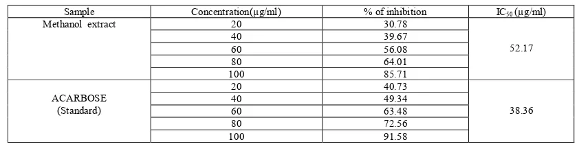
(3) Somnath De et al. Int. Res. J. Pharm. 2016, 7 (12) Table 1: In vitro anti-inflammatory activity of methanolic extract of Cardanthera difformis on protein denaturation (Fresh egg albumin) Treatment Methanolic extract of Cardanthera difformis. Concentration (µg/ml) 100 200 400 800 1000. Percentage of inhibition (%) 172.50 175.00 195.00 223.75 225.00. Table 2: In vitro anti-inflammatory activity of Diclofenac sodium on protein denaturation (Fresh egg albumin) Treatment Diclofenac sodium. Concentration (µg/ml) 100 200 400 800 1000. Percentage of inhibition (%) 180.00 197.50 211.25 233.13 234.37. Table 3: In vitro anti-inflammatory activity of Diclofenac sodium & methanol extract of Cardanthera difformis on sodium on protein denaturation (Fresh egg albumin) Sl.No. 1 2 3 4 5. Concentration (µg/ml) 100 200 400 800 1000. % inhibition Diclofenac sodium 180.00 197.50 211.25 233.13 234.37. % inhibition Cardanthera difformis 172.50 175.00 195.00 223.75 225.00. In vitro antidiabetic activity In vitro α-amylase inhibition assay Table 4: In vitro antidiabetic activity of alpha- amylase inhibition method of standard(Acarbose) methanol extract of Cardanthera difformis Sample Methanol extract. ACARBOSE (Standard). Concentration(µg/ml) 20 40 60 80 100 20 40 60 80 100. % of inhibition 23.62 33.89 39.66 55.63 61.31 33.75 49.22 59.20 67.22 73.97. IC50(µg/ml) 74.88. 46.46. In vitro α-glucosidase inhibition assay Table 5: In vitro anti-diabetic activity of alpha-glucosidase inhibition method of standard and methanol extract of Cardanthera difformis Sample Methanol extract. ACARBOSE (Standard). Concentration(µg/ml) 20 40 60 80 100 20 40 60 80 100. DISCUSSION Protein denaturation is a process in which protein lose their tertiary structure and secondary structure by application of external stress or compound such as strong acid or base concentration inorganic salt, an organic solvent or heat most biological protein lose their biological function when denaturation. Denaturation of protein is a well-documented cause of inflammation. As a part of the investigation on the mechanism of the anti- inflammatory and anti-arthritic activity, ability of plant extract to inhibit protein denaturation was. % of inhibition 30.78 39.67 56.08 64.01 85.71 40.73 49.34 63.48 72.56 91.58. IC50 (µg/ml) 52.17. 38.36. studied. Several anti-inflammatory drugs have showed dose dependent ability to inhibit thermally induced protein denaturation26. Denaturation of protein is a well document cause of inflammation in condition like Rheumatoid arthritis. Diabetes mellitus (DM) is a common endocrine system disease that causes metabolic disorders and which leads to multiple organ damage syndrome. Clinical admiral diabetes is divided into two types, with more than 90% of patients having Type II diabetes27. The number of diabetes cases was 171 million in 2000 and is expected to rise to 366 million in 2030 .Inhibition of. 58.
(4) Somnath De et al. Int. Res. J. Pharm. 2016, 7 (12) and α-amylase, enzymes involved in the α-glucosidase digestion of carbohydrates, can significantly decrease the postprandial increase of blood glucose after a mixed carbohydrate diet and therefore can be an important strategy in the management of postprandial blood glucose level in type 2 diabetic patients and borderline patients28. Intestinal α – glucosidase is a glucosidase acting as a key enzyme for carbohydrate digestion, located at the epithelium of the small intestine. α-glucosidase has been recognized as a therapeutic target for the modulation of postprandial hyperglycemia, which is the earliest metabolic abnormality that occurs in Type II DM29. Several natural α-glucosidase and α-amylase inhibitors including acarbose, voglibose and miglitol are clinically used as a treatment, but their prices are high and clinical side effects occur 30,31. Natural products are still the most available source of α-glucosidase inhibitors. Therefore, screening of alpha-amylase and glucosidase inhibitors in medicinal plants has received much attention. Therefore, in the present study we investigated α-amylase and α-glucosidase inhibitory activity by using the methanolic extract of Cardanthera difformis. In vitro anti-diabetic studies demonstrated that Cardanthera difformis extract h as both α-glucosidase and α-amylase inhibitory activity. The percentage of inhibition at 100, 80, 60, 40 and 20 μg/ml concentrations of plant extract showed a concentration-dependent reduction in percentage inhibition. Acarbose like drugs, that inhibit α-glucosidase and amylase present in the epithelium of the small intestine, have been demonstrated to decrease post-prandial hyperglycaemia32 and improve impaired glucose metabolism without promoting insulin secretion in NIDMM patients33. These medications are most useful for people who have just been diagnosed with type 2 diabetes and who have blood glucose levels only slightly above the level considered serious for diabetes. They also are useful for people taking sulfonylurea medication or metformin, who need an additional medication to keep their blood glucose levels within a safe range. Therefore, the retardation and delay of carbohydrate absorption with a plant-based α-glucosidase inhibitor offers a prospective therapeutic approach for the management of type 2 diabetes mellitus and borderline patients34,35. The results of this study indicate that the administration of Cardanthera difformis an probably manage the postprandial blood glucose levels and confirm the usage of these plants. ACKNOWLEDGEMENT We would like to express our heartfelt thanks to Dr. Pulakesh Bera, Department of Chemistry, Panskura Banamali College for providing the adequate facilities to carry out my project work. REFERENCES 1. Majithia V, Geraci S A , Am. J. Med 2007; 120(11) : 936. 2. Westwood O M , Nelson P N , Hay F C , Rheumatology (Oxford) 2006; 45 (4) : 379. 3. Chandra S, Chatterjee P, Dey P, Bhattacharya S. Evaluation of Anti-Inflamatory Effect of Ashwagandha: A Preliminary Study in Vitro. Pharmacog J 2012; 4(29):47-9. 4. Brown J H , Mackey H K , Proc Soc Exp Biol Med 1968; 128 : 225. 5. Grant N H , Alburn H E , Kryzanauskas C , Biochem Pharmacol 1970; 19 : 715. 6. Tripathi KD. Essentials of medical pharmacology. 6th ed. New Delhi: Jaypee Brother’s Medical Publishers (P) Ltd.; 2008. 7. Bennett PN, Brown MJ. Clinical pharmacology. New Delhi : Churchill Livingstone; 2005.. 8. Apparao C, Kameswararao B, Kesavulu MM. Evaluation of antidiabetic effect of Momordica cymbalaria fruit in alloxan diabetic rats. Fitoterapia 2003; 74: 7-13. 9. Modak M, Dixit P, Londhe J, Ghaskadbi S, Devasagayam TPA. Indian herbs and herbal drugs used for the treatment of diabetes. J Clin Biochem Nutr 2007; 40:163-73. 10. Rao P, Jamil K. Pharmacological evaluation of herbal extracts for their in vitro hypoglycemic activity. International Journal of Phytopharmacology 2011; 2 ( 1) : 15-21. 11. Wadkar KA, Magdum CS, Patil SS, Naikwade NS. Antidiabetic potential and Indian medicinal plants. Journal of Herbal Medicine and Toxicology 2008; 2 (1) : 45-50. 12. Tarling CA, Woods K, Zhang R, Brastianos HC, Brayer GD, Andersen RJ, Withers SG. The Search for Novel Human Inhibitors: High-Throughput Pancreatic α-Amylase Screening of Terrestrial and Marine Natural Product Extracts. Chem BioChem 2008; 9: 433-438. 13. Cheng AYY, Fantus IG. Oral antihyperglycemic therapy for type 2 diabetes mellitus. Canadian Medicinal Association Journal 2005; 172 ( 2) : 213-226. 14. Mukherjee PK, Maiti K, Mukherjee K, Houghton PJ. Leads from Indian medicinal plants with hypoglycemic potentials. J Ethnopharmacol. 2006; 106 ( 1) : 1-28. 15. Sudha P, Zinjarde SS, Bhargava SY, Kumar AR. Potent αamylase inhibitory activity of Indian Ayurvedic medicinal plants. BMC Complementary and Alternative Medicine 2011; 11( 5) : 1-10. 16. Ponnusamy S, Ravindran R, Zinjarde S, Bhargava S, KumarAR. Evaluation of traditional Indian antidiabetic medicinal plants for human pancreatic amylase inhibitory effect in vitro.Evidence-Based Complementary and Alternative Medicine 2011; 1-10. 17. Das D C, De S, Bhattacharya S, Das M. Antibacterial and phytochemical analysis of Cardanthera difformis Druce leaf extracts from West Bengal, India. Int. J phytomedicine 2013;5(4) :446-451. 18. De S, Das D C, Mondal T, Das M, Ivestigation of the Antibacterial and Antifungal activity of Cardanthera difformis Druce whole plant extract against some human clinical International Journal of Bioassays 2014; 3(11) :3464-3468. 19. De S , Das D C , Mandal T, Investigation of Antioxidant Properties of Cardanthera difformis Druce Whole Plant Extract. Indian Journal of Applied Research 2015; 5(7) : 161-163. 20. De S , Das D C , Mandal T, In-vitro anthelmintic activity of Cardanthera difformis Druce whole plant methanolic extract in Indian adult earthworm. Journal of Pharmacognosy and Phytochemistry 2016; 5(1): 203-205. 21. Sharmila N, Gomathi N, International Journal of Phytomedicine 2011; 3: 151-156. 22. Saleem T K, Azeem A K, Dilip C, Sankar C, Prasanth N V , Duraisami R, Anti-inflammatory activity of leaf extracts of Gendaruss vulgaris Nees. Asian Pacific Journal of Tropical Biomedicine 2011; 147-149. 23. Chandra S, Chatterjee P , Dey P, Bhattacharya S, Evaluation of anti- inflammatory effect of ashwagandha: a preliminary study in vitro. Pharmacognosy Journal. 2012; 4 (29): 47-49. 24. Jyothi KSN, Hemalathr P, Calla S. Evaluation of alpha amylase inhibitory potential of three medicinally important traditional wild food plants of India. International Journal of Green Pharmacy 2011; 95-99. 25. Tadera K, Minaki Y, Takamatsu K, Matsuoka T. Inhibition of alpha glucosidase and alpha amylase by flavonoids. J Nutr Sci Vitaminol 2006; 52: 149-153. 26. Padmanaban P, Jangle S N , Evaluation of antiinflammatory activity of herbal preparations combination of. 59.
(5) Somnath De et al. Int. Res. J. Pharm. 2016, 7 (12). 27. 28. 29.. 30.. 31. 32.. four medicinal plants. International journal of basic and applied medicinal sciences. 2012; 2(1): 109-116. Wang Y, Zhang X. Study progress on α-glucosidase inhibitors.Strait Pharmaceut 2009; 21: 4-5. Yao Y, Sang W, Zhou M, Ren G. Antioxidant and alphaglucosidase inhibitory activity of colored grains in China. J. Agric.Food Chem 2010; 58: 770-774. Karthic K, Kirthiram KS, Sadasivam A, Thayumanavan B. Identification of amylase inhibitors from Syzygium cumini Linn seeds. Indian Journal of experimental biology 2008; 46: 677-680. Karthic K, Kirthiram KS, Sadasivam A, Thayumanavan B. Identification of amylase inhibitors from Syzygium cumini Linn seeds. Indian Journal of experimental biology 2008; 46: 677-680. Kim JS, Kwon CS, Son KH. Inhibition of α-glucosidase and amylase by Luteolin, a flavonoid. Biosci Biotech Biochem. 2000; 64: 2458–2461. Sima AAF, Chakrabarti S. Long-term suppression of postprandial hyperglycaemia with acarbose retards the. development of neuropathies in the BB/W-rat. Diabetologia 2004; 35: 325–330. 33. Carrascosa JM, Molero JC, Fermin Y, Martinez C, Andres of chronic treatment with A, Satrustegui J. Effects acarbose on glucose and lipid metabolism in obese diabetic wistar rats. Diab Obes Metab 2001; 3: 240–248. 34. McCue P, Vattem D, Shetty K. Inhibitory effect of clonal oregano extracts against porcine pancreatic amylase in vitro. Asia Pac J Clin Nutr 2004; 13: 401–408. 35. Subramanian R, Asmawi MZ, Sadikun A. In vitro αglucosidase and α-amylase enzyme inhibitory effects of Andrographis paniculata extract and and rographolide. Acta Biochimica Polonica 2008; 55 (2) : 391–398. Cite this article as: Somnath De, Dulal Chandra Das, Tanusri Mandal. In vitro antiinflammatory and anti-diabetic activity of methanolic extract of Cardanthera difformis Druce. Int. Res. J. Pharm. 2016;7(12):5660 http://dx.doi.org/10.7897/2230-8407.07121467. Source of support: Nil, Conflict of interest: None Declared Disclaimer: IRJP is solely owned by Moksha Publishing House - A non-profit publishing house, dedicated to publish quality research, while every effort has been taken to verify the accuracy of the content published in our Journal. IRJP cannot accept any responsibility or liability for the site content and articles published. The views expressed in articles by our contributing authors are not necessarily those of IRJP editor or editorial board members.. 60.
(6)
Figure
Related documents
This calculation, for the case of chaotic systems, combined the tendency for orbit density to increase exponentially with period with the tendency of
During the control period (without PB) cholate was the predominant bile salt in the peripheral blood, whereas chenodeoxycholate was predomi- nant in the total bile salt pool..
Comparing Results of Artificial Neural Network Solution with Simplex Method The neural Network was able to optimize soap production and the maximum profit for each. month can be
The heading angles calculated by WalkCompass mostly follow the direction of the walk along the corridor without being affected by the random changes in the orientation of the
4 DataToMem IN 32 Data ready to be written to Output memory, after being processed by the Function Execute module. Delivered to the Memory
The purpose of this study was to compare the biomechanical effect of a supplemental S1 long iliosacral screw versus a transsacral screw in an unstable type C vertically oriented
We will speak in this search for Data Mining, which has become one of the topics interesting, which aims to find useful information from large data sets At the present time is
Neben der Antikörperbildung durch B-Lymphozyten spielt die Aktivierung von T-Lymphozytensubpopulationen im Rahmen einer systemischen Inflammation durch den
