ARTICLE
Determination of Genetic Predisposition to Patent
Ductus Arteriosus in Preterm Infants
John M. Dagle, MD, PhDa, Nathan T. Lepp, MDa, Margaret E. Cooper, MS, MSISb, Kendra L. Schaa, BSa, Keegan J. P. Kelsey, BSa, Kristin L. Orr, BSc,
Diana Caprau, MDa, Cara R. Zimmerman, BSc, Katherine M. Steffen, BAc, Karen J. Johnson, RNa, Mary L. Marazita, PhDb,d, Jeffrey C. Murray, MDa
aDepartment of Pediatrics andcCarver College of Medicine, University of Iowa, Iowa City, Iowa;bCenter for Craniofacial and Dental Genetics anddDepartment of Human Genetics, Graduate School of Public Health, University of Pittsburgh, Pittsburgh, Pennsylvania
The authors have indicated they have no financial relationships relevant to this article to disclose.
What’s Known on This Subject
PDA is a significant cardiac complication seen in preterm infants. Twin studies have suggested that some component of PDA is inherited. There have been limited investi-gations of the specific genetic variations that might predispose preterm infants to PDA.
What This Study Adds
We found that common sequence variations in 3 genes (TFAP2B,TRAF1, andPTGIS) are associated with PDA in preterm infants. This type of information could be used to help determine medical management of this condition.
ABSTRACT
OBJECTIVE.Patent ductus arteriosus is a common morbidity associated with preterm birth. The incidence of patent ductus arteriosus increases with decreasing gestational
age to⬃70% in infants born at 25 weeks’ gestation. Our major goal was to determine
if genetic risk factors play a role in patent ductus arteriosus seen in preterm infants.
METHODOLOGY.We investigated whether single-nucleotide polymorphisms in genes that regulate smooth muscle contraction, xenobiotic detoxification, inflammation, and other processes are markers for persistent patency of ductus arteriosus. Initially, 377 single-nucleotide polymorphisms from 130 genes of interest were evaluated in DNA
samples collected from 204 infants with a gestational age of⬍32 weeks. A
family-based association test was performed on genotyping data to evaluate overtransmis-sion of alleles.
RESULTS.Pvalues of⬍.01 were detected for genetic variations found in 7 genes. This prompted additional analysis with an additional set of 162 infants, focusing on the 7
markers with initialPvalues of⬍.01, and 1 genetic variant in the angiotensin II type
I receptor previously shown to be related to patent ductus arteriosus. Of the initial
positive signals, single-nucleotide polymorphisms in the transcription factor AP-2
and tumor necrosis factor receptor–associated factor 1 genes remained significant. Additional haplotype analysis revealed genetic variations in prostacyclin synthase to be associated with patent ductus arteriosus. An angiotensin II type I receptor poly-morphism previously reported to be associated with patent ductus arteriosus after prophylactic indomethacin administration was not associated with the presence of a patent ductus arteriosus in our population.
CONCLUSIONS.Overall, our data support a role for genetic variations in transcription
factor AP-2 , tumor necrosis factor receptor–associated factor 1, and prostacyclin
synthase in the persistent patency of the ductus arteriosus seen in preterm infants.
Pediatrics2009;123:1116–1123
P
ATENT DUCTUS ARTERIOSUS(PDA) is a common complication occurring in pre-term infants and has been associated with the development of chronic lung disease, necrotizing enterocolitis, and intraventricular hemorrhage (IVH). The ductus arteriosus (DA) is an important in utero vascular connection between the aorta andpulmonary artery. At birth, the pulmonary artery pressures are high and there is limited shunting from the aorta to the pulmonary arteries via the ductus arteriosus. As the pulmonary artery pressures begin to fall during the first hours to days of life, significant left to right shunting of blood from the systemic circulation to the pulmonary circulation occurs. This shunting causes alterations in systemic blood flow and decreases flow to skin, muscle,
kidneys, and the gastrointestinal tract.1In addition, pulmonary overcirculation caused by the left to right shunting
across the PDA can lead to pulmonary edema and a worsening of the infant’s already compromised respiratory
status.2 Finally, the presence of PDA has recently been shown to have a negative effect on cerebral perfusion,
resulting in impaired oxygen delivery to an already vulnerable premature brain.3
www.pediatrics.org/cgi/doi/10.1542/ peds.2008-0313
doi:10.1542/peds.2008-0313
Drs Dagle and Lepp contributed equally to this work.
Dr Caprau’s current affiliation is Department of Pediatrics, University of Utah School of Medicine, Salt Lake City, UT.
Key Words
patent ductus arteriosus, genetic predisposition, premature infants
Abbreviations DA— ductus arteriosus PDA—patent ductus arteriosus IVH—intraventricular hemorrhage SNP—single-nucleotide polymorphism
AGTR1—angiotensin II type I receptor
TFAP2B—transcription factor AP-2
TRAF1—tumor necrosis factor receptor– associated factor 1
PTGIS—prostacyclin synthase FBAT—Family-Based Association Test
CETP— cholesteryl ester transfer protein, plasma
CYP2D6— cytochrome P450, family 2, subfamily D, polypeptide 6
CRHR1— corticotrophin-releasing hormone receptor 1
LIPC— hepatic lipase
Postnatal closure of the DA occurs in 2 phases. First,
after birth, increasing PaO2and a decreasing amount of
circulating prostaglandins allow the smooth muscle of the ductus arteriosus to contract, functionally limiting luminal blood flow. After this physiologic occlusion has occurred, hypoxia of the medial layer of the DA occurs resulting in the elaboration of inflammatory mediators and growth factors. These compounds subsequently in-duce fibrosis, resulting in permanent anatomic closure of
the DA, creating the ligamentum arteriosum.1
Notably, not all preterm infants develop PDA. The most recent data from the Vermont Oxford Network (2006) of nearly 40 000 preterm infants with a birth weight 501 to 1500 g show the overall incidence of PDA
to be 37.2%.4 The incidence of PDA in infants with
gestational ages of 24, 25, and 26 weeks was 76.9%, 69.5%, and 61.5%, respectively.
There have been recent efforts to delineate a genetic cause of PDA. However, these studies have focused pri-marily on PDAs associated with syndromes in small co-horts of patients, and have generally excluded preterm
infants.5–7In 1 such analysis, mutations in theMYH11, a
gene encoding smooth muscle myosin heavy chain, were found to be causative for 2 kindreds who presented
with thoracic aortic aneurysm and a PDA.8 Mutations
associated with Char syndrome, a disorder characterized by facial dysmorphism, hand anomalies, and PDA, were
found in transcription factor AP-2  (TFAP2B), a gene
encoding a transcription factor found in neural crest-derived cells.5,7
One study has addressed PDA in preterm infants and their response to medical management by examining 159 infants born before 33 weeks’ gestation who re-ceived prophylactic indomethacin. The authors investi-gated whether a polymorphism in the angiotensin II
type I receptor (AGTR1) gene (rs5186) influenced PDA
closure with indomethacin. Infants with the CC geno-type of this locus were found to have a lower risk of PDA than those infants with AA or CA genotypes after early
indomethacin administration.9
In this study, we evaluated the hypothesis that com-mon variants in candidate genes might play a role in the development of a PDA. The infants in this analysis are a defined cohort from a larger study population in which possible genetic contributions to preterm delivery were investigated. All genotypes available were analyzed by
using a hypothesis-generating strategy, although our a priori hypothesis was that genes regulating pathways controlling smooth muscle contraction or those associ-ated with syndromes including PDA would be most likely to be associated with PDA in preterm infants.
METHODS
We used a 2-phased approach in this study. The first analysis of 204 infants was a data-mining, candidate gene survey using genotype information from a larger study investigating genetic contributions to prematurity. Using a family-based association test, we chose 7
single-nucleotide polymorphisms (SNPs) withPvalues of⬍.01
for additional study. A second phase of study was then performed on these genes of interest by adding an addi-tional 162 infants to the study population and repeating the statistical analysis on the total sample of 366 infants.
Sample Population
Since 2000, blood or buccal swabs from infants (and their parents) admitted to the NICU at the University of Iowa Children’s Hospital have been collected and banked. This institutional review board-approved pro-gram is designed to generate samples for use in genetic studies evaluating diseases of the infant. A second insti-tutional review board approval was obtained to access these samples and the associated clinical information necessary for this study. This population is a subgroup of 1 that has been described previously (as have some of the SNPs used in this study), but evaluated for preterm labor as an end point with respect to the progesterone
receptor10 and genes affecting cholesterol metabolism.11
Approximately 90% of the sample population was white by parental report.
A total of 366 infants of ⬍32 weeks’ gestation was
included for analysis. The first analysis, in which 377 SNPs were screened for possible genes of interest, included 204 infants (collected between 2000 and 2005). The second
analysis, which encompassed those markers withPvalues
of⬍.01 from the initial screen, included an additional 162
infants (collected between 2005 and 2007). In addition, a few new genes and SNPs were investigated in the second analysis that were not under consideration during the first
phase of the study (eg,EPAS1andAGTR1). The gestational
ages for the study population are presented in Fig 1. Clas-0
10 20 30 40 50 60
22 23 24 25 26 27 28 29 30 31
N
o
. of
inf
ant
s
Gestational age, wk
FIGURE 1
sification of disease state was performed by a chart re-view of each infant. PDA screening was performed
ac-cording to our NICU clinical guidelines. All infants⬍28
weeks’ gestational age were examined by a pediatric cardiologist by using echocardiography between days 5
and 7 of life, regardless of symptoms. Infantsⱖ28 weeks’
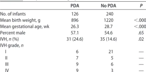
gestation were examined by echocardiography between day 5 and 7 of life if a murmur suggestive of a PDA was present. All preterm infants were examined if they had symptoms of congestive heart failure any time after day 3 of life. There was no attempt to stratify PDA by stage. No infant received prophylactic indomethacin (ie, in the first 2 days of life). In the first phase of analysis, PDA was present in 69 (33.8%) of the study infants and absent in 135 (66.2%). Of the 69 infants diagnosed with a PDA, 16 underwent surgical ligation for closure of the PDA, all after failure of closure with indomethacin. Of the 162 infants added into the second phase of the study, 57 (35.2%) were diagnosed with a PDA and 24 underwent surgical ligation, again after failure of the PDA to close after indomethacin treatment. Table 1 lists the demo-graphics of the infants in the study. On average, infants with PDAs were born 2 weeks earlier and were 300 g
lighter than infants without a PDA (P⬍.0001,ttest with
a pooled variance method). The proportion of male in-fants in each group was similar. Consistent with their earlier gestational age, infants with a PDA were more likely to develop an intraventricular/periventricular
hemorrhage than those without a PDA (P ⫽.02). The
group of infants with a PDA included 91 singletons, 30 twins, and 5 triplets. Those without PDA included 171 singletons, 58 twins, 7 triplets, and 4 quadruplets.
DNA Processing and Genotyping
DNA was extracted from cord blood for the infants in the analysis and from venous blood, buccal swabs, or saliva from parents. Allelic variation was determined by using the TaqMan genotyping system (Applied Biosystems,
Foster City, CA), as previously described.10Allele scoring
was performed by using the Sequence Detection Systems 2.2 software (Applied Biosystems). The genotype data were uploaded into a Progeny database (Progeny Soft-ware, LLC, South Bend, IN), also containing phenotypic data, for subsequent statistical analysis.
Candidate Genes
Candidate genes were chosen based on a review of the current literature as well as hypotheses of biological
plausibility. Genes in the smooth muscle pathway, such as the prostaglandin synthases as well as those that had been associated with PDAs in syndromes, were specifi-cally chosen for analysis of PDA in these preterm infants. Because part of this study used a data mining approach, some of the genes evaluated were those that had been previously reported or hypothesized to be associated with preterm birth. SNPs within each gene were selected by using data from the International HapMap project (www.hapmap.org), balancing the greatest coverage of the gene with the fewest number of tagging and/or known functional SNPs required. In general, a minor allele frequency of 0.1 was chosen as a lower cutoff for a SNP to ensure that an adequate number of individuals within the population would be carriers of the minor allele. Genes selected for analysis are listed in Table 2. A complete listing of the SNPs with their dbSNP identifi-cation (“rs”) numbers is published as supporting infor-mation at www.pediatrics.org/content/full/123/4/1116.
Statistical Analysis
Genotyping data for each SNP were assessed by using the program PedCheck for any departures from mendelian
inheritance patterns.12 Alleles at each marker were
tested for association with PDA by using the
Family-Based Association Test (FBAT).13–15In addition, a
haplo-type FBAT was performed for sliding windows of 2, 3, and 4 SNPs across genes when appropriate.
Each infant was part of a trio of father, mother, and child. If 2 infants in a family shared the same affection status (ie, both with or both without PDA) then the nuclear family was the analysis unit. Otherwise, the infants with differing PDA status formed 2 separate trios with the same parents for the subgroup analyses. Given the multiple testing of these 377 SNPs in the first phase
of the study, for statistical significance at an ␣level of
.05, the most conservative correction (Bonferroni)
would requirePvalues of⬍.0001 to be considered
for-mal evidence of association. To effectively explore the possible association between PDA and candidate genes while avoiding false-negative results, we chose those
SNPs with P values of ⬍.01 as criteria for additional
consideration in subsequent phases of the study in which all of the PDA cases were analyzed together.
RESULTS
In the initial phase of the analysis, 7 markers with a P
value of ⬍.01 were found to be positively associated
with the development of a PDA in our study population. One marker in each of the following genes was
identi-fied: TNF receptor-associated factor 1 (TRAF1),
cho-lesteryl ester transfer protein, plasma (CETP),
cyto-chrome P450, family 2, subfamily D, polypeptide 6 (CYP2D6), prostaglandin I2 (prostacyclin) synthase (
PT-GIS), corticotrophin-releasing hormone receptor 1
(CRHR1), hepatic lipase (LIPC), and TFAP2B. P values and SNP identification can be found in Table 3. A plot of all genetic variants considered in the analysis and the
negative log of theirPvalue is shown in Fig 2.
In the second phase of our analysis, we evaluated the
TABLE 1 Study Infant Demographics
PDA No PDA P
No. of infants 126 240
Mean birth weight, g 896 1220 ⬍.0001
Mean gestational age, wk 26.3 28.7 ⬍.0001
Percent male 57.1 54.6 .65
IVH,n(%) 31 (24.6) 35 (14.6) .02
IVH grade,n
I 6 21 —
II 7 5 —
III 9 6 —
7 SNPs that had Pvalues ⬍.01 in the initial candidate gene phase with an additional 57 infants with PDA. We
found that thePvalues for 5 of the 7 markers increased,
suggesting initial false-positives. Notably, thePvalues of
2 of the initial 7 markers remained significant, suggest-ing that they are truly associated with the presence of a
PDA in our population:TFAP2B(rs987237: G allele;P⫽
.003) andTRAF1(rs1056567: T allele;P⫽.005).
Inter-estingly, we also found that the A allele of the TFAP2B SNP rs987237 was positively associated with the absence of a PDA. As is the case with most candidate gene association studies, additional replication in a larger, dis-tinct population is needed to further support our find-ings.
An attempt was also made to delineate markers that were associated with the failure to respond to medical management and the subsequent need for surgical liga-tion. Although the number of infants requiring surgical ligation (after failing indomethacin therapy) was rather
small (n⫽40), we did find a borderline positive
associ-ation with 2 of the studied polymorphisms: TFAP2B,
rs987237 (P ⫽ .04 with 10 informative families) and
EPAS1, rs1867785 (P ⫽ .03 with 13 informative fami-lies). These results also need to be replicated studying a larger population of infants with persistent PDAs after nonsteroidal antiinflammatory drug therapy.
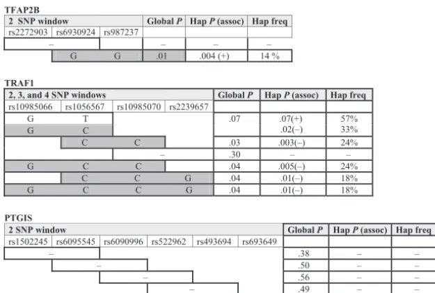
Haplotype analysis of the genotyped SNPs was per-formed to examine whether combinations of alleles might be positively associated with the presence of the
PDA. In this analysis, 1 gene (TFAP2B) showed an
asso-ciation with the presence of a PDA, whereas 2 genes (TRAF1andPTGIS) were associated with the absence of a PDA, as shown in Fig 3. With respect to TFAP2B, 14% of our population had a G allele at both rs6930924 and rs987237, and this combination was associated with the
presence of a PDA (P⫽.004). Conversely, the presence
of a G allele at rs6930924 and an A allele at rs987237
was associated with the absence of a PDA (P ⫽ .02).
Haplotype analysis of bothTRAF1andPTGISgenes
dem-onstrate a negative association with PDA, suggesting a protective effect of specific allele combinations. As
shown in Fig 3 middle (TRAF1), 24% of the study
pop-TABLE 2 Genes Included in the PDA Analysis
ABCA1 CYP1A2 FMO3 IL1RN NQO1 SFTPD
ABCC11 CYP1B1 FOLR1 IL4 NR3C1 SHMT1
ADRB2 CYP2C19 GART IL5 OPRM1 SLC19A1
AKR1C3 CYP2C9 GP1BA IL6 OXT SLC24A5
ALDH2 CYP2D6 GP6 IL8 OXTR TBX1
APOA1 CYP2E1 GPR51 ITGA2 PGR TBX5
APOA4 DARC GSTT1 ITGB3 PLA2G4A TFAP2B
APOA5 DDC HLA-B KCNN3 PLUNC TGFB1
APOB DEFB1 HLA-G LCAT PTEN TGFBR1
APOC2 DHCR24 HMGCR LDLR PTGER1 TGFBR2
APOC3 DHCR7 HPGD LIPC PTGER2 TLR4
APOE DHFR HSD11B1 LPL PTGER4 TNFA
BIRC2 EDN1 HSD11B2 MBL2 PTGES TNFAIP3
BIRC3 EDN2 HSD3B1 MMP1 PTGES2 TNFRSF1A
CETP EP300 IFNG MMP9 PTGFR TNFRSF1B
CFH EPHX1 IGF1 MTHFD1 PTGIS TRADD
CHD7 ESR1 IGF2 MTHFR PTGS1 TRAF1
CHRNA4 F13 IL10 MTR PTGS2 TRAF2
CRHR1 F2 IL10RA MTRR PTPN11 VEGF
CYP11A1 F5 IL1A NAT2 RPAIN ZFHX1B
CYP17A1 FAS IL1B NNMT SERPINA6
CYP1A1 FMO1 IL1R2 NOS2A SFTPB
TABLE 3 SNPs Associated With the Presence of PDA in Infants of<32 Weeks’ Gestation
Gene SNP P
PDA-1a PDA-2b PDAc No PDA Prematurity
PTGIS rs493694 0.005 0.24 0.36 0.08 0.05
TRAF1 rs1056567 0.005 0.03 0.005 0.70 0.03
CETP rs711752 0.006 0.19 0.16 0.73 0.23
CRHR1 rs173365 0.007 — 0.28 0.07 0.03
CYP2D6 rs28360521 0.007 0.31 0.014 0.34 0.02
LIPC rs1973028 0.007 0.82 0.061 0.71 0.39
TFAP2B rs987237 0.007 0.38 0.003 0.10 0.52
— indicates⬍10 informative families. aInitial phase: 69 infants with PDA.
bSecond phase: 57 infants with PDA.
ulation had a C allele at both rs1056567 and rs10985070, and this combination was negatively
asso-ciated with the presence of a PDA (P⫽.003). OurTRAF1
results are consistent in that the T allele of rs1056567 had a positive association with the presence of a PDA in the FBAT analysis, whereas the C allele showed a neg-ative association with the presence of a PDA in the
haplotype FBAT analysis. Likewise, in the PTGIS gene
(Fig 3 bottom), 47% of the study population had a G allele at rs49394 and an A allele at rs693649, and this combination was also negatively associated with the
presence of a PDA (P⫽.01). No other genes analyzed in
this way had haplotype blocks that were associated with the presence or absence of PDA.
Because decreasing gestational age is a major risk factor for the development of a PDA, our data set was analyzed to determine if the genetic polymorphisms that were positively associated with PDA were significant due
to a positive association with preterm delivery at ⬍32
weeks. None of the genes identified as being associated with PDA had a positive association when delivery at
⬍32 weeks’ gestation was used as the affected status
(P⬎.01; see Table 3). We further addressed this issue by
performing a regression analysis taking gestational age
into account. After the regression, TRAF1 (rs1056567)
remained positively associated with PDA (P ⫽ .003),
whereas TFAP2B (rs987237) did not (P ⫽ .33). This
negative result must be interpreted with caution, be-cause PDA and gestational age are linearly related, sug-gesting a latent trait. In addition, the number of infor-mative families in the TFAP2B analysis was rather small
(n ⫽ 25), decreasing the power of the regression
analysis.
Finally, we attempted to replicate findings from the only previous report linking genetic sequence variations
with PDA.9We investigated 6 genetic variations covering
the AGTR1 gene (including the previously studied
AGTR1-A1166C polymorphism, rs5186) for association with PDA. By using FBAT analysis, we were unable to detect a distortion in allele transmission at any loci in triads containing a preterm infant with PDA. Thus, no significant association was found between these poly-0.0
0.5 1.0 1.5 2.0 2.5
P = .01
CRHR1 CYP2D6
TFAP2B PTGIS
LIPC CETP
TRAF1
SNP
-lo
g(
P
val
ue
)
FIGURE 2
Results of the FBAT for genetic association with PDA. The negative log of thePvalue is plotted for each SNP associ-ation with PDA. SNP signals reaching about the dotted line representPvalues of⬍.01.
TFAP2B
2 SNP window Global P Hap P (assoc) Hap freq
rs2272903 rs6930924 rs987237
– – – –
G G .01 .004 (+) 14 %
TRAF1
2, 3, and 4 SNP windows Global P Hap P (assoc) Hap freq
rs10985066 rs1056567 rs10985070 rs2239657 G T
G C .07 .07(+).02(–) 57%33% C C .03 .003(–) 24%
– .30 – –
G C C .04 .005(–) 24% C C G .04 .01(–) 18% G C C G .04 .01(–) 18%
PTGIS
2 SNP window Global P Hap P (assoc) Hap freq
rs1502245 rs6095545 rs6090996 rs522962 rs493694 rs693649
– .38 – –
– .50 – –
– .56 – –
– .49 – –
G A .07 .01 (–) 47%
FIGURE 3
Haplotype analysis for association with PDA in theTFAP2B,
TRAF1, andPTGISgenes. Haplotypes for 3 genes were
ana-lyzed by using a 2-SNP window (TFAP2BandPTGIS) or a com-bination of 2-, 3-, and 4-SNP windows (TRAF1). Allele combi-nations yielding an individual haplotype with aPvalue of ⬍.05 are shaded gray. The sign in parentheses after theP
morphisms, including rs5186 (P ⫽ .48), and the pres-ence of a PDA.
DISCUSSION
Previous studies have begun to address the role of ge-netic factors influencing common neonatal morbidities. A retrospective, multicenter study of 450 twin pairs found a genetic contribution for several morbidities of the preterm infant such as bronchopulmonary dysplasia,
necrotizing enterocolitis, and IVH.16Retinopathy of
pre-maturity has also been found to have a strong genetic
predisposition.17
Our approach was to evaluate sequence polymor-phisms within candidate genes for association with iso-lated (nonsyndromic) PDA in high-risk preterm infants. We identified 7 SNPs associated with the development of
a PDA with aPvalue of⬍.01. Another 16 markers were
found to have aPvalue between .01 and .05. We chose
aPvalue of .01 to provide a first-level cutoff for markers
that might be of interest and warranted additional study in the second sample population. How to appropriately analyze large numbers of samples and hypotheses from a statistical standpoint represents a significant challenge that is now arising in large genome-wide association studies where hundreds of thousands of markers are
tested on thousands of cases.18
Sequence variations in 1 SNP (rs987237) inTFAP2B,
the gene mutated in Char syndrome, was found to be associated with both PDA and failure of the PDA to close with indomethacin in our population of normal preterm infants. This gene is expressed in neural crest derivatives and is generally involved with development, cell-cycle
control, and apoptosis.19,20Embryologic studies using the
chick model system have identified cardiac neural crest cells in the wall of the ductus arteriosus, further sup-porting a role for a gene controlling differentiation of neural crest derivatives in the persistent patency of the
ductus arteriosus.21 Recently, TFAP2B was found to be
expressed in the smooth muscle of the mouse DA, play-ing a key role in ductal closure by participatplay-ing in a transcriptional network that regulates ductal smooth muscle development (Ronald Clyman, MD, personal
written communication, 2007). The specific TFAP2B
marker of interest (rs987237) is located in a highly con-served region between exons 3 and 4 containing a num-ber of putative transcription factor binding sites. The SNP rs987237 is present in a haplotype block that, in white persons, extends from the intronic region between ex-ons 1 and 2 to beyond the end of the gene. This block encompasses exons 2, 4, and 5, where mutations
re-ported to cause Char syndrome are located.7The actual
genetic variation(s) responsible for PDA in preterm in-fants could lie anywhere within this haplotype block. Detailed mapping of this region and confirmation of the finding in other preterm populations is necessary to further define the actual etiologic polymorphism.
A second SNP (rs1056567), in a haplotype block
ad-jacent to theTRAF1gene, was also found to be associated
with PDA in our population. This protein mediates the activity of NF-kappaB and plays a role in modulating
both the inflammatory and apoptotic pathways.22,23Both
apoptosis and inflammation are known to play an im-portant role in the second (more permanent) phase of ductal closure, fibrosis after muscular contraction, and hypoxia. The SNP rs1056567 is also in a haplotype block
within a second gene, PHF19 (PHD finger protein 19).
PHF19, which is a human homolog of the Drosophila polycomb-like gene, encodes a protein that binds to DNA and is postulated to play a role in transcriptional regulation.24
Haplotype analysis determines whether combinations of neighboring polymorphisms, rather than individual sequence variations alone, are positively associated with a phenotype. In addition to a 2-SNP window in the
TRAF1gene, a second 2-SNP combination was found to
be associated with PDA in thePTGISgene. This gene is
responsible for the production of prostaglandin I2 (pros-tacyclin), a potent vasodilator that plays a critical role in
fetal patency of the ductus arteriosus.25It is possible that
alterations in the regulation of this gene could lead to abnormal postnatal closure of the DA in a preterm in-fant, because prostacyclin levels are elevated in preterm infants, and the concentration of a prostacyclin
metab-olite (6-ketoprostaglandin F1-␣) has been shown to
cor-relate with ductal diameter.26Recent reports suggest that
haplotype structure may be more important in deter-mining the effect of a variant on function rather than the
individual SNP.27Although none of the genetic variants
that we identified is likely to be a causative factor for PDA, sequence variations located within the identified haplotype blocks they represent may be etiologic. Our approach has identified a region of interest for additional fine mapping for potential genetic contributors to PDA. One limitation of a hypothesis-generating (ie, data-mining), candidate gene approach such as ours is the large number of observations that are produced by the analysis. Because of these multiple observations, it
be-comes difficult to delineate whatPvalue represents an
appropriate level of significance. We recognize the im-portance of this issue in the present study. Our use of
P⫽.01 as a first-phase criteria was a compromise to limit
false-positives, but may have also led to us to miss some significant genes where our power to detect an affect was limited by sample size. In fact, the second phase of our study demonstrated that 5 of the 7 original positive signals were likely false-positives rather than truly asso-ciated with PDA. Employing the traditional Bonferroni correction for multiple comparisons in the initial phase of the study would have led to a very stringent level for statistical significance (.00013), eliminating all candidate genes in the study. This degree of stringency is likely not appropriate when investigating common complex hu-man diseases where an individual gene, along with en-vironmental forces, may play a variable, but important, role in the development of a disease.
It is very likely that extreme prematurity is the envi-ronmental condition on which a genetic predisposition to PDA becomes manifest, a hypothesis supported by the very low incidence of PDA in term infants. The cardio-vascular environment of a preterm neonate is very dif-ferent from that of a term infant in a number of ways
positive-pressure ventilation, etc) that may promote per-sistent patency of the DA. Additional conditions, such as birth via cesarean section and lower hematocrit at birth, have been associated with failure of the PDA to close
with indomethacin treatment.28It has also been
demon-strated that preterm infants of mothers who received indomethacin tocolysis have an increased incidence of
symptomatic PDA.29 Prematurity and the abnormal
anatomy and physiology that goes along with it, coupled with a genetic predisposition, may lead to the develop-ment of a PDA.
There are practical benefits in delineating genetic factors associated with PDA. It is common practice to attempt closure with nonsteroidal antiinflammatory medications such as indomethacin or ibuprofen. The number of such attempts before surgical closure is un-dertaken varies by institution. Medical treatment of the PDA, however, is not without potential risks and adverse effects. Although early closure of the PDA has been shown to reduce incidence of IVH, no reduction in
bron-chopulmonary dysplasia has been seen.30 If an infant’s
genotype is associated with failure of medical closure and could reliably predict the need for surgical ligation, we may one day use genotype data to decide whether to subject the infant to medical therapy before definitive closure of the PDA surgically. In this study, we did see a
borderline significant association of alleles in theTFAP2B
andEPAS1genes with the subsequent need for surgical ligation that, if replicated, might afford an opportunity for such early predictions. Finally, identifying genes and pathways that play a role in PDA closure can advance our understanding of developmental cardiovascular physiology in humans.
ACKNOWLEDGMENTS
This work was supported by a grant from the Children’s Miracle Network, University of Iowa GCRC, National Institutes of Health (grants R01 HD052953-01 and P30 ES05605), and the March of Dimes (grant FY2005-126). Dr Lepp’s fellowship has been supported by a National
Institutes of Health T32 training grant (grant
HD041922). Ms Steffen was supported by a Doris Duke Fellowship, and Ms Orr was supported by a National Institutes of Health Medical Student Research Fellow-ship.
We thank the families who graciously agreed to par-ticipate in this study. Special thanks go to our research nurses Gretchen Cress, RN, BSN, Nancy Krutzfield, RN, BSN, Ruthann Schrock, RN, BSN, and Laura Knosp, RN, BSN. Thanks also go to Jan Crew, Emily Marcus, Susan Berends, Jamie L’Heureux, Megan Bjork, Sara Welter, and Nikki Ehn for technical support.
REFERENCES
1. Hermes-DeSantis ER, Clyman RI. Patent ductus arteriosus: pathophysiology and management.J Perinatol.2006;26(suppl 1):S14 –S18
2. Clyman RI. Mechanisms regulating the ductus arteriosus.Biol Neonate.2006;89(4):330 –335
3. Lemmers PM, Toet MC, van Bel F. Impact of patent ductus arteriosus and subsequent therapy with indomethacin on
ce-rebral oxygenation in preterm infants.Pediatrics.2008;121(1): 142–147
4. Horbar JD. The Vermont-Oxford Neonatal Network: integrat-ing research and clinical practice to improve the quality of medical care.Semin Perinatol.1995;19(2):124 –131
5. Satoda M, Pierpont ME, Diaz GA, Bornemeier RA, Gelb BD. Char syndrome, an inherited disorder with patent ductus ar-teriosus, maps to chromosome 6p12–p21. Circulation. 1999; 99(23):3036 –3042
6. Mani A, Meraji SM, Houshyar R, et al. Finding genetic contri-butions to sporadic disease: a recessive locus at 12q24 com-monly contributes to patent ductus arteriosus.Proc Natl Acad Sci U S A. 2002;99(23):15054 –15059
7. Mani A, Radhakrishnan J, Farhi A, et al. Syndromic patent ductus arteriosus: evidence for haploinsufficient TFAP2B mu-tations and identification of a linked sleep disorder.Proc Natl Acad Sci U S A. 2005;102(8):2975–2979
8. Zhu L, Vranckx R, Khau Van Kien P, et al. Mutations in myosin heavy chain 11 cause a syndrome associating thoracic aortic aneurysm/aortic dissection and patent ductus arteriosus. Nat Genet.2006;38(3):343–349
9. Treszl A, Szabo M, Dunai G, et al. Angiotensin II type 1 recep-tor A1166C polymorphism and prophylactic indomethacin treatment induced ductus arteriosus closure in very low birth weight neonates.Pediatr Res.2003;54(5):753–755
10. Ehn NL, Cooper ME, Orr K, et al. Evaluation of fetal and maternal genetic variation in the progesterone receptor gene for contributions to preterm birth. Pediatr Res. 2007;62(5): 630 – 635
11. Steffen KM, Cooper ME, Shi M, et al. Maternal and fetal variation in genes of cholesterol metabolism is associated with preterm delivery.J Perinatol.2007;27(11):672– 680
12. O’Connell JR, Weeks DE. PedCheck: a program for identifica-tion of genotype incompatibilities in linkage analysis. Am J Hum Genet.1998;63(1):259 –266
13. Horvath S, Xu X, Laird NM. The family based association test method: strategies for studying general genotype-phenotype associations.Eur J Hum Genet.2001;9(4):301–306
14. Laird NM, Horvath S, Xu X. Implementing a unified approach to family-based tests of association. Genet Epidemiol. 2000; 19(suppl 1):S36 –S42
15. Rabinowitz D, Laird N. A unified approach to adjusting asso-ciation tests for population admixture with arbitrary pedigree structure and arbitrary missing marker information. Hum Hered.2000;50(4):211–223
16. Bhandari V, Bizzarro MJ, Shetty A, et al. Familial and genetic susceptibility to major neonatal morbidities in preterm twins.
Pediatrics.2006;117(6):1901–1906
17. Bizzarro MJ, Hussain N, Jonsson B, et al. Genetic susceptibility to retinopathy of prematurity. Pediatrics. 2006;118(5): 1858 –1863
18. Christensen K, Murray JC. What genome-wide association studies can do for medicine. N Engl J Med. 2007;356(11): 1094 –1097
19. Hilger-Eversheim K, Moser M, Schorle H, Buettner R. Regula-tory roles of AP-2 transcription factors in vertebrate develop-ment, apoptosis and cell-cycle control.Gene. 2000;260(1–2): 1–12
20. Moser M, Pscherer A, Roth C, et al. Enhanced apoptotic cell death of renal epithelial cells in mice lacking transcription factor AP-2beta.Genes Dev.1997;11(15):1938 –1948
21. Waldo KL, Kirby ML. Cardiac neural crest contribution to the pulmonary artery and sixth aortic arch artery complex in chick embryos aged 6 to 18 days.Anat Rec.1993;237(3):385–399 22. Zhang B, Wang Z, Li T, Tsitsikov EN, Ding HF. NF-kappaB2
mutation targets TRAF1 to induce lymphomagenesis.Blood.
23. Oyoshi MK, Barthel R, Tsitsikov EN. TRAF1 regulates recruit-ment of lymphocytes and, to a lesser extent, neutrophils, my-eloid dendritic cells and monocytes to the lung airways follow-ing lipopolysaccharide inhalation. Immunology. 2007;120(3): 303–314
24. Wang S, Robertson GP, Zhu J. A novel human homologue of
Drosophilapolycomblike gene is up-regulated in multiple can-cers.Gene.2004;343(1):69 –78
25. Schneider DJ, Moore JW. Patent ductus arteriosus.Circulation.
2006;114(17):1873–1882
26. Kluckow M, Evans N, Leslie G, Rowe J. Prostacyclin concen-trations and transitional circulation in preterm infants requir-ing mechanical ventilation.Arch Dis Child Fetal Neonatal Ed.
1999;80(1):F34 –F37
27. Nackley AG, Shabalina SA, Tchivileva IE, et al. Human cate-chol-O-methyltransferase haplotypes modulate protein
ex-pression by altering mRNA secondary structure.Science.2006; 314(5807):1930 –1933
28. Kalis NN, Pieper C, van der Merwe PL, Nel ED. Factors influ-encing successful closure with indomethacin of the patent ductus arteriosus in premature infants.Cardiovasc J S Afr.2001; 12(5):268 –272
29. Hammerman C, Glaser J, Kaplan M, Schimmel MS, Ferber B, Eidelman AI. Indomethacin tocolysis increases postnatal patent ductus arteriosus severity. Pediatrics. 1998;102(5). Available at: www.pediatrics.org/cgi/content/full/102/5/ e56
30. Schmidt B, Roberts RS, Fanaroff A, et al. Indomethacin pro-phylaxis, patent ductus arteriosus, and the risk of bronchopul-monary dysplasia: further analyses from the Trial of Indometh-acin Prophylaxis in Preterms (TIPP). J Pediatr.2006;148(6): 730 –734
TILL CHILDREN DO US PART
“Half a century ago, the conventional wisdom was that having a child was the surest way to build a happy marriage. Women’s magazines of that era promised that almost any marital problem could be resolved by embarking on parenthood. Over the past 2 decades, many researchers have concluded that three’s a crowd when it comes to marital satisfaction. More than 25 separate studies have established that marital quality drops, often quite steeply, after the transition to parenthood. And forget the ‘empty nest’ syndrome: when the children leave home, couples report an increase in marital happiness. But does the arrival of children doom couples to a less satisfying marriage? Not necessarily. Two researchers at the University of California at Berkeley, Philip and Carolyn Cowan, report in a forthcoming briefing paper for the Council on Contemporary Families that most studies finding a large drop in marital quality after childbirth do not consider the very different routes that couples travel toward parenthood. Some couples plan the conception and discuss how they want to conduct their relationship after the baby is born. Others disagree about whether or when to conceive, with one partner giving in for the sake of the relationship. And sometimes, both partners are ambivalent. The Cowans found that the average drop in marital satisfaction was almost entirely accounted for by the couples who slid into being parents, disagreed over it or were ambivalent about it. Couples who planned or equally wel-comed the conception were likely to maintain or even increase their marital satisfaction after the child was born.”
DOI: 10.1542/peds.2008-0313
2009;123;1116
Pediatrics
Karen J. Johnson, Mary L. Marazita and Jeffrey C. Murray
Kelsey, Kristin L. Orr, Diana Caprau, Cara R. Zimmerman, Katherine M. Steffen,
John M. Dagle, Nathan T. Lepp, Margaret E. Cooper, Kendra L. Schaa, Keegan J.P.
Infants
Determination of Genetic Predisposition to Patent Ductus Arteriosus in Preterm
Services
Updated Information &
http://pediatrics.aappublications.org/content/123/4/1116
including high resolution figures, can be found at:
References
http://pediatrics.aappublications.org/content/123/4/1116#BIBL
This article cites 29 articles, 11 of which you can access for free at:
Subspecialty Collections
sub
http://www.aappublications.org/cgi/collection/fetus:newborn_infant_
Fetus/Newborn Infant
following collection(s):
This article, along with others on similar topics, appears in the
Permissions & Licensing
http://www.aappublications.org/site/misc/Permissions.xhtml
in its entirety can be found online at:
Information about reproducing this article in parts (figures, tables) or
Reprints
http://www.aappublications.org/site/misc/reprints.xhtml
DOI: 10.1542/peds.2008-0313
2009;123;1116
Pediatrics
Karen J. Johnson, Mary L. Marazita and Jeffrey C. Murray
Kelsey, Kristin L. Orr, Diana Caprau, Cara R. Zimmerman, Katherine M. Steffen,
John M. Dagle, Nathan T. Lepp, Margaret E. Cooper, Kendra L. Schaa, Keegan J.P.
Infants
Determination of Genetic Predisposition to Patent Ductus Arteriosus in Preterm
http://pediatrics.aappublications.org/content/123/4/1116
located on the World Wide Web at:
The online version of this article, along with updated information and services, is
http://pediatrics.aappublications.org/content/suppl/2009/04/09/123.4.1116.DC1
Data Supplement at:
by the American Academy of Pediatrics. All rights reserved. Print ISSN: 1073-0397.