an atypical presentation mimicking
temporomandibular joint disorder
Andrea Jensen,
BSc, DC*Paul S Nolet,
DC, MS, FCCSS(C)**Murtaza A Diwan,
MD, BSc, FRCS(C)†A 50-year-old female presented to a chiropractic clinic with left jaw pain consistent with temporomandibular joint disorder. Examination revealed a large ulcerated mass on the posterolateral margin of the tongue which was later diagnosed as squamous cell carcinoma. Squamous cell carcinoma is the most common of the oral cancers. These cancers are often detected late making treatment more complicated and reducing the chance of survival. In the early stages squamous cell carcinoma can be asymptomatic. Symptoms can be similar to that of temporomandibular joint disorder making examination of the patient’s mouth important to rule out oral cancers. Oral cancers should be considered when patients present to a chiropractor with pain in the area of the temporomandibular joint. Risk factors such as chronic tobacco and alcohol use should raise concern in these patients. Suspicious lesions should be referred
immediately for further investigation. (JCCA 2004; 48(4):266–272)
k e y wo r d s : squamous cell carcinoma; cancer; temporomandibular joint disorder; chiropractic; diagnosis, differential.
Une femme de 50 ans se présente à une clinique chiropratique. Elle souffre de douleur à la mâchoire gauche correspondant à un trouble de l’articulation temporo-mandibulaire. L’examen révèle une grande masse ulcérée sur l’angle postérolatéral de la langue. Cette masse a été diagnostiquée comme étant un
carcinome épidermoïde. Ce type de carcinome est la plus fréquente forme de cancer de la bouche. Le traitement du cancer buccal est difficile puisqu’il est souvent
diagnostiqué trop tard. Aussi, la chance de survie en est réduite. Au stade précoce, le carcinome épidermoïde peut être asymptomatique. Ses symptômes sont similaires à ceux d’une pathologie de l’articulation temporo-mandibulaire et il est donc primordial de procéder à un examen buccal afin d’éliminer toute possibilité de cancer de la bouche. La possibilité d’un cancer buccal doit être prise en considération lorsqu’un patient se présente à la clinique et qu’il souffre de douleur dans la région de l’articulation temporo-mandibulaire. Les facteurs de risque tels que le tabagisme et l’abus d’alcool doivent être une source de préoccupations pour ce patient. Les lésions suspectes doivent immédiatement être soumises à un examen approfondi.
(JACC 2004; 48(4):266–272)
m o t s c l é s : carcinome épidermoïde; cancer; pathologie de l’articulation temporo-mandibulaire; chiropratique; diagnostic; différentiel.
** Private practice, Toronto, Ontario
** Private practice, Guelph, Ontario. Masters of Public Health candidate, Faculty of Professional Schools, Lakehead University.
*† Private practice, Guelph, Ontario.
Correspondence may be sent to Dr. Paul S. Nolet, e-mail: [email protected]
Introduction
Temporomandibular joint disorder (TMD) is a condition that presents to a chiropractors office on a somewhat reg-ular basis (1–2 times per month).1 Chiropractors should
be expected to do a thorough history and examination when accessing TMD. One of the less common causes of pain in the area of the temporomandibular joint are oral cancers. The most common oral cancer is squamous cell carcinoma.2 These cancers often present in the later
stag-es of the disease where the treatment is more complicated and survival less likely. It is important that any chiroprac-tor treating TMD be aware of oral cancers to help detect them in the early stages of the disease. We present the case of a 50-year-old female patient with squamous cell carcinoma on the posterolateral margin of the tongue who presented with persistent left sided jaw pain consist-ent with TMD.
Case report
A 50-year-old female was referred to a chiropractic office by an ears-nose-throat specialist (ENT) for assessment of a left temporomandibular joint problem secondary to arthritis that was possibly provoked by two assaults over 20 years earlier. At that time, she stated that the jaw pain had started after a bout of the flu. She felt that her jaw pain had been aggravated by coughing. The patient de-layed following through with the chiropractic consulta-tion until 6 months later.
She presented to the chiropractic office with left jaw pain that was worsening (10/10 on a numerical rating scale) with radiation to the left ear. The pain was de-scribed as continuous and was aggravated by chewing and lying on her left side. She reported having recently broken a lower left molar. Within the last month she had started having difficulty swallowing. She was a chronic smoker (1–2 packs a day) for the last 34 years and a so-cial drinker. There was a history of depression and she was currently taking Zoloft. She had been treated for pe-ripheral vascular disease with vascular surgery being pre-formed on the left inner thigh. She had tested positive for hepatitis B infection. The jaw pain was being managed by acetaminophen.
The patient was a women who appeared older than her stated years. She had poor dental health and was missing several teeth. Examination revealed full cervical range of motion with mild pulling on right lateral flexion. Postural
evaluation revealed mild anterior head carriage. Jaw movements aggravated her pain and she was reluctant to open her mouth to the extreme. Palpation of the left tem-poromandibular joint elicited tenderness although there was more sensitivity to palpation in the submandibular re-gion. The patient became apprehensive on intraoral exam-ination reporting that she had broken a tooth 3 weeks earlier which was irritating her tongue. Upon inspection of the area a large ulcerated mass was observed near the base of the tongue on the left. The mass was 3 to 4 times the thickness of her tongue. When questioned about the mass she related it to her broken tooth. Due to her poor dental health it was difficult to determine whether her tooth was in fact broken. The ENT specialist was immediately called and an appointment booked the following week. The patient was not seen by the chiropractor subsequent to her initial visit. Three days after seeing the ENT specialist a biopsy was performed which was positive for squamous cell (T3) carcinoma at the left posterior oral tongue.
The patient was referred to a regional cancer centre where she was seen 13 days after the biopsy. She was ex-amined by a head-neck surgeon who prescribed pain medication and scheduled her for surgery 14 days later. CT imaging of the head and neck showed lymphadenopa-thy ipsilateral to the mass which was not palpable on ex-amination. Radiation therapy was recommended after the surgery.
Surgery consisted of a left commando resection via a lower lip splitting incision, two incisions in the left neck, a mandibulectomy and adjacent large glossectomy and reconstruction with a radial forearm osteocutaneous free flap. She was fitted with a percutaneous endoscopic gas-trostomy tube.
Two days post surgery the patient had a major myocar-dial infarction and was transferred to an intensive care unit. She made a slow but full recovery over several weeks. Upon discharge from the hospital she was still on a feeding tube with a weight gain of 3 to 4 pounds. She was able to make verbal sounds but her speech was limit-ed with about 25% of her words being discernable.
of her nutrition with a feeding tube. Examination at this time revealed a soft tissue proliferative mass at the junc-tion of the free-flap and remaining left base of the tongue. The patient died within nine months of the recur-rence of the intraoral lesion.
Discussion
Cancer of the oral cavity accounts for only 4% of all can-cers, squamous cell carcinoma being the most frequent. Oral cancer is on the rise and worldwide is estimated to be the sixth most common cancer. It is primarily a dis-ease occurring after 45 years of age and is most likely to affect the tongue, lips and floor of the mouth in 90–95% of cases.3,4 Squamous cell carcinoma presents
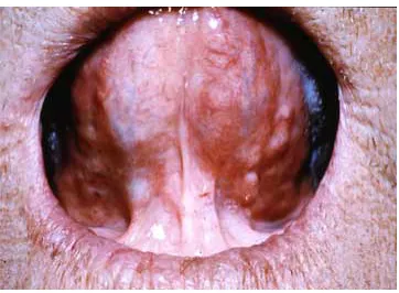
intra-oral-ly as a non-healing ulcer which appears as a white (Fig-ure 1, 2) or red patch.5 Oral cancer is more prevalent in
males and is associated with tobacco use in all forms and alcohol drinking.6,5 Most authors agree that either
sub-stance alone can induce oral cancers, however, most cas-es rcas-esult from a combination of smoking and drinking.3–6
The Canadian Cancer Society along with the National Cancer Institute of Canada publishes yearly statistics on cancer in Canada.7 They have estimated for 2003 that
there will be 139,900 new cases of cancer diagnosed and 67,400 deaths that year due to cancer. Oral cancers will account for 3100 of these new diagnoses of which 2100 will be in males and 1000 in females. Deaths due to oral cancers will occur in approximately 730 males and 360 females. The lifetime probability of developing oral can-cer is 1:185.2 in males and 1:400 in females. The impor-tance of different types of cancers can be measured with the death to case ratio. In Canada the death to case ratio for oral cancer is .36 which is listed as a fairly good prog-nosis (a ratio of over .5 being a poor progprog-nosis and under .3 being a good prognosis).
Patients diagnosed with intraoral cancers tend to have unfavourable outcomes when detected in later stages. Ex-amination of the oral cavity can be easily performed and will readily reveal abnormal changes; therefore, early di-agnosis should be possible and outcomes for these pa-tients should improve.8 Unfortunately, only 30–40% of
patients survive an average of 5 years despite recent sur-gical advances.7 The short survival time is largely due to
late detection and treatment.9,8 Shira8 found that the
aver-age time lapse between symptoms and consultation is 4.6 months, while Bruun9 found it to be 4.9 months. Both
Bruun and Shira9,8 discovered that it was another 5.6
months before these patients were referred for further ex-amination and treatment.
This delay in diagnosis and treatment of oral cancers can be attributed to three things. First, public awareness of oral cancer is low compared with other forms of can-cer; therefore, patients generally ignore early signs and symptoms.6 Second, health professionals like dentists
and specialists who routinely diagnose more common complaints of the oral cavity may overlook the risk of a possible malignancy. On the other hand, health profes-sionals not as familiar with diagnosis and treatment of oral complaints realize the need for referral sooner.8
Third, oral squamous cell carcinoma presents in a variety of ways, and most early lesions are asymptomatic.6 One
study found that 15% of patients with intraoral squamous cell carcinoma did not exhibit any subjective symptoms even when the tumor was greater than 4 cm in diameter.10
Furthermore, there is usually a lack of “red flag” symp-toms such as persistent ulcers and neurologic disturbanc-es.8 According to Bruun and Shira,9,8 the most frequent
complaint of oral cancer was swelling followed by pain. These are generally banal complaints and may occur with much more common disorders.9,8
Temporomandibular joint disorder (TMD) refers to a collection of painful symptoms affecting the joint formed by the mandible and the temporal bone of the skull. It has been estimated that approximately 85–90% of the popu-lation will show at least one temporomandibular joint (TMJ) symptom in their lifetime. TMD is more common in women but has no predilection for age.11
TMD can be a difficult disorder to diagnose and treat for four reasons. First, it is not a well defined condition. TMD tends to be a multi factorial disorder that includes structural, functional and psychosocial factors.12 TMD
may be due to any number of underlying causes. Most commonly it is associated with dysfunction and myofa-cial pain as a result of teeth clenching, jaw grinding, or repetitive motions like gum chewing. Less common caus-es include joint disorders like osteoarthrosis, and me-chanical problems.13,14 Many authors have suggested that
cervical spine dysfunction can give rise to TMD; howev-er, one study using an anamnestic self-administered ques-tionnaire did not support this theory.12,15 Therefore, not
before
after
Figure 1
T2 squamous cell carcinoma on the ventral tongue.
Figure 2
The patient after radiation therapy to the squamous cell carcinoma.
Second, it rarely signals a more serious disease.16 The
classic presentation of TMD is pain in front of the tragus with radiation to the ear, lower jaw, cheek and temple.14
Interestingly, patients with intraoral malignancies may also present with similar complaints of pain.
Third, TMD presents with a myriad of symptoms. These include acute and/or chronic pain and/or tender-ness of the muscles of mastication, facial muscles, and the cervical-thoracic musculature, ear pain with or with-out radiation to the jaw and cheeks that worsens with chewing, a feeling of fullness in the ear, neck and/or jaw stiffness, ringing in the ear, clicking and popping sounds, malocclusion and worn teeth, mouth and tooth pain espe-cially upon chewing, vertigo, numbness, inadequate jaw opening (less than a 3 finger opening), and headaches that are usually temporal in location.17 With such a wide
array of symptoms, disorders of surrounding structures may be easily mistaken as TMD.
Fourth, there is a lack of people seeking intervention for TMD. According to Walling,14 only 5% of adults with
TMJ symptoms seek medical help. Hawk and as-sociates18 found that out of those people seeking
chiro-practic intervention for musculoskeletal conditions (which is the predominant reason for office visits to a chiropractor), only 0.46% were treated for TMJ pain as their chief complaint. Consequently, there is a lack of ex-perience in dealing with TMD. This may lead to an inac-curate diagnosis.
When a patient is suspected of having TMD, the diag-nosis is often made by ruling out other conditions. Conse-quently, a detailed physical history is imperative. It should include a description of the pain and related symptoms as well as information relating to the dental and medical history. This is essential in uncovering any underlying conditions.16 A thorough examination of the
temporomandibular joint requires checking the jaw range of motion during opening and closing, palpating the joint for tenderness, clicks or pops, and examining the ear, teeth, mouth, neck, jaw and salivary glands including the surrounding nerves, muscles and soft tissue.16,14 This
means looking into the oral cavity and observing any le-sions. Since the oral mucosa is easily examined and changes can be readily observed, suspicious lesions should be immediately referred for further evaluation. Only then can the clinician rule out conditions that may mimic TMD.
Unfortunately, a significant number of studies on TMD did not discuss typical differential diagnoses. Only one article was found that discussed neoplasms of the tempo-romandibular joint that could mimic TMD. No studies were found that mentioned intraoral cancer as a differen-tial diagnosis for TMD. Furthermore, very few studies emphasized the need for intraoral examinations in order to rule out more sinister causes of facial, jaw and ear pain.
Consequently, the difficulty in diagnosing and treating TMD combined with the relatively low prevalence of oral cancers has contributed to a poor understanding of how pain from disorders of the oral cavity can mimic TMD. In turn, this may lead to an inaccurate diagnosis of TMD.
It is essential to understand the neuroanatomy of the head and neck in order to realize how intraoral malignan-cies can lead to an inaccurate diagnosis of TMD. The principal innervation of the TMJ is the auriculotemporal nerve.19 It is a branch of the posterior trunk of the
man-dibular nerve which originates from the trigeminal nerve, one of the largest cranial nerves.20 The trigeminal nerve
is a mixed nerve (motor and sensory) that provides senso-ry innervation to the face and mucous membranes of the nasal and oral cavities, and motor innervation to the mus-cles of mastication.21
The sensory root of the trigeminal nerve exits the pons with the motor root and expands to form the trigeminal ganglion or the gasserian ganglion. At this point it gives rise to three trunks; the ophthalmic, maxillary and man-dibular divisions. The manman-dibular trunk joins the motor root of the trigeminal nerve to form the mandibular nerve. After leaving the skull via the foramen ovale it travels in the infratemporal fossa and divides into four main terminal branches. These are the motor branches, the lingual nerve, the inferior dental branches and the mental branches. The lingual nerve is of interest since it conveys sensation from the anterior 2/3 or oral part (the body) of the tongue and lower gums. The mandibular nerve also supplies the skin of the lower lip, jaw, chin, tympanic membrane, auditory meatus and upper ear.21
Sensory information including noxious stimuli from these structures terminate on the spinal nucleus of the trigeminal nerve within the brainstem. The information is then conveyed to the thalamus (predominantly the ventral posteromedial nuclei) via the trigeminothalamic tract.21
The patient’s lesion was located on the left posterior oral tongue; however, her chief complaint was severe left jaw pain with radiation to the left ear. Since the spinal nucleus of the trigeminal nerve receives nociceptive information from the jaw, ear and oral tongue via the mandibular nerve, pain may be perceived in any of these areas when the origin was actually the anterior 2/3 of the tongue.20
Examination of the oral cavity is important in any pa-tient presenting with symptoms of TMD. Chiropractors should perform a thorough history and examination. His-tory is important to elicit risk behaviors, hisHis-tory of head and neck radiotherapy, family history of head and neck cancer and personal history of cancer. The examination should include visual inspection and palpation of the head, neck, oral and pharyngeal regions. Visual inspec-tion should be done with a good light source. Mirrors can be useful in seeing area of the mouth that are difficult to normally see. Palpation should include the nodes of the neck and floor of the mouth and tongue. Protraction of the tongue with gauze can help visualize the posterolater-al tongue and tongue base.22
Conclusion
Patients initially suspected of TMD requires a thorough history of the presenting complaint including a recent ex-perience of TMJ pain and /or sudden mandibular restric-tion. The clinical exam must include TMJ palpation and palpation of the masticatory muscles to duplicate the chief complaint of pain. Joint sounds and mandibular range of motion are secondary signs since they can be asymptomat-ic. It is vital to examine other systems including adjacent musculoskeletal systems, neurologic, vascular and oto-laryngoglic systems to rule out other causes of pain.19
As TMD is continually researched and re-defined, we may find that traditional signs and symptoms like TMJ joint sounds and mandibular restriction will lose their rel-ative importance. Simpler determinants including a histo-ry of pain in the muscles of mastication and palpable muscle and joint tenderness will define TMD.19 This
way, it may become easier to catch more sinister causes of head/neck/face pain like squamous cell carcinoma of the tongue.
This case study emphasizes the need for health practi-tioners to perform intraoral exams when investigating any head or neck complaint. Patients with a history of
smok-ing and drinksmok-ing and who are over 40 years of age should be screened for the possibility of oral cancer. This is es-pecially important for health care providers who routine-ly deal with more common conditions. Screening for oral cancer is simple since lesions of the oral mucosa are readily visible and at most requires a good light source and a little extra time.5 Health practitioners who
encoun-ter patients with a suspicious oral lesion inconsistent with the presenting head/neck/face complaint, must include malignancy as part of their differential diagnosis. Imme-diate referral to a specialist is warranted so that oral can-cer can be detected early. Only then can outcomes for these patients improve.
Oral cancers are not common despite their increasing prevalence but should be made more publically aware as part of a prevention program. Since most cases of oral cancer result from the combined effect of smoking and drinking, tobacco and alcohol abuse cessation programs should be aggressively promoted.20 Chiropractors can
di-rect patients in most provinces to the Canadian Cancer Society smokers’ helpline (1-877-513-5333) and other quitlines for free information, advice and support on smoking sessation.23 Nutritional education may also play
a role in prevention. Chiropractors have long understood the relationship of treating the whole person and not just the part that is diseased. Patients with a history of alcohol and tobacco abuse should receive nutritional evaluation and advise. Numerous studies support the hypothesis that beta-carotene and vitamin C have a significant protective effect against oral cancers.13 Therefore, increased
con-sumption of fruits and vegetables in the daily diet is help-ful in the prevention of oral cancer. Chiropractors have a unique opportunity to promote health and wellness in their practices and should include improved nutrition as part of their treatment plan. This could make a significant impact for the patients at risk of intraoral malignancies.
References
1 Christensen M, Morgan D (eds). Job Analysis of Chiropractic: A Project Report, Survey Analysis and Summary of the Practice of Chiropractic within the United States. Greeley, CO: National Board of Chiropractic Examiners, 1993.
3 Araujo Filho VJ, De Carlucci D, Sasaki SU, Montag E, Azato FN, Cordeiro AC, Ferraz AR. Incidence of oral cancer profile at a general hospital in Sao Paulo. Rev Hosp Clinc Fac med Sao Paulo 1998; 53(3):110–113.
4 Carvalho MB, Lenzi J, Lehn CN, Fava AS, Amar A, Kanda JL, WalderMenezes MB, Franzi SA, Magalhaes MR, Curioni OA, Marcel R, Szeliga S, A Sobrinho JD, Rapoport A. Clinical and epidemiological characteristics of oral squamous cell carcinoma in women. Rev Assoc Med Bras 2001; 47(3):208–214.
5 Zakrzewska JM. Oral cancer. BMJ 1999; 318:1051–1054. 6 Blot W, McLaughlin J, Winn D, Austin D, Greenberg R,
Preston-Martin S, Bernstein L, Schoenberg J, Stemhagen A, Fraumeni J. Smoking and drinking in relation to oral and pharyngeal cancer. Cancer Research 1988; 48:3282–3287.
7 Canadian Cancer Society, 2003. Canadian Cancer Statistics 2003. URL: http://www.cancer.ca
8 Shira RB. Time lapse by diagnosis of oral cancer. Oral Surgery 1996; 42(2):139–149.
9 Bruun JP. Time lapse by diagnosis of oral cancer. Oral Surg Oral Med Oral Pathol 1976; 42(2):139–149.
10 Rindum J, Pindborg JJ. Intraoral cancer: 100 consecutive cases. Tandlaegebladet 1989; 93(13):504–505.
11 Saghafi D, Curl, DD. Chiropractic manipulation of anteriorly displaced temporomandibular disc with adhesion. JMPT 1995; 18(2):98–104.
12 De Wijer A, de Leeuw JR, Steenks MH, Bosman F. Temporomandibular and cervical spine disorders. Spine 1996; 21:1638–1646.
13 Hart AK, Karakla DW, Pitman KT, Adams JF. Oral and oropharyngeal squamous cell carcinoma in young adults: A report on 13 cases and review of the literature. Otolaryngology-Head and Neck Surgery 1999; 120(6):828–833.
14 Walling AD. Review of diagnosis and treatment of TMJ disorders. American Family Physician 1998; 58(8):1841–1842.
15 Knutson GA, Moses J. Possible manifestation of temporomandibular joint dysfunction on chiropractic cervical x-ray studies. JMPT 1999; 22(1):32–37. 16 Harvard Women’s Health Watch 1996;4(4):2–3. 17 Birkner K. TMJ/Temporomandiubular joint syndrome.
MMRC Health Educator Reports 1997:1–4. 18 Hawk C, Long CR, Boulanger KT. Prevalence of
nonmusculoskeletal complaints in chiropractic practice: Report from a practice-based research program. JMPT 2001; 24(3):157–169.
19 Kraus SL. Temporomandibular Disorders 2nd ed. Churchill Livinstone Inc., 1994.
20 Moore KL. Clinically Oriented Anatomy 2nd ed. Williams and Wilkins, 1985.
21 Brazis P, Masdeu J, Biller J. Localization in Clinical Neurology 4th ed. Williams and Wilkins, 2001. 22 Barker G, Baughman R, Burzynski N, et al. 1996 Oral
Cancer Conference: Background Papers, URL: http:// www.cdc.gov/oralhealth/conferences/oc-bgpapers.htm 23 Canadian Cancer Society, 2003. Smokers Helpline and
other Quitlines. URL: http://www.cancer.ca/ccs/internet/ standard/0,3182,3172_49465673__lan gId-en,00.html