Article
1
Comparative Transcriptomics Identifies Novel Genes
2
and Pathways Involved in Post-Traumatic Osteoarthritis
3
Development and Progression
4
Aimy Sebastian1, Jiun C. Chang1,2, Melanie E. Mendez1,2, Deepa K. Murugesh1, Sarah Hatsell3,
5
Aris N. Economides3, Blaine A. Christiansen4 and Gabriela G. Loots1,2*
6
1 Lawrence Livermore National Laboratories, Physical and Life Sciences Directorate, Livermore, CA.
7
2 UC Merced, School of Natural Sciences, Merced, CA.
8
3 Regeneron Pharmaceuticals, Tarrytown, NY.
9
4 UC Davis Medical Center, Department of Orthopedic Surgery, Sacramento, CA.
10
* Correspondence: [email protected]; Tel.: +01-925-423-0923
11
12
Abstract: Injuries to the anterior cruciate ligament (ACL) often result in post-traumatic osteoarthritis
13
(PTOA). To better understand the molecular mechanisms behind PTOA development following
14
ACL injury, we profiled ACL injury-induced gene expression changes in knee joints of three mouse
15
strains with varying susceptibility to OA: STR/ort (highly susceptible), C57BL/6 (moderately
16
susceptible) and super-healer MRL/MpJ (not susceptible). Right knee joints of the mice were
17
injured using a non-invasive tibial compression injury model that closely mimics ACL rupture in
18
humans and global gene expression was quantified before and at 1-day, 1-week, and 2-weeks
post-19
injury using RNA-seq. Following injury, STR/ort displayed severe cartilage degeneration while
20
MRL/MpJ had little cartilage damage. Gene expression analysis suggested that prolonged
21
inflammation and elevated catabolic activity in STR/ort injured joints, compared to the other two
22
strains may be responsible for the severe PTOA phenotype observed in this strain. MRL/MpJ had
23
the lowest expression values for several inflammatory cytokines and catabolic enzymes activated in
24
response to ACL injury. Furthermore, we identified several genes highly expressed in MRL/MpJ
25
compared to the other two strains including B4galnt2 and Tpsab1 which may contribute to enhanced
26
healing in the MRL/MpJ. Overall, this study has increased our knowledge of early molecular
27
changes associated with PTOA development.
28
Keywords: Osteoarthritis, RNA-seq, STR/ort, C57BL/6, MRL/MpJ, ACL injury, PTOA, regeneration,
29
inflammation, B4galnt2.
30
________________________________________________________________________________________
31
1. Introduction
32
Osteoarthritis (OA) is a painful degenerative joint disease that causes disability and diminishes
33
the quality of life for millions of people worldwide [1]. Joint injury, particularly injuries to the anterior
34
cruciate ligament (ACL), often result in post-traumatic osteoarthritis (PTOA) within 1-2 decades from
35
the injury [2]. PTOA accounts for about 12% of all OA cases, yet the mechanisms contributing to
36
PTOA after joint injury are not well understood and currently there are no effective treatments
37
available for PTOA [3]. Many people developing injury- or age- related OA do not show any
38
symptoms until significant joint damage has occurred, and joint pain is not always indicative of OA
39
[4]. For many diagnosed with OA the only available treatment options are joint replacement surgery
40
and/or pain management. Therefore, there is a dire need for the discovery of biomarkers that can
41
facilitate early detection of the disease and new therapeutic strategies for the prevention of PTOA.
42
While many factors can influence the development of OA, injury mediated OA holds the greatest
43
Preprints (www.preprints.org) | NOT PEER-REVIEWED | Posted: 14 August 2018 doi:10.20944/preprints201808.0244.v1
© 2018 by the author(s). Distributed under a Creative Commons CC BY license.
promise for the development of effective pharmacologic interventions because a treatment can be
44
administered at the time of surgery, or immediately post injury.
45
Acute joint trauma triggers several molecular events over the course of the first 1-2 weeks
post-46
injury, which directly or indirectly contribute to the subsequent cartilage damage characteristic of
47
OA. An understanding of these early molecular events provides a basis for identifying potential
48
biologic targets for intervention to prevent subsequent joint degeneration [5]. Characterization of
49
gene expression changes during OA development and progression at the whole genome level will
50
provide novel mechanistic insights that could be translated into disease-modifying therapies.
51
Numerous studies have used human biopsy samples to gain new insights about joint OA
52
pathogenesis [6-8] however; there are limitations in terms of the types of studies that can be
53
conducted using human subjects. Human samples are usually obtained during knee replacement
54
surgery therefore, they represent late stages of the disease. To overcome this gap in knowledge,
55
animal models allow us to investigate OA development longitudinally, and are particularly well
56
suited to studying early molecular events to derive new insights into the key factors contributing to
57
disease progression. Using a non-invasive tibial compression (TC) injury model [9] that closely
58
mimics anterior cruciate ligament (ACL) rupture in humans we recently profiled the genes
59
expression in knee joints from C57BL/6 mice during the onset and progression of PTOA and
60
identified the molecular changes that characterize early and late stages of PTOA [10], including
61
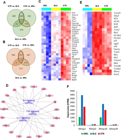
enhanced inflammatory responses at early timepoints and cartilage and bone remodeling at both
62
early and late timepoints. We also noted that majority of the transcriptional changes happen within
63
the first few weeks post-injury.
64
In this study, we used the TC injury model to study early molecular events associated with
65
PTOA development in three mouse strains with varying susceptibility to OA: STR/ort (highly
66
susceptible), C57BL/6 (moderately susceptible) and MRL/MpJ (not susceptible). The STR/ort mice
67
develop OA spontaneously early in life and show many human OA characteristics, including
68
proteoglycan loss, extracellular matrix (ECM) degradation and subchondral sclerosis [11]. They also
69
exhibit osteophyte formation, a phenotype more readily seen in animal models where the joint is not
70
stabilized. The MRL/MpJ is a mouse strain with exceptional abilities to heal wounds made in multiple
71
tissues without the production of a fibrotic scar [12]. MRL/MpJs are protected from PTOA and do not
72
develop degenerative joint changes following articular fracture [13]. It has been suggested that
73
MRL/MpJ mice possess an intrinsic ability to regenerate articular cartilage, yet the molecular
74
mechanisms responsible for this phenotype have yet to be revealed [14]. To characterize genes that
75
contribute to increased OA susceptibility in STR/ort and resistance to PTOA in MRL/MpJ, and to
76
understand the molecular changes associated with early stages of PTOA development in these mice,
77
we profiled gene expression in the knee joints of MRL/MpJ, C57BL/6 and STR/ort by RNA sequencing
78
(RNA-seq), at 0-day, 1-day, 1-week and 2-weeks post-injury. Understanding the molecular and
79
genetic basis of enhanced OA susceptibility in STR/ort and resistance to PTOA in MRL/MpJs will
80
improve our understanding of PTOA pathogenesis and may highlight new treatment options for
81
PTOA or identify biomarkers that track disease progression.
82
This study identified 944, 2330 and 2702 genes differentially regulated in MRL/MpJ, C57BL/6
83
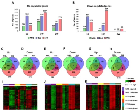
and STR/ort respectively, in response to ACL injury, including 553 genes which were shared by all
84
strains. We identified increased, persistent inflammation, elevated catabolic activity and elevated
85
apoptosis as significant contributors to PTOA development. This study also identified several genes
86
that may contribute to enhanced healing and tissue regeneration including B4galnt2 and Tpsab1.
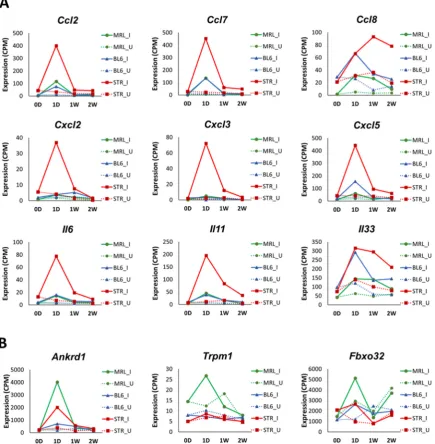
87
Furthermore, this study identified several potential OA biomarkers including Mamdc2 and Pxdn.
2. Results
90
2.1 Evaluation of PTOA development and progression in STR/ort, C57BL/6 and MRL/MpJ mice following
91
ACL injury
92
Injured and uninjured contralateral joints of STR/ort, C57BL/6 and MRL/MpJ mice were
93
phenotyped using histology and/or micro-computed tomography (μCT) at 6- and 12-weeks
post-94
injury to assess tissue morphology. Gene expression was profiled by RNA-seq before injury, 1-day,
95
1-week, and 2-weeks post-injury (Figure 1A). The joint damage was assessed by measuring the extent
96
of osteophyte formation and the severity of cartilage degradation. Osteophyte formation was
97
observed in all three strains by 6-weeks post-injury; MRL/MpJ injured joints had significantly less
98
ectopic bone than the other strains (Figure 1B, C). In all three strains, injured joints lost significant
99
subchondral bone volume in the femoral epiphysis relative to the uninjured contralateral joints
100
(Figure 1D). Trabecular bone volume fraction (BV/TV) was significantly higher in STR/ort compared
101
to the other two strains (Figure 1D), and STR/ort had a significantly higher bone mass than the other
102
two strains, consistent with prior publications [15]. At 12-weeks post-injury, STR/ort contralateral
103
joints displayed significant proteoglycan loss and cleft down below the superficial and into the mid
104
zone of the tibial cartilage whereas MRL/MpJ and C57BL/6 contralateral joints had healthy cartilage
105
(Figure 1E). Injured joints of C57BL/6 and STR/ort exhibited severe cartilage erosion at 12-weeks
106
post-injury (Figure 1E, F). In contrast, the MRL/MpJ displayed an insignificant proteoglycan loss,
107
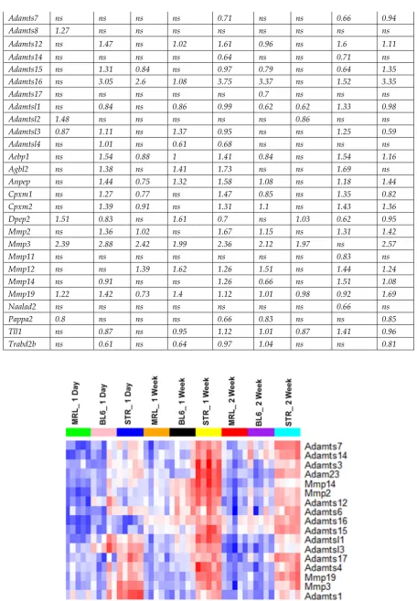
suggesting that MRL/MpJ are protected from ACL injury induced cartilage damage. MRL/MpJ
108
injured joints were significantly different than C57BL/6 and STR/ort injured joints, but no statistical
109
difference was found between C57BL/6 and STR/ort injured joints (Figure 1F).
110
111
Figure 1. ACL injury leads to PTOA in C57BL/6 and STR/ort, but not in MRL/MpJ mice. Knee joints were
112
injured at 10-weeks of age. A) Timeline for histology, μCT and RNA-seq sample collection [0-day (0D), 1-day
113
post-injury (1D), 1-week post-injury (1W), 2-weeks post-injury (2W), 6-weeks post-injury (6W) and 12-weeks
114
post-injury (12W)]. B) μCT representation of injured and uninjured joints at 6-weeks post-injury. Darker regions
115
Preprints (www.preprints.org) | NOT PEER-REVIEWED | Posted: 14 August 2018 doi:10.20944/preprints201808.0244.v1
in the injured scans depict osteophytes. C) Osteophyte volume quantification (dark regions in the injured scans
116
in B) at 6-weeks post-injury. D) Epiphyseal trabecular bone volume ratio of the distal femur was quantified and
117
analyzed between injured and uninjured joints at 6-weeks post-injury. E) Histological assessment of uninjured
118
and injured joints at 12-weeks post-injury using Safranin-O and Fast Green staining (5X magnification). F)
119
OARSI scoring of histological sections of injured and uninjured joints at 12-weeks post-injury. Scale bar = 1mm.
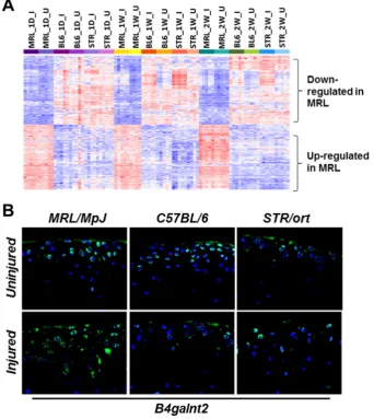
120
*p<0.05, **p<0.01, ***p<0.001, ns not significant.
121
2.2 Characterizing genes related to osteoarthritis susceptibility
122
To identify transcripts that correlate with OA risk, we profiled the knee joints of 10-weeks old
123
uninjured MRL/MpJ, C57BL/6 and STR/ort mice and examined all pair-wise comparisons (Figure 2A,
124
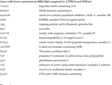
B, Table S1). Four hundred ninety-seven genes were up-regulated in STR/ort compared to both
125
C57BL/6 and MRL/MpJ (Figure 2A). This included 33 ‘inflammatory response’ related genes (Il1b, Il6,
126
Ccl2, Ccl7, Cxcl1, Cxcl2 etc.) (Figure 2C) and 78 genes associated with ‘apoptotic process’ including
127
Egr1, Id3, Cebpb, Fos and Jun (Figure S1A). Genes up-regulated in STR/ort compared to the other two
128
strains also showed enrichment for ontology terms ‘response to wounding (40 genes)’, ‘response to
129
oxidative stress (24 genes including Ptgs2, Rhob, Duox1, Areg and Plk3)’, ‘response to hypoxia (19
130
genes)’, and ‘ossification (25 genes)’. Enriched signaling pathways associated with these genes
131
include ‘TNF signaling pathway (15 genes)’, ‘IL17 signaling pathway (14 genes)’ and ‘IL6-mediated
132
signaling events (8 genes)’ (Figure 2D). Genes down-regulated in STR/ort compared to the other two
133
strains showed enrichment for biological processes such as ‘leukocyte activation (29 genes)’,
134
‘hemopoiesis (27 genes)’ and ‘immune response (39 genes including Tlr1, Tlr5, Itgam, Itgad, Cd3d and
135
Cd8a)’.
137
Figure 2. Genes associated with osteoarthritis susceptibility. A) Overlap between genes up-regulated in STR/ort
138
(STR) compared to C57BL/6 (BL6) and MRL/MpJ (MRL) mice and in BL6 compared to MRL, at 0-day. Genes
up-139
regulated in STR compared to both BL6 and MRL are shown in black ovals and genes down-regulated in MRL
140
[up-regulated in both STR and BL6 compared to MRL] are shown in red ovals. B) Overlap between genes
down-141
regulated STR compared to BL6 and MRL and in BL6 compared to MRL, at 0-day. Genes down-regulated in STR
142
compared to both BL6 and MRL are shown in black ovals and genes up-regulated in MRL [down-regulated in
143
both STR and BL6 compared to MRL] are shown in green ovals. C) Inflammatory response related genes highly
144
expressed in STR compared to BL6 and MRL at 0-day. D) TNF, IL17 and IL6- signaling pathway associated genes
145
with high expression in STR compared to both BL6 and MRL. E) Interferon signaling pathway associated genes
146
with low expression in MRL compared to both STR and BL6. F) Matrix metalloproteinases (MMPs) with low
147
expression in MRL compared to both STR and BL6.
148
MRL/MpJ had the lowest expression values for the majority of ‘inflammatory response’ related
149
genes (Figure 2C). Genes with lowest expression in MRL/MpJ compared to other two strains (839
150
genes) also showed enrichment for ontology terms including ‘extracellular matrix organization (69
151
genes)’, ‘collagen catabolic process (18 genes)’, ‘response to wounding (71 genes)’, ‘immune response
152
(98 genes)’ and ‘cytokine production (50 genes)’. Expression of interferon signaling pathway
153
Preprints (www.preprints.org) | NOT PEER-REVIEWED | Posted: 14 August 2018 doi:10.20944/preprints201808.0244.v1
associated genes such as Ifitm1, Ifit3, Ifi27, Ifi35, Trim2 and Trim5 and matrix metalloproteinases
154
(MMPs) including Mmp2, Mmp3, Mmp14 and Mmp19 were also significantly lower in MRL/MpJ
155
compared to the other two strains (Figure 2E, F). Several genes involved in wound healing including
156
Postn, Fn1, Dcn, Ctgf, Col3a1, Col5a1, Plat and Hbegf also exhibited lowest expression levels in
157
MRL/MpJ (Figure S1B). Four hundred sixty-nine genes were up-regulated in MRL/MpJ compared to
158
both STR/ort and C57BL/6 (Figure 2B). This included 21 genes associated with ‘T cell activation (Cd3d,
159
Cd4, Jak3 etc.)’. Other enriched ontology terms associated with genes up-regulated in MRL/MpJ
160
included ‘lymphocyte aggregation (21 genes)’, ‘defense response (49 genes)’, ‘heme metabolic process
161
(7 genes)’ and ‘cellular ion homeostasis (23 genes)’. Enriched pathways associated with the
up-162
regulated genes included ‘Heme biosynthesis (4 genes)’ and ‘Complement cascade (6 genes)’.
163
2.3 Early molecular changes associated with PTOA development in STR/ort, C57BL/6 and MRL/MpJ mice
164
To identify the early molecular changes associated with PTOA development in STR/ort, C57BL/6
165
and MRL/MpJ mice, we examined gene expression changes in injured joints compared to uninjured
166
contralateral joints at 1-day, 1-week and 2-weeks post-injury. At 1-day post-injury, we identified 441
167
(326 up; 115 down), 1590 (986 up; 604 down), and 735 (359 up; 376 down) differentially expressed
168
genes (>1.5 fold) in the injured joints of MRL/MpJ, C57BL/6 and STR/ort, respectively, including 201
169
genes commonly changed in all three strains (Figure 3A-D, Table S2). Enriched biological processes
170
associated with the up-regulated genes included ‘extracellular matrix organization’, ‘vasculature
171
development’, ‘cell migration’, ‘angiogenesis’, ‘response to wounding’ and ‘inflammatory response’
172
for all three strains and ‘granulocyte migration’, ‘reactive oxygen species metabolic process’,
173
‘cytokine secretion’ and ‘response to tumor necrosis factor’ for STR/ort alone. At 1-day post-injury,
174
59, 78 and 45 ‘inflammatory response’ related genes were up-regulated in STR/ort, C57BL/6 and
175
MRL/MpJ respectively. Several inflammatory cytokines including Ccl2, Ccl7, Ccl8, Cxcl5, Il6 and Il33
176
were up-regulated in all three genotypes in response to injury (Table 1). The majority of these
177
transcripts showed significantly higher expression levels in injured STR/ort compared to the other
178
two strains (Figure 4A). Furthermore, several immune/inflammatory response genes up-regulated at
179
1-day remained elevated in STR/ort at later time points compared to uninjured controls but, their
180
expression returned to uninjured control level in C57BL/6 and MRL/MpJ strains by 1-2 weeks
post-181
injury (Table 1). Only few injury-induced genes including Ankrd1, Trpm1 and Fbxo32 showed highest
182
expression in MRL/MpJ compared to the other two strains (Figure 4B). Genes down-regulated in
183
STR/ort injured joints compared to uninjured controls showed enrichment for ‘biomineral tissue
184
development’, ‘muscle structure development’ and ‘glycosaminoglycan metabolic process’ whereas
185
genes downregulated in MRL/MpJ injured joints compared to contralateral joints showed enrichment
186
for ‘pyruvate metabolic process’, ‘positive regulation of fatty acid oxidation’ and ‘nucleotide
187
phosphorylation’.
189
Figure 3. Early molecular changes associated with PTOA development. Genes up- (A) and down-regulated (B)
190
at 1-day [1D], 1-week [1W] and 2-weeks [2W] post-injury in STR/ort (STR), C57BL/6 (BL6) and MRL/MpJ (MRL).
191
Overlap between genes up- (C) and down-regulated (D) at 1-day post-injury. Overlap between genes up- (E)
192
and down-regulated (F) at 1-week post-injury. Overlap between genes up- (G) and down-regulated (H) at
2-193
weeks post-injury. Genes up-regulated in all three strains at 1-day (I), 1-week (J) and 2-weeks (K) post-injury.
194
Majority of these genes showed highest expression in injured STR.
195
Table 1: Cytokines up-regulated in knee joints after ACL injury. Fold up-regulation (log2 scale) in injured joints
196
compared to uninjured joints are shown in the table. ns: not significantly differentially expressed.
197
MRL BL6 STR
Gene 1D 1W 2W 1D 1W 2W 1D 1W 2W
Ccl2 3.61 ns ns 3.23 ns ns 3.53 1.26 1.14
Ccl7 4.52 2.27 ns 3.73 1.97 ns 4.21 1.58 2.02
Ccl8 2.52 2.9 ns 1.66 1.81 ns 1.04 1.23 1.84
Ccl17 ns ns ns ns ns ns ns ns 1.99 Ccl19 ns ns ns 0.67 ns ns ns ns ns
Ccl20 ns ns ns 3.24 ns ns 4.73 4.27 ns
Ccl28 ns ns ns 0.72 ns ns ns ns ns
Csf1 0.69 ns ns ns ns ns 0.59 ns ns
Cxcl1 2.8 ns ns 2.59 ns ns 3.02 1.51 1.87
Cxcl2 ns ns ns ns ns ns 2.86 2.13 ns
Cxcl3 ns ns ns 3.23 ns ns 4.17 1.81 ns
Preprints (www.preprints.org) | NOT PEER-REVIEWED | Posted: 14 August 2018 doi:10.20944/preprints201808.0244.v1
Cxcl5 1.53 ns ns 2.48 ns ns 3.06 1.18 ns Cxcl14 1.52 0.59 ns 0.97 ns ns 1.87 ns 1.18
Cxcl16 1.06 0.91 ns 1.72 0.88 ns 0.83 1.17 0.78
Il1b ns ns ns ns ns ns 0.64 ns ns
Il5 ns ns ns ns 2.03 ns ns ns ns
Il6 2.29 ns ns 2.29 ns ns 3.24 1.73 1.95
Il11 2.7 1.68 ns 2.82 1.67 2.02 4.01 2.28 2.82
Il17d ns 1.15 ns ns 1.01 ns ns ns ns
Il33 1.17 1.5 ns 1.71 1.06 1.2 1.28 1.58 1.25
Lif 1.16 ns ns ns ns ns 1.16 ns ns
Tnf ns ns ns ns ns ns 0.77 ns ns
Tnfsf9 ns ns ns 1.27 ns ns ns ns ns
Tnfsf15 ns ns ns ns 1.52 ns ns 1.04 ns
Tnfsf18 ns ns ns ns ns ns 1.58 2.26 1.52
At 1-week post-injury, 654 (617 up; 37 down), 1203 (958 up; 245 down) and 2048 (1194 up; 854
198
down) genes were differentially regulated in injured joints of MRL/MpJ, C57BL/6 and STR/ort,
199
respectively, relative to respective uninjured controls (Figure 3A, B, E, F). This included 269 genes
200
commonly changed in all three genotypes (Table S2). At 2-weeks post-injury, 873 (778 up; 95 down)
201
and 514 (501 up; 13 down) genes were differentially regulated in injured joints of STR/ort and
202
C57BL/6, respectively whereas only 221 (215 up; 6 down) genes were found to be differentially
203
regulated in injured joints of MRL/MpJ, and 183 of these genes overlapped with genes differentially
204
expressed in the other two strains (Figure 3A, B, G, H). Also, most of the genes differentially
205
expressed at 2-weeks were up-regulated, with less than 11% of differentially expressed genes being
206
significantly down-regulated, in any strain (Table S2).
207
Although a large number of genes differentially expressed in response to injury were common
208
to all three strains, STR/ort exhibited the highest expression values for majority of these genes (Figure
209
3I-K). Several genes associated with ‘extracellular matrix organization’, ‘vasculature development’,
210
‘response to wounding’, ‘osteoblast differentiation’, ‘ossification’ and ‘collagen catabolic processes’
211
were up-regulated at 1-week and/or 2-weeks post-injury in all three strains. A number of catabolic
212
enzymes including Mmp2, Mmp3, Mmp19, Adamts1 and Adamts4 were up-regulated in all three strains
213
at 1-week and/or 2-weeks post-injury and the expression values of majority of these catabolic
214
enzymes were significantly higher in STR/ort compared to the other two strains (Table 2, Figure 5).
215
At 1-week post-injury, genes associated with chondrocyte differentiation including Sox9 and Runx1
216
were up- and several muscle related genes including Myh7, Myl2, Myl3, Myoc, Acta1, Actc1
down-217
regulated exclusively in STR/ort and C57BL/6 (Table S2). Several memebers of Wnt signaling, a major
218
signaling pathway involved in skeletal development and bone metabolism, including Wnt receptor
219
Fzd2 and Wnt pathway inhibitors Sfrp1 and Sfrp2 were up-regulated in all three strains at 1- and
2-220
weeks post-injury (Table S2). It is likely that these genes play a significant role in cartilage and bone
221
remodelling following ACL injury.
223
Figure 4. Expression profiles of inflammatory cytokines. A) Selected inflammatory cytokines with significantly
224
higher expression in injured STR/ort (STR) compared to C57BL/6 (BL6) and MRL/MpJ (MRL), at 1-day
post-225
injury. B) Selected injury-induced genes with significantly higher expression in injured MRL compared to the
226
other two strains, at 1-day post-injury. I: Injured; U: Uninjured; 0D: 0-day; 1D: 1-day; 1W: 1-week; 2W: 2-weeks.
227
Table 2: Metallopeptidases up-regulated in knee joints after ACL injury. Fold up-regulation (log2 scale) in
228
injured joints compared to uninjured joints are shown in the table. ns: not significantly differentially expressed.
229
MRL BL6 STR
Gene 1D 1W 2W 1D 1W 2W 1D 1W 2W
Adam5 ns ns ns ns ns ns ns 0.78 ns
Adam9 ns ns ns 0.96 ns ns ns 0.66 ns
Adam12 0.65 0.73 ns 0.96 1.11 ns 0.72 1.6 0.73
Adam23 ns 1.1 0.96 1.01 1.04 1.13 ns 1.55 1.66
Adamts1 0.71 ns ns 1.02 ns ns 1.31 1.01 ns
Adamts3 ns ns ns ns 0.83 ns ns 0.99 0.73
Adamts4 1.81 1.21 ns 1.66 1.56 ns 1.64 1.73 1.43
Adamts6 ns 0.67 ns ns 0.97 ns ns 0.63 0.77
Preprints (www.preprints.org) | NOT PEER-REVIEWED | Posted: 14 August 2018 doi:10.20944/preprints201808.0244.v1
Adamts7 ns ns ns ns 0.71 ns ns 0.66 0.94
Adamts8 1.27 ns ns ns ns ns ns ns ns
Adamts12 ns 1.47 ns 1.02 1.61 0.96 ns 1.6 1.11
Adamts14 ns ns ns ns 0.64 ns ns 0.71 ns
Adamts15 ns 1.31 0.84 ns 0.97 0.79 ns 0.64 1.35
Adamts16 ns 3.05 2.6 1.08 3.75 3.37 ns 1.52 3.35
Adamts17 ns ns ns ns ns 0.7 ns ns ns
Adamtsl1 ns 0.84 ns 0.86 0.99 0.62 0.62 1.33 0.98
Adamtsl2 1.48 ns ns ns ns ns 0.86 ns ns
Adamtsl3 0.87 1.11 ns 1.37 0.95 ns ns 1.25 0.59
Adamtsl4 ns 1.01 ns 0.61 0.68 ns ns ns ns
Aebp1 ns 1.54 0.88 1 1.41 0.84 ns 1.54 1.16
Agbl2 ns 1.38 ns 1.41 1.73 ns ns 1.69 ns
Anpep ns 1.44 0.75 1.32 1.58 1.08 ns 1.18 1.44
Cpxm1 ns 1.27 0.77 ns 1.47 0.85 ns 1.35 0.82
Cpxm2 ns 1.39 0.91 ns 1.31 1.1 ns 1.43 1.36
Dpep2 1.51 0.83 ns 1.61 0.7 ns 1.03 0.62 0.95
Mmp2 ns 1.36 1.02 ns 1.67 1.15 ns 1.31 1.42
Mmp3 2.39 2.88 2.42 1.99 2.36 2.12 1.97 ns 2.57
Mmp11 ns ns ns ns ns ns ns 0.83 ns
Mmp12 ns ns 1.39 1.62 1.26 1.51 ns 1.44 1.24
Mmp14 ns 0.91 ns ns 1.26 0.66 ns 1.51 1.08
Mmp19 1.22 1.42 0.73 1.4 1.12 1.01 0.98 0.92 1.69
Naalad2 ns ns ns ns ns ns ns 0.66 ns
Pappa2 0.8 ns ns ns 0.66 0.83 ns ns 0.85
Tll1 ns 0.87 ns 0.95 1.12 1.01 0.87 1.41 0.96
Trabd2b ns 0.61 ns 0.64 0.97 1.04 ns ns 0.81
230
231
Figure 5. Matrix degrading enzymes showed highest expression in injured STR/ort. Expression of selected
232
metalloproteinases in injured STR/ort (STR), C57BL/6 (BL6) and MRL/MpJ joints.
2.4 Potential candidate genes associated with enhanced healing and articular cartilage regeneration in
235
MRL/MpJ
236
Compared to STR/ort and C57BL/6, 204 genes were up- and 217 were down-regulated in both
237
injured and uninjured joints of MRL/MpJ, at all timepoints examined (Table S3). ACL injury had little
238
effect on the expression of majority of these genes (Figure 6A). Using microarrays, Cheng et al.
239
profiled genes involved in digit amputation response in MRL/MpJ and C57BL/6 at 0-day
[pre-240
amputation], 3-days, 1-week and 2-weeks post-amputation and identified genes differentially
241
expressed in digits at various timepoints post-amputation compared to 0-day in both strains as well
242
as genes differentially expressed between strains [16]. To characterize the genes that may contribute
243
to enhanced healing and/or regeneration in MRL/MpJ, we examined the overlap between genes
244
differentially expressed in MRL/MpJ compared to the other strains in both knee joints and digits at
245
0-day, 1-week and 2-weeks post-injury. Our analysis identified 5 genes up-regulated in MRL/MpJ
246
including B4galnt2, Tpsab1, Vwa5a, and Aox4 and 16 genes down-regulated in MRL/MpJ including
247
Mamdc2, Capg, Myoc and Trim12a compared to the other strains, in both datasets, at all timepoints
248
examined (Figure S2, Table 3). We further experimentally validated the differential expression of
249
B4galnt2, a gene associated with muscle regeneration [17], in knee joints of all three strains.
250
Immunohistochemistry confirmed that B4galnt2 (Figure 6B) was highly expressed in both injured and
251
uninjured joints of MRL/MpJ compared to other two strains.
252
253
Figure 6: Candidates genes associated with enhanced healing in MRL/MpJ. A) Genes differentially expressed in
254
injured and uninjured MRL/MpJ (MRL) knee joints compared to STR/ort (STR) and C57BL/6 (BL6) knee joints,
255
at all timepoints examined. B) Elevated IHC expression of B4galnt2 in injured and uninjured joints of MRLs.
256
Preprints (www.preprints.org) | NOT PEER-REVIEWED | Posted: 14 August 2018 doi:10.20944/preprints201808.0244.v1
Table 4: Genes differentially expressed in MRL/MpJ compared to both C57BL/6 and STR/ort in knee joints and
257
compared to C57BL/6 in digits.
258
Gene symbol Gene name
Genes with higher expression in MRL/MpJ compared to C57BL/6 and STR/ort
Tpsab1 tryptase alpha/beta 1
Ccdc38 coiled-coil domain containing 38
Aox4 Aldehyde oxidase 4
B4galnt2 Beta-1,4-N-acetyl-galactosaminyl transferase 2
Vwa5a von Willebrand factor A domain containing 5A
Genes with lower expression in MRL/MpJ compared to C57BL/6 and STR/ort
Trim12a tripartite motif-containing 12A
Mamdc2 MAM domain containing 2
Serpina3b serine (or cysteine) peptidase inhibitor, clade A, member 3B
Rab6b RAB6B, member RAS oncogene family
Capg capping protein (actin filament), gelsolin-like
Myoc myocilin
Fam171b family with sequence similarity 171, member B
H2-D1 histocompatibility 2, D region locus 1
Slc15a2 solute carrier family 15 (H+/peptide transporter), member 2
Ccdc109b Coiled-coil domain containing 109B
Thnsl2 Threonine synthase-like 2
Pccb propionyl Coenzyme A carboxylase, beta polypeptide
Gpx3 glutathione peroxidase 3
Ezh1 enhancer of zeste 1 polycomb repressive complex 2 subunit
Acsf2 Acyl-CoA synthetase family member 2
Pycard PYD and CARD domain containing
259
3. Discussion
260
STR/ort, C57BL/6 and MRL/MpJ respond differently to knee joint injury; here we have
261
introduced the first report that directly compares molecular and histological outcomes to a
262
noninvasive ACL injury induced joint damage in these three strains. All three strains had deficits in
263
epiphyseal trabecular bone in the injured joints and exhibited considerable osteophyte formation.
264
STR/ort mice had degeneration in the contralateral joint and severe degeneration in the injured joint
265
whereas C57BL/6 mice had severe degeneration in the injured joint but not in the contralateral joint.
266
In contrast, MRL/MpJ mice were almost completely protected from articular cartilage degeneration
267
in this model. Consistent with previous reports [18], STR/ort showed higher trabecular bone volume
268
fraction (BV/TV) compared to the other two strains. However, previous studies suggested that
269
cartilage degeneration is independent of the underlying bone mass [15], a hypothesis with diverging
270
opinions in the field.
271
It has been suggested that the inflammatory response resulting from joint injury may be a
272
significant factor in the progression of PTOA [19]. Studies have shown that STR/ort mice, a model for
273
spontaneous osteoarthritis, exhibit elevated levels of both local and systemic inflammatory markers.
274
Serum analysis showed elevated expression of several cytokines including Il1β, Ccl4 and Il5 in
275
STR/ort mice compared with that of CBA mice [20]. There is also evidence that MRL/MpJ mice have
276
reduced inflammation which may play a role in protecting these mice from PTOA [21]. Compared to
C57BL/6 mice, MRL/MpJ mice had lower mRNA expression of Tnfα and Il1b in the synovial tissue
278
and lower protein levels of Il1a and Il1b in the synovial fluid, serum, and joint tissues [21]. Consistent
279
with these observations, our RNA-seq analysis showed that uninjured STR/ort joints express elevated
280
inflammatory markers including Il1b, Il6, Ccl2 and Cxcl1 and MRL/MpJ have the lowest expression
281
values for these genes (Figure 2C). Joint injury further amplified the expression of inflammatory
282
response related genes at 1-day post-injury, which was greater in STR/ort than in the other two
283
strains, and this inflammatory response persisted, while many of these genes reversed to pre-injury
284
levels in C57BL/6 and MRL/MpJ shortly thereafter (Table 1, Figure 4A). This elevated, persistent
285
inflammation may contribute significantly to the enhanced PTOA phenotype observed in STR/ort.
286
We observed that a number of genes associated with ‘T cell activation’ were highly expressed in
287
MRL/MpJ super-healer mice. However, immunodeficient MRL.RAG1 knockout mice were able to
288
show complete ear hole closure, indicating that the regenerative response is not dependent on T or B
289
cells in the ear [22]; it remains to be determined whether the same holds true for injured joints. We
290
also observed higher expression of mast cell protease Tpsab1 in both injured and un-injured joints of
291
MRL/MpJ compared to the other two strains at all timepoints (Figure S2), which correlates elevated
292
mast cells with the enhanced healing observed in this mouse strain. Another gene highly expressed
293
in MRL/MpJ compared to the other two strains, B4galnt2, has been shown to play a role in skeletal
294
muscle growth in response to acute muscle injury [17] (Figure 6B). We hypothesize that B4galnt2 also
295
contributes to enhanced healing in MRL/MpJ, and future studies will address the role of this gene in
296
PTOA. The regenerative healing characteristics of the MRL/MpJ strain can also be attributed to
297
reduced expression of apoptosis associated genes. STR/ort had the highest expression of several
298
apoptosis associated genes including Fos, Jun and Id3 whereas MRL/MpJ had the lowest expression
299
(Figure S1A).
300
In the mammalian adult, default response to injury involves inflammation, replacement of
301
mature cells and the formation of scar tissue. Healing in the MRL/MpJ appears more fetal-like with
302
the formation of a blastema, healing without scarring, and with the replacement of lost tissue by
303
functionally and architecturally normal tissue [23]. Remodeling and degradation of the ECM by
304
MMPs is a key step in wound healing. Gourevitch et al. have shown that Mmp2 and Mmp9 protein
305
levels were up-regulated in the MRL/MpJ healing ear hole tissue compared with the C57BL/6 and,
306
the MMP activity correlated with blastema formation in the regenerating ear holes [23]. It has also
307
been shown that MRL/MpJ mice exhibit elevated levels of Mmp2, -9, and -14 in the retina compared
308
to C57BL/6 and this elevated MMP expression creates a permissive environment for retinal
309
regeneration in MRL/MpJ mouse [24]. Contrary to these prior findings, we determined that mRNA
310
levels of Mmp2, Mmp3 and Mmp14 were significantly lower in the knee joints of MRL/MpJ compared
311
to C57BL/6 and STR/ort joints (Figure 5, Figure S3). Although the expression of these catabolic
312
enzymes elevated in response to injury in all three strains, the expression of these genes remained
313
significantly lower in the injured MRL/MpJ joints compared to injured C57BL/6 and STR/ort joints.
314
These results may point out fundamental differences due to anatomical location and function. MMPs
315
are critical for cartilage remodeling in joints and elevated levels of these molecules in the joint are
316
usually correlative of enhanced cartilage catabolic activity or degradation in OA [25, 26]. Flannelly et
317
al. have shown that mRNA levels of Mmp2, Mmp3, Mmp7, Mmp9, Mmp13 and Mmp14 were higher in
318
STR/ort than in age-matched CBA mice, at various ages [27]. Consistent with their findings, we found
319
higher levels of MMPs and other matrix degrading enzymes such as Adamts1, Adamts3 and Adamts4
320
in injured STR/ort relative to the other two injured strains (Figure 5), suggesting that elevated levels
321
of these matrix degrading enzymes may contribute to enhanced cartilage degradation in STR/ort, and
322
lower levels in MRL/MpJ may be one mechanism by which this strain is resistant to PTOA.
323
Ankrd1, a transcriptional repressor of MMP13 [28], was highly up-regulated in MRL/MpJ at
1-324
day post-injury whereas only moderately changed in C57BL/6 and STR/ort (Figure 4B). Global
325
deletion of Ankrd1 resulted in delayed excisional wound closure [29]. Deletion of Ankrd1 also resulted
326
in moderate down-regulation of Mmp2 and Mmp14 [28]. Ankrd1 also plays an anti-inflammatory role
327
Preprints (www.preprints.org) | NOT PEER-REVIEWED | Posted: 14 August 2018 doi:10.20944/preprints201808.0244.v1
through feedback inhibition of NF-κB transcriptional activity [30]. These findings suggest that Ankrd1
328
may play a role in protecting MRL/MpJ against injury induced cartilage damage, possibly by keeping
329
the MMP expression at a low level. MAM Domain Containing 2 (Mamdc2), a gene encoding an ECM
330
protein, had extremely low expression in MRL/MpJ but, was moderately expressed in the other two
331
strains (Figure S2). Mamdc2 was up-regulated in injured STR/ort joints compared to uninjured
332
contralateral joints, at 2-weeks post-injury. Interestingly, we also found Mamdc2 to be up-regulated
333
in C57BL/6 at 6- and 12-weeks post-injury [10]. Furthermore, MAMDC2 was significantly
up-334
regulated in human OA samples [31], which positions MAMDC2 an ideal candidate biomarker for
335
PTOA. Peroxidasin (Pxdn) is another potential OA biomarker identified in this study which was also
336
up-regulated in human OA [31]. Very little is known about the functions of these genes, and they
337
warrant further investigation.
338
One limitation of our study is that we sequenced the whole joints instead of individual tissues
339
of the joint, which makes it difficult to tease out the cellular source of the gene expressions observed.
340
These challenges may be overcome by examining candidate proteins for their tissue specific
341
expression using other techniques such as immunohistochemistry. Another limitation is that we used
342
the contralateral joint as controls instead of age matched sham injured joints; this may have caused
343
us to underestimate changes mediated by the injury that had systemic effects on both joints.
344
Regardless, this study identified hundreds of genes and several new pathways that may contribute
345
to PTOA pathogenesis and should be further evaluated in forthcoming studies. Our study provides
346
novel insights into genes and molecular pathways involved in the early stages of PTOA development
347
and identified several putative candidate genes that may contribute to enhanced healing observed in
348
MRL/MpJ. In addition, the data generated in this study could help facilitate future research in the
349
identification and development of novel approaches to treat PTOA.
350
4. Materials and Methods
351
4.1 Animals and tibial compression (TC) joint injury
352
Right knee joints of 10 weeks old STR/ort, C57BL/6 and MRL/MpJ mice were injured using a
353
compressive load of 10-12N, as previously described [9, 10]. Mice were anesthetized via isoflurane
354
inhalation and placed in a prone position with right tibias vertically aligned between two platens for
355
tibial compression. ACL rupture was produced via a single dynamic axial compressive load at 1 mm/s
356
using an electromagnetic material testing machine (ElectroForce 3200, TA Instruments, New Castle,
357
DE). Buprenorphine analgesia was administered immediately post-injury (0.01 mg/kg). All animal
358
experimental procedures were completed in accordance with the institutional animal care and use
359
committee (IACUC) guidance at Lawrence Livermore National Laboratory under an approved
360
protocol.
361
4.2 Histological Assessment of Articular Cartilage and Joint Degeneration
362
Injured and uninjured (contralateral) joints were collected at 6- and 12- weeks (n>5 per group)
post-363
injury. Joints were dissected, fixed in 4% paraformaldehyde, decalcified using 0.5M EDTA, infiltrated
364
in increasing concentrations of isopropanol, equilibrated into mineral oil, and embedded into paraffin
365
wax. 6μm paraffin sections were stained on glass slides using 0.1% Safranin-O and 0.05% Fast Green
366
using standard procedures (IHC world) and imaged using a Leica DM5000 microscope. Three blind
367
reviewers independently assessed OA severity using a modified OARSI [scale to examine the medial
368
compartment of injured and uninjured joints (sagittal views) (grade scale 0–0.5 normal; 1–2 mild; 3–
369
4 moderate; 5–6 severe cartilage damage).
370
4.3 Micro-computed tomography analysis and osteophyte quantification
372
Whole knee joints (n>5 per group) were scanned using μCT (SCANCO μCT 35, Brüttisellen,
373
Switzerland) according to rodent bone structure analysis guidelines (X-ray tube potential=55 kVp,
374
intensity=114 μA, 10 μm isotropic nominal voxel size, integration time=900 ms) [32]. Trabecular bone
375
in the distal femoral epiphysis was analyzed by manually drawing contours on 2D transverse slices.
376
The distal femoral epiphysis was designated as the region of trabecular bone enclosed by the growth
377
plate and subchondral cortical bone plate. Osteophyte volume in joints was quantified by drawing
378
contours around all heterotopic mineralized tissue attached to the distal femur and proximal tibia, as
379
well as the entire patella, fabellae, and menisci; the patella, fabellae, and menisci of contralateral limbs
380
were also contoured. Total mineralized osteophyte volume was then determined as the volumetric
381
difference in mineralized tissue between injured and uninjured joints. Statistical analysis was
382
performed using a paired t-test to compare injured and contralateral knees.
383
4.4 RNA sequencing and data analysis
384
Injured and contralateral joints (n>4 per group) were dissected and cut at the base edges of femoral
385
and tibial joint regions with small traces of soft tissues to preserve the intact knee joint. The RNA
386
was isolated and sequenced as previously described [10]. RNA-seq data quality was checked using
387
FastQC (version 0.11.5) software. Sequence reads were aligned to the mouse reference genome
388
(mm10) using TopHat (version 2.0.11) [33, 34]. After read mapping, ‘featureCounts’ from Rsubread
389
package (version 1.22.2) [35] was used to perform summarization of reads mapped to RefSeq genes
390
and gene-wise read counts were generated. Genes were filtered from downstream analysis if they
391
did not have read count of at least 2 in at least five libraries. RUVseq [36] was used to normalize data
392
using 25 housekeeping genes (Supplementary table 4). Differentially expressed genes were identified
393
using edgeR (version 3.14.0) [37]. A gene was considered significantly differentially expressed when
394
its false discovery rate (FDR) corrected p-value was less than 0.05 and fold change was greater than
395
1.5. Heatmaps were generated using heatmap.2 function in R package ‘gplots’. Human OA RNA-seq
396
data was obtained from Steinberg et al [31].
397
4.5 Immunohistochemistry
398
Sagittal serial sections were stained utilizing primary antibodies against B4galnt2 (Novus Biologicals,
399
Colorado, USA). Trypsin/EDTA was used for antigen retrieval for 25 minutes at 37°C. Antibody
400
staining was performed as previously described [38]. Negative control slides were incubated with
401
secondary antibody-only. Stained slides were mounted with Prolong Gold with DAPI (Molecular
402
Probes). ImagePro Plus V7.0 Software and a QIClick CCD camera were used for imaging and photo
403
editing.
404
4.6 Functional Annotation
405
Gene ontology analysis was performed using ToppGene [39] and enriched gene ontology terms and
406
pathways (p-value<0.01) were identified. Cytoscape was used for pathway visualization [40].
407
5. Conclusions
408
Our data suggest that prolonged inflammation and enhanced expression of matrix degrading
409
enzymes may contribute to a severe PTOA phenotype. This study identified many new potential
410
therapeutic targets including B4galnt2 and potential OA biomarkers including Mamdc2 and Pxdn.
411
This study also highlights several candidate genes that may contribute to enhanced healing and/or
412
tissue regeneration.
413
Supplementary Materials: Supplementary materials can be found online.
414
Preprints (www.preprints.org) | NOT PEER-REVIEWED | Posted: 14 August 2018 doi:10.20944/preprints201808.0244.v1
Author Contributions: Study design: G.G.L; Data acquisition: J.C.C., A.S, M.E.M, D.K.M., B.A.C, S.H. and A.N.E.
415
Data analysis and interpretation: A.S., B.A.C, and G.G.L. A.S, B.A.C and G.G.L wrote the manuscript.
416
Funding: AS, JCC, BAC and GGL were supported by DOD grant OR130220 and LLNL grant 16-ERD-007. BAC
417
was supported by NIH/NIAMS grant AR062603.
418
Acknowledgments: This work was performed under the auspices of the U.S. Department of Energy by Lawrence
419
Livermore National Laboratory under Contract DE-AC52-07NA27344.
420
Conflicts of Interest: The authors declare no conflict of interest.
421
Abbreviations
422
ACL Anterior cruciate ligament
423
ECM Extracellular matrix
424
MMP Metalloproteinase
425
OA Osteoarthritis
426
PTOA Post-traumatic osteoarthritis
427
RNA-seq RNA sequencing
428
μCT Microcomputed tomography
429
430
References
431
1. Hunter, D.J., D. Schofield, and E. Callander, The individual and socioeconomic impact of
432
osteoarthritis. Nat Rev Rheumatol, 2014. 10(7): p. 437-41.
433
2. Lohmander, L.S., et al., The long-term consequence of anterior cruciate ligament and meniscus
434
injuries: osteoarthritis. Am J Sports Med, 2007. 35(10): p. 1756-69.
435
3. Thomas, A.C., et al., Epidemiology of Posttraumatic Osteoarthritis. J Athl Train, 2017. 52(6): p.
436
491-496.
437
4. Hannan, M.T., D.T. Felson, and T. Pincus, Analysis of the discordance between radiographic
438
changes and knee pain in osteoarthritis of the knee. J Rheumatol, 2000. 27(6): p. 1513-7.
439
5. Anderson, D.D., et al., Post-traumatic osteoarthritis: improved understanding and opportunities for
440
early intervention. J Orthop Res, 2011. 29(6): p. 802-9.
441
6. Ramos, Y.F., et al., Genes involved in the osteoarthritis process identified through genome wide
442
expression analysis in articular cartilage; the RAAK study. PLoS One, 2014. 9(7): p. e103056.
443
7. Lambert, C., et al., Gene expression pattern of cells from inflamed and normal areas of osteoarthritis
444
synovial membrane. Arthritis Rheumatol, 2014. 66(4): p. 960-8.
445
8. Klinger, P., et al., The Transient Chondrocyte Phenotype in Human Osteophytic Cartilage: A Role
446
of Pigment Epithelium-Derived Factor? Cartilage, 2013. 4(3): p. 249-55.
447
9. Christiansen, B.A., et al., Musculoskeletal changes following non-invasive knee injury using a novel
448
mouse model of post-traumatic osteoarthritis. Osteoarthritis Cartilage, 2012. 20(7): p. 773-82.
449
10. Chang, J.C., et al., Global molecular changes in a tibial compression induced ACL rupture model of
450
post-traumatic osteoarthritis. J Orthop Res, 2017. 35(3): p. 474-485.
451
11. Staines, K.A., et al., The STR/ort mouse model of spontaneous osteoarthritis - an update.
452
Osteoarthritis Cartilage, 2017. 25(6): p. 802-808.
453
12. Heydemann, A., The super super-healing MRL mouse strain. Front Biol (Beijing), 2012. 7(6): p.
454
522-538.
13. Ward, B.D., et al., Absence of posttraumatic arthritis following intraarticular fracture in the
456
MRL/MpJ mouse. Arthritis Rheum, 2008. 58(3): p. 744-53.
457
14. Fitzgerald, J., et al., Evidence for articular cartilage regeneration in MRL/MpJ mice. Osteoarthritis
458
Cartilage, 2008. 16(11): p. 1319-26.
459
15. Osterberg, A., et al., Subchondral bone sclerosis and cancellous bone loss following OA induction
460
depend on the underlying bone phenotype. Joint Bone Spine, 2017. 84(1): p. 71-77.
461
16. Cheng, C.H., et al., Keratin gene expression profiles after digit amputation in C57BL/6 vs.
462
regenerative MRL mice imply an early regenerative keratinocyte activated-like state. Physiol
463
Genomics, 2013. 45(11): p. 409-21.
464
17. Xu, R., et al., Deletion of Galgt2 (B4Galnt2) reduces muscle growth in response to acute injury and
465
increases muscle inflammation and pathology in dystrophin-deficient mice. Am J Pathol, 2015.
466
185(10): p. 2668-84.
467
18. Pasold, J., et al., High bone mass in the STR/ort mouse results from increased bone formation and
468
impaired bone resorption and is associated with extramedullary hematopoiesis. J Bone Miner Metab,
469
2013. 31(1): p. 71-81.
470
19. Sokolove, J. and C.M. Lepus, Role of inflammation in the pathogenesis of osteoarthritis: latest
471
findings and interpretations. Ther Adv Musculoskelet Dis, 2013. 5(2): p. 77-94.
472
20. Kyostio-Moore, S., et al., STR/ort mice, a model for spontaneous osteoarthritis, exhibit elevated levels
473
of both local and systemic inflammatory markers. Comp Med, 2011. 61(4): p. 346-55.
474
21. Lewis, J.S., Jr., et al., Genetic and cellular evidence of decreased inflammation associated with reduced
475
incidence of posttraumatic arthritis in MRL/MpJ mice. Arthritis Rheum, 2013. 65(3): p. 660-70.
476
22. Heber-Katz, E. and R.K. Naviaux, The MRL Mouse: A Model of Regeneration and Cancer, in
477
Murine Models, Energy Balance, and Cancer, N.A. Berger, Editor. 2015, Springer International
478
Publishing: Cham. p. 47-64.
479
23. Gourevitch, D., et al., Matrix metalloproteinase activity correlates with blastema formation in the
480
regenerating MRL mouse ear hole model. Dev Dyn, 2003. 226(2): p. 377-87.
481
24. Tucker, B., et al., Elevated MMP Expression in the MRL Mouse Retina Creates a Permissive
482
Environment for Retinal Regeneration. Invest Ophthalmol Vis Sci, 2008. 49(4): p. 1686-95.
483
25. Burrage, P.S., K.S. Mix, and C.E. Brinckerhoff, Matrix metalloproteinases: role in arthritis. Front
484
Biosci, 2006. 11: p. 529-43.
485
26. Troeberg, L. and H. Nagase, Proteases involved in cartilage matrix degradation in osteoarthritis.
486
Biochim Biophys Acta, 2012. 1824(1): p. 133-45.
487
27. Flannelly, J., et al., Metalloproteinase and tissue inhibitor of metalloproteinase expression in the
488
murine STR/ort model of osteoarthritis. Osteoarthritis Cartilage, 2002. 10(9): p. 722-33.
489
28. Almodovar-Garcia, K., et al., ANKRD1 acts as a transcriptional repressor of MMP13 via the
AP-490
1 site. Mol Cell Biol, 2014. 34(8): p. 1500-11.
491
29. Samaras, S.E., et al., Global deletion of Ankrd1 results in a wound-healing phenotype associated with
492
dermal fibroblast dysfunction. Am J Pathol, 2015. 185(1): p. 96-109.
493
30. Liu, X.H., W.A. Bauman, and C. Cardozo, ANKRD1 modulates inflammatory responses in C2C12
494
myoblasts through feedback inhibition of NF-kappaB signaling activity. Biochem Biophys Res
495
Commun, 2015. 464(1): p. 208-13.
496
31. Steinberg, J., et al., Integrative epigenomics, transcriptomics and proteomics of patient chondrocytes
497
reveal genes and pathways involved in osteoarthritis. Sci Rep, 2017. 7(1): p. 8935.
498
Preprints (www.preprints.org) | NOT PEER-REVIEWED | Posted: 14 August 2018 doi:10.20944/preprints201808.0244.v1
32. Bouxsein, M.L., et al., Guidelines for assessment of bone microstructure in rodents using
micro-499
computed tomography. J Bone Miner Res, 2010. 25(7): p. 1468-86.
500
33. Trapnell, C., L. Pachter, and S.L. Salzberg, TopHat: discovering splice junctions with RNA-Seq.
501
Bioinformatics, 2009. 25(9): p. 1105-11.
502
34. Kim, D., et al., TopHat2: accurate alignment of transcriptomes in the presence of insertions, deletions
503
and gene fusions. Genome Biol, 2013. 14(4): p. R36.
504
35. Liao, Y., G.K. Smyth, and W. Shi, featureCounts: an efficient general purpose program for assigning
505
sequence reads to genomic features. Bioinformatics, 2014. 30(7): p. 923-30.
506
36. Risso, D., et al., Normalization of RNA-seq data using factor analysis of control genes or samples.
507
Nat Biotechnol, 2014. 32(9): p. 896-902.
508
37. Robinson, M.D., D.J. McCarthy, and G.K. Smyth, edgeR: a Bioconductor package for differential
509
expression analysis of digital gene expression data. Bioinformatics, 2010. 26(1): p. 139-40.
510
38. Chang, J.C., et al., SOST/Sclerostin Improves Post Traumatic Osteoarthritis and Inhibits MMP2/3
511
Expression After Injury. J Bone Miner Res, 2018.
512
39. Chen, J., et al., ToppGene Suite for gene list enrichment analysis and candidate gene prioritization.
513
Nucleic Acids Res, 2009. 37(Web Server issue): p. W305-11.
514
40. Shannon, P., et al., Cytoscape: a software environment for integrated models of biomolecular
515
interaction networks. Genome Res, 2003. 13(11): p. 2498-504.