1
The effect of ageing on the standing up movement analysed using the
Capacity, Reserve, Movement Objectives, and Compensation (CaReMoOC)
framework
Keywords: Ageing, Mobility Impairments, Capacity, Reserve, Compensation, Biomechanics, Geriatrics, Modelling, Rehabilitation, Sit-to-Stand, Sit-to-walk, Timed-up-and-go
Eline van der Kruk*, Department of Bioengineering, Imperial College London, London, UK –e.van-der-kruk@imperial.ac.uk
Imperial College London|Bessemer Building |London SW7 2AZ
Anne K. Silverman, Department of Mechanical Engineering, Colorado School of Mines, Golden, USA - asilverm@mines.edu Peter Reilly, Department of Orthopaedics, Imperial College Healthcare, London UK - p.reilly@imperial.ac.uk
Anthony M.J. Bull, Department of Bioengineering, Imperial College London, London, UK - a.bull@imperial.ac.uk
* corresponding author
2 Abstract
3 1. Introduction
Prolonging independence for older adults is a major concern for our ageing societies (1). One of the more important movements in daily-life is standing up, for example, getting up from a chair, getting out of bed, or leaving the toilet. When standing up can no longer be performed independently, then in-home care or moving to a care facility is required. The timed-up-and-go test (TUG), which times patients while standing up from a chair, walking 6m, and sitting back on the chair, is used when quantifying frailty as part of a validated scale (2), demonstrating the importance of this task. The mechanisms that contribute to mobility impairments in standing up are complex, overlapping, and interdependent. Consequently, the fields of biomechanics, motor control, and physiology must be combined to understand these mechanisms.
The recently-introduced CaReMoOC general framework describes mechanisms of movement limitations in the context of strict definitions of neuromusculoskeletal capacity, reserve, movement objectives, and compensation and their combinations (3). Neuro-musculoskeletal (NMSK) capacity is defined as the physiological abilities of the neuro-musculoskeletal system. Capacity accumulates due to genetic and/or environmental factors up to a point around mid-twenties at which healthy age-related decline sets in. Reserve is the task-specific difference between capacity and task demand. Within the redundancy of capacity and reserve, humans both consciously and unconsciously decide on movement strategies. To reach a movement goal, in this case standing up, there are several feasible strategies within this capacity to reach this goal each with their own task demands, for example standing up with or without using arms. The applied motion strategy of humans considers metabolic energy consumption, speed, safety (e.g. stability margins), and/or pain avoidance, which are jointly referred to as the movement objectives.
4
task, humans have NMSK reserve, so that, if NMSK capacity reduces, the task is not necessarily impaired. Moreover, humans have multiple compensation strategies to meet the task goal. These compensation strategies can be based on Compensation by Selection, which is a changed movement trajectory, for example using arms to stand up, or Compensation by Reorganisation, which is an altered muscle recruitment, for example using greater levels of co-contraction. There are two reasons for compensation; Compensation for Capacity, when capacity does not meet the task demands, or Compensation for Movement Objectives, due to a change in psychological priorities, for example due to an increased fear of falling.
5 2. Methods
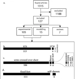
A literature search was performed in the search engine Scopus, using the following keywords (and their synonyms): strateg* (approach, technique, way) AND, sit-to-walk, OR sit-to-stand, OR rise (raise, arise, stand, stand-up) AND chair (seat) (1st October 2019). Inclusion criteria were: biomechanics and motor control on sit-to-stand (STS) or sit-to-walk (STW) in healthy and frail adults (<60y) and elderly (>60y), osteoarthritis patients, and full papers. Non-English articles were excluded. Although the review was targeted at healthy ageing, osteoarthritis patients were also included, as this pathology is highly prevalent (over 50% of adults over 65-year of age (4)) in elderly people and research on this topic is extensive. The reference lists were reviewed for any missing articles from the database search. 1315 articles were found, of which 127 fulfilled the inclusion criteria. These comprised 105 experimental, 7 review, and 15 modelling papers (Figure 1).
6
7 3. STEP 1: Compensation strategies
Step 1 identifies the compensation strategies that have been considered in literature, where compensation strategies are defined as movement alterations to compensate for a lack of capacity or a change in weighting of movement objectives in relation to a baseline, for example a previous state or a control group. The age-related compensation strategies identified for standing up include: trunk movements, arm movements, pacing (related to a stop in between standing and walking), and asymmetry.
Several aspects of these compensation strategies in standing up are poorly reflected in existing research. As the design of an experiment is a trade-off between replication of daily practice to allow for clinical translation and standardization of the protocol to improve repeatability, robustness, and comparability, most prior studies have typically used standardized experimental protocols, restricting aspects of compensation. These experimental protocols facilitate comparison between groups and studies but limits their translation to characterising mobility of the elderly in their homes, communities, and clinics.
3.1 Arm and trunk strategies
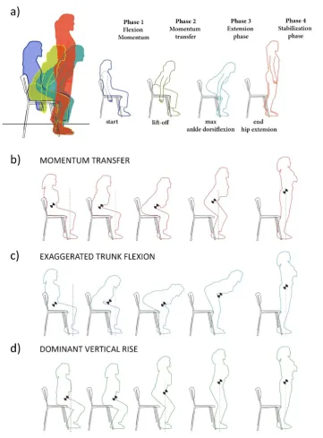
When arm movement is restricted, sit-to-stand strategies are described by four variables: trunk flexion, velocity of the centre of mass (COM), and the distance between the COM and the base of support (BOS). With these variables, three observed strategies have been described: momentum transfer (MT), exaggerated trunk flexion (ETF), and dominant vertical rise (DVR) (Figure 2).
8
and Scarborough et al. found that this strategy was used by 50% (11/22) and 68% (65/95) of their participants, respectively (Table 1).
The ETF strategy is described as an exaggerated trunk flexion prior to lift-off, frequently followed by further trunk flexion that places the COM over the BOS during lift-off and results in delayed trunk extension during the final transition to erect posture (5). This strategy uses little horizontal momentum, relying mostly on the knee musculature to extend the knee. Hughes et al. described this
strategy as the ‘stabilization strategy’ with maximum horizontal COM velocities of less than 7.5 cm/s
and reducing the COM/BOS distance more than 5cm prior to lift-off. This group used several other preparatory movements to position themselves for rising; generally, participants moved the buttocks forward and feet backward, moved the trunk slowly forward and then extending to the standing position. The peak horizontal accelerations of the COM for the ETF strategy are less than half of the MT strategy (7). ETF was used by 18% (4/22) and 17% (16/95) of the participants in the studies of Hughes and Schenkman and Scarborough et al., respectively (Table 1).
The dominant vertical rise (DVR) strategy shows a stagnation of forward trunk flexion immediately at lift-off, followed by dominant vertical COM displacement and knee-hip extension, with trunk extension movement delayed until after knee-hip extension is complete (5). Scarborough et al. reports that 15% (14/95) of their participants used this strategy. The strategy was not described by Hughes and Schenkman (1996); they only described the ‘combined category group’ with participants who did
not fall in the MT or ETF category (32% of the participants; 7/22; Table 1). This combined category group reduced the COM to BOS distance prior to lift-off, but still required momentum to achieve the standing position.
To date, no direct relationship has been found between NMSK capacity and these compensation strategies (5).
9
necessity of using arms (mostly related to Compensation for Capacity), and the preference of using arms (mostly related to Compensation for Movement Objectives). With a seat at knee height almost half of the healthy elderly population (48%) were unable to stand up without the use of arms (8). In a study on osteoarthritis (OA) patients, more than 80% of the participants was unable to stand-up without the use of arms (9). In repetitive trials looking into preferred strategies in standing up, adults use inconsistent strategies over the trials (10). Healthy adults frequently used their arms of which 20% pushed off on the chair, 60% pushed off on the knees, and 50% used an arm swing in one or more trials (11) (Table 1). In OA patients, 83% used an arm push-off in one or more trials (50% of the OA patients used a knee push-off and 42% used a chair push off in one or more trials (11).
These observational studies show that unrestricted arms in experimental protocols would better reflect
10
Figure 2 a) The common way to describe standing-up in four phases (6). Phase 1 - flexion-momentum phase: begins with initiation of the movement and ends just before the buttocks leave the chair (lift-off); note that prior to this visible onset of the STS movement, anticipatory actions can be noted (16,17). The head-arms-trunk segments are the main contributors to the body’s forward propulsion
11
Table 1 Overview of studies categorizing STS and STW strategies while standing up from a chair
Ref. Se
at h e ig h t (% r e fe rs to kn e e h e ig h t)
Task Nu
mb e r o f p ar ti ci p a n ts A ge ( ye ar s) N o ar ms A rm sw in g P u sh o ff ar ms Un ab le to r ise
MT ETF DVR Co. AC AK AR
Hughes and Schenkman (1996)
six heights: 43.2-55 cm
STS 22 72 (65-105) 100% 50% 18% - 32% - - - - Scarborough et al.
(2007)
100% STS 95 MT: 75.34±7.32 ETF: 74.02±6.66 DVR: 75.59±5.62
100% 68% 17% 15% - - - -
Mazza et al. (2004) 80% STS 131 78.1±8 41% .. .. .. .. 13% 13% .. .. .. 33% 100% STS 131 78.1±8 52% .. .. .. .. 8% 18% .. .. .. 22% 120% STS 131 78.1±8 58% .. .. .. .. 12% 27% .. .. .. 3% Komaris et al. (2018) 100%* STW 10 46±7.4 40% .. .. .. .. 50% 70% 20% 60% - -
100%* STW 12 OA:70±5.3 17% .. .. .. .. 58% 83% 42% 50% - - Dolecka et al. (2015)*** 46 cm STS 10 79.5 (69–90) 20% .. .. .. .. .. 80% - 37% 43% -
46 cm** STS 10 79.5 (69–90) 23% 77% 40% 37%
Shown are the percentages of participants that used a specific strategy. In Hughes and Schenkman and Scarborough et al.
the use of arms was restricted (arms were crossed over chest). In Mazza et al. the participants were asked to perform a
sit-to-stand task, without arms, if unable to rise then swinging arms was allowed, if still unable to rise participants could push
using their arms (on thighs or seat). In Komaris et al. an armless seat was used, and there were no restrictions on the use of
arms. Their participants performed several trials, noted is the percentage of participants that used a certain strategy in any
of these trials. MT = Momentum Transfer, ETF = Exaggerated Trunk Flexion, DVR = Dominant Vertical Rise, Co. = Combined;
AC = Arms on seat of chair push-off; AK = Arms through knee push-off; AR = armrest
*Participants performed one to five trials (dependent on their capabilities). If a participant was unable to rise to a standing
position, the chair was re-adjusted to 115% of knee height.
** table in front
*** percentage of trials not participants. Each participant performed three trials.
- this strategy was not allowed in the experiment
12
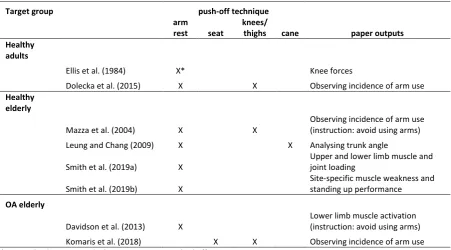
Table 2 Compensation strategies for studies that permitted unrestricted arm movements in sit-to-stand or sit-to-walk.
Target group push-off technique
arm
rest seat
knees/
thighs cane paper outputs
Healthy adults
Ellis et al. (1984) X* Knee forces
Dolecka et al. (2015) X X Observing incidence of arm use
Healthy elderly
Mazza et al. (2004) X X
Observing incidence of arm use (instruction: avoid using arms) Leung and Chang (2009) X X Analysing trunk angle Smith et al. (2019a) X
Upper and lower limb muscle and joint loading
Smith et al. (2019b) X
Site-specific muscle weakness and standing up performance
OA elderly
Davidson et al. (2013) X
13 3.2 Assumed symmetry
The second main compensation strategy identified in the literature is asymmetry. With biomechanical asymmetry we refer to asymmetric movement or force applied by contralateral limbs in any of the three movement planes. In the reviewed studies, bilateral symmetry was often assumed (and constrained) by restricting arm movements and analysing a single plane, usually the sagittal-plane, while there is also movement in the frontal plane (19). Foot positioning of the participants was mostly (111/120) fixed in a symmetrical position (Figure 1), shoulder width apart, with the knee angle at 90° and moving the feet during the trial was restricted. A fixed position of the feet at 90° knee angle influences the COM velocity at seat off (20). Anterior foot placement requires high velocities at seat off, directly followed by a backward deceleration, whereas in posterior foot placement close to the centre of mass, such large velocities are unstable (7,21,22). The sequence of movement also differs with foot placement; with an anterior placement the trunk extends first followed by the hip joint and then the knee joint, whereas posterior placement results in a pattern in which the knee joint extends first, followed by the trunk and the hip joint (21,22). Also muscle recruitment varies with foot position (21,22).
14
The effects and possible benefits of biomechanical asymmetry strategies on joint loading throughout
the body, and potential implications for long-term joint degeneration, have therefore not yet been
quantified. Restricting asymmetry and preferred foot placement in the experimental protocol limits the
available compensation movement strategies of the participants, and therefore may not reflect daily
15 3.3 Restricted pacing: fixed end-goal
Most studies evaluated standing-up with stand as an end-goal (113/120) (Figure 1), rather than STW with walking as an end-goal. However, in daily life people often do not pause between a standing up motion and walking. Typical of sit-to-walk strategies is that the first step is initiated before the body is fully extended, and people consistently use the same foot to initiate swing (16,26). The motor control is also different: STW requires merging of a discrete task (standing up from a chair) with a rhythmic task (walking) and these two tasks overlap near the instant of seat-off. The mechanics of STS and STW are different: the vertical ground reaction force is significantly different between the two feet in the STW task, but not in STS (26). In the anterior-posterior direction both STS and STW show a propulsive impulse followed by a braking impulse during standing-up. However, during the rising phase of STW, this impulse rapidly transitions to propulsion again to initiate walking (26).
With these differences in timing, control and mechanics, the ageing process causes difficulty in merging the rising and walking tasks (16,27). This difficulty in merging implies that a pause in between tasks could be a compensation for the lack of NMSK reserve. No studies were found analysing the absence of merging in relation to reserve, although one study reports that there was no significant effect of the isokinetic strength (muscular capacity) of the knee flexors/extensors and dorsiflexors on the basis of the existence of a sudden stop in between rising and walking in the elderly (18). The lack of fluidity of the motion has been found in stroke patients (28,29) and it has been hypothesized that poor balance is one of the reasons why stroke patients are unable to begin walking fluently from the sitting position (28). Clinical advice is to perform STW in a fluent motion, but in the case of instability caused by dizziness due to neurological or cardiovascular conditions, people may learn to compensate by pausing during the stabilization phase before continuing with walking or other activities (30).
As STS is a different task than STW both in mechanics and control, the lack of research in this latter
movement limits the practical translation of research output to daily life practice. Research should aim
16 4. STEP 2: Compensation for Capacity
Step 1 showed the possible compensation strategies and limitations of experimental protocols. Appreciating these existing limitations, Step 2 in applying the CaReMoOC framework is to capture the current knowledge on Compensation for Capacity. This form of compensation relates to task-performance enhancing recruitment of NMSK resources in response to a relatively high task demand. Compensation for Capacity occurs when (part of the) capacity does not meet the task demands of the lowest cost strategy (global minimum). As a result, the next best strategy will be selected (local minimum).
4.1 Muscular capacity
Muscle strength and power reduces with age, which is one of the contributors to an inability to stand up. Muscle loss is site specific and likely related to daily life activities (Figure 3). Several methods have been used to quantify muscle strength in relation to standing-up: 1) isometric or isokinetic strength tests (9,18,23,31–36), 2) handgrip strength measures (37,38), 3) reducing muscle strength through exercise (39), 4) modelling (40–42), and 5) adding mass to change the load/capacity ratio (43–45). Many studies have found clear relationships between strategies in standing up and isometric and/or isokinetic strength tests. Higher isokinetic strength of the knee extensors was associated with smaller trunk flexion during STW (18), although this relation was not found for the isometric strength of the knee extensors during STS (23). In addition, isometric hip extension strength did not show a correlation with trunk flexion in STS (23), however, when the hip extensor strength was determined by the maximum weight that could be lifted no more than one time with acceptable form, more hip extensor strength was associated with a more upright trunk at lift-off in STS (31). This latter study has its limitations in how the hip strength was derived, so this conclusion may not be as robust.
17
extension phase, the maximum angular velocity of the trunk was higher and the vertical velocity lower for people with lower handgrip strength. However, as relative muscular decline varies between people (31), these correlative results might not be relevant for large numbers of subjects.
Muscle strength reduction has been studied via exercise-induced muscle damage (39). The study showed that the duration of STS and maximum trunk flexion angle increased with reduced muscle strength, and peak ankle dorsiflexion, and knee extension and knee flexion decreased (due to a reduced knee joint range of motion). Also, the vertical ground reaction force decreased after induced muscle damage, which was reflected in the knee and hip moment and power reduction. As muscle damage can also result in pain, compensation strategies found in this study were likely altered to reduce pain as well due to muscle strength reduction.
Some studies studied muscle weakness by adding mass to change the load/capacity ratio (43–45). However, these cannot be directly applied to the change of load/capacity as observed with ageing. Muscle strength capacity in elderly people declines at different rates between muscle groups (31). Therefore, these experimental protocols do not allow for realistic compensation strategies reflective of ageing.
18
of whether introducing larger muscle strength reductions and muscle group specific reductions would have led to the (in)ability of arm-restricted rising of this model.
One study recently analysed the joint and muscle forces of standing up with and without the use of armrests using upper- and lower limb musculoskeletal models in sit-to-stand for young, middle-aged, and older adults (41). Lacking actual strength measurements, the strength profiles of the musculoskeletal models were adjusted based on average values from literature: 25±5% force reduction for the upper limb, and 37±9.7% force reduction for lower limb. The peak glenohumeral joint reaction force while using the armrests was significantly higher in older compared to young adults, despite no difference of the hand reaction forces measured on the armrests. This result could be the result of Compensation by Reorganisation, and/or a difference in shoulder anatomy. Their results confirm that older adults compensate in standing up without arms by reducing the knee joint and extensor loading and increased use of hip extensors and plantarflexors. Another study by the same group showed that selective muscle weakness of the serratus anterior was a key determinant of mobility in STS, by simulating muscle weakness with an upper limb musculoskeletal model (42). By removing muscles from the model one by one, serratus anterior weakness proved to limit the movement most and required most compensatory actions from the other muscles, leading to high upper body joint reaction forces. Since the motion in this model is prescribed (measured kinematics), the model does not allow for compensation by selection (e.g. changing the movement slightly to compensate for the muscle weakness).
19
older adults than in young, which emphasizes the need to further evaluate the upper limb muscle strength in relation to compensation in standing up. Their model indicates that serratus anterior weakness is the most debilitating upper limb muscle weakness for standing up.
Both experimental and modelling studies restricted compensation. Moreover, the studies investigating
effects of muscle strength mostly evaluated (lower-limb) strength as an isolated variable, neglecting
20
Figure 3 : a) The loss of skeletal muscle mass is site-specific and is likely associated with the patterns of muscle
activations that occur in daily life activity (31,48). b) Using a three-dimensional inverse musculoskeletal model, Caruthers et al. (2016) determined the muscle forces and recruitment, and their individual contributions to the body centre of mass (COM) accelerations in the vertical and horizontal direction during STS without arm aid in healthy young adults. The gluteus maximus (GMAX) (hip extension), the quadriceps (specifically the vastus lateralis; knee extension), and the soleus (SOL; ankle plantarflexion) have the highest muscle forces in STS (24). The vertical and forward accelerations are driven by the GMAX, biceps femoris long head, adductor magnus, and the plantarflexors. The vertical lift and stabilization/slow forward motion is controlled by the quadriceps and tibialis anterior (TA). The monoarticular hip and knee extensors shorten while active and thus contribute to the extending moments of these joints, as do the biarticular hamstring muscles (HAM) (49). However, the biarticular rectus femoris (RF) is active but has a very low shortening velocity, acting almost isometrically. It has been proposed that the function of RF demonstrates how muscles can act as tendons, transporting moments from the hip to the knee (50). The gastrocnemius (GAS) muscle may also transfer the moments from the knee to the ankle joint. This makes the co-contraction of the antagonist muscle an efficient way of
21 4.2 Neural capacity
The neural system consists of the (peripheral) nervous system and the sensory feedback system, which can be divided into the visual, auditory, vestibular systems, and proprioception. In the standing-up movement, individual aspects from the neural system have been determined to test their effect on standing up. Age-related changes in the peripheral nervous system and hearing have not been widely studied in complex motor tasks and in the standing-up motion; this review found no studies that incorporated them in their experimental set-up. The effect of neural changes in these parts of the neural system on the compensation in standing-up remains, therefore, unknown.
Hurley et al. (1998) incorporated a measure of proprioception combined with a STW test (stand-up and walk along a level for 15.5m) in young, middle-aged and elderly groups. In the proprioceptive test, participants were asked to reproduce a previously positioned knee angle. Joint position sense in elderly subjects was worse than in the middle-aged and young, and as age increased the acuity decreased. The elderly also took significantly more time to perform the STW test, suggesting that there may be relationship between decline in joint position sense and movement performance in standing up, however there are certainly many other confounding factors, such as loss of muscle strength.
Older adults with poor binocular visual acuity show higher average foot pressures and absolute accelerations of the centre of pressure (indication of variation) during standing up compared to older adults with good visual binocular acuity (55). This result may indicate challenges with balance or a fear of falling in the poor vision acuity group. Fear of falling has previously been associated with poor vision acuity (56). However, no significant differences in standing up kinematics have been found in adults between eyes open and eyes closed conditions (57,58).
Proprioception and visual acuity have been shown to play a role in compensation for stability
22
restricted experimental set-ups, other relevant factors that might interact with the compensation
strategies identified in these studies cannot be ruled out.
4.3 Skeletal capacity
Compensation for skeletal capacity in complex motor tasks is not often considered. Although the changed mechanics of bone might not directly translate to the motor control of complex motor tasks, the consequences of change will affect movement. As the consequences of a fall are more severe in the elderly (increased fracture risk), there is an increased fear of falling. This greater fear results in greater emphasis on stability as a movement objective in the motor control of movement, which will be discussed in Section 5.
A combination of muscle stiffness and skeletal changes results in a reduction of joint range of motion (ROM) with ageing. The age-related reduced hip flexion ROM approaches the maximal angle used in STS (59). Elderly people also locate their feet more forward than young participants, which is more prevalent in elderly with OA than in healthy elderly participants and may be due to a reduced ROM in the knee and/or ankle joint (60).
Wear of the joints, or osteoarthritis (OA), results in pain with loading, which puts more emphasis on pain avoidance in selecting a movement strategy. The most prominent compensation strategy of unilateral knee and hip OA patients is asymmetry of movement (Compensation by Selection) (9,61–
23
with slower knee angular velocities. Moderate knee OA patients try to maintain the same movement trajectory as healthy individuals in standing up, resulting in increased antagonist activation, which is identified as Compensation by Reorganisation (9,34,53). This increased activation may increase knee stability, thus providing relief from pain and discomfort.
Especially in OA patients (which is about 50% of the older population over 65 years), asymmetry,
altered muscle recruitment, and foot positioning play an important role in compensation and should
be incorporated in research studies. Altered trunk flexion strategies, which previously were shown to
be related to muscle weakness, may also be related to pain avoidance in knee OA patients, and to the
24 5. STEP 3: Compensation for Movement Objectives
Step 3 in applying the CaReMoOC framework to the standing up literature is to look at Compensation for Movement Objectives, which relates to the emergence of different movement strategies due to a shift in the weighting of movement objectives. The applied motion strategy of humans considers metabolic energy, speed, safety (e.g. stability margins), and/or pain avoidance. As different strategies assign a different weighting to each of these objectives, so the resulting movement strategies can vary profoundly (compensation) (3). Whilst appreciating that there are limitations in the experiments presented in the literature, this section describes the current knowledge on Compensation for Movement Objectives in standing up.
5.1 Energy
25 5.2 Speed
In some movements and situations, such as in sports, speed is the main movement objective. In contrast, in daily life we rarely think about the speed we move at. Speed is selected unconsciously, probably weighting different movement objectives. Although the self-selected speed of movement in older adults is usually slower than in young adults, older adults can move at a faster velocity when required. In walking, several studies have investigated the relationship between walking velocity, energy expenditure, and self-selected speed (71,72). However, these relationships are less clear for the standing up movement.
As the speed of the standing-up movement increases, the onset and maximum value of vertical momentum increases, whereas the horizontal momentum maintains the same timing and has a disproportionally smaller increase from slow to fast speeds (73,74). Limiting the peak horizontal momentum at different speeds is a movement strategy to maintain equilibrium at the end of the task (standing balance) and was found in both young and older adults (75,76). The quadriceps and erector spinae muscle activate earlier in fast movement conditions, while the tibialis anterior does not change in onset timing. Notably, this might be very different for the STW task, yet no studies have investigated this.
5.2 Stability
Due to a known increased incidence and more severe consequences of a fall, many older adults develop a fear of falling. This fear changes the way they move. In terms of movement objectives, more emphasis is put on stability.
26
progression of the COM into walking (27). This strategy could be an indication of stability considerations or could also be related to the inability to generate this horizontal momentum. Older adults activate all involved ankle plantarflexors prior to the time of seat off, while young adults activate these muscles later (31,41). This difference in muscle activation timing may represent a stabilization strategy in the elderly. The peak magnitude of the muscle activations tends to be greater for older adults than for the young, and significantly different for the rectus femoris (31,51) and the hamstrings (51). These studies together suggest that elderly people activate their muscles at higher levels to accomplish the standing up movement. This higher activation is partly because older adults have strength deficits, and thus the relative effort is higher for older adults in the same movement. In addition, the higher activations in the hamstrings and quadriceps are likely also due to co-contraction, which is thought to increase stiffness and joint stability, but also increase muscle fatigue (51). This is a clear example of Compensation by Reorganisation.
Fallers (elderly people with a history of falling) have different movement strategies in standing-up compared to non-fallers; fallers have a longer rise time and a lower vertical ground reaction force in both STS and STW (79,80). Also, STW fallers demonstrated Compensation by Selection by taking smaller step-lengths at stance-off and a more anterior COM position at lift-off (80). Where non-fallers had a dorsiflexor moment at both lift-off and swing-off in STW, fallers had a plantar-flexor moment. These differences could be either related to reduced dorsiflexor strength in adults with a history of falls (81), or due to the ETF strategy applied by fallers to improve stability compared to the MT strategy used by younger adults (54). In STS, fallers showed Compensation by Reorganisation with an increased activation of the gastrocnemius lateralis. This activation is likely related to a compensation technique to improve stability.
27
slow rapid knee flexion is important to avoid a fall. Although the mechanisms of a fall after standing-up were similar between a grostanding-up of young and older adults, the standing-standing-up strategy was diferent between groups. Older adults had difficulty increasing the vertical momentum of their centre of mass at lift-off with less kinetic energy prior to this instant (76). This difference in strategy may cause elderly to respond with insufficient knee extensor support to avoid a fall (52).
The studies mentioned in this section evaluated standing up strategies without the use of arms. As the
use of arms can support adults in stability (15), more research on this compensation strategy for stability enhancement will be of value for daily life. Also, many of these studies did not look at STW,
yet stability will play a significant role in the control of the transfer between standing and walking.
5.4 Pain
Pain is difficult to quantitively determine in experimental protocols. The most commonly applied methods are the WOMAC questionnaire (33,59,61,63,64) and the visual analogue scale (VAS) (32,61,62,82,83). The presence of pain substantially affects movement strategies. Quantification of pain has been used to categorize participants (59,64), to correlate the pain level to rise-time (33,61), for asymmetries (62,63), and to correlate with the trunk flexion angle (61). Pain avoidance can lead to a compensation long after the pain is mitigated or eliminated in standing up (32). Former stressors or pain in participants might therefore still influence current movement strategies, which the researcher should be aware of when analysing movement strategies in standing up.
Incorporating a pain measure for previous or current experienced pain is therefore advised when
28
6. Discussion and conclusions
An inability to stand-up or falling during standing-up can significantly diminish quality of life. This is the first time that the Capacity, Reserve, Movement Objectives, and Compensation framework (CaReMoOC) has been used to analyse the effect of ageing on an important functional task. Strategies in standing-up are determined by the trunk movements, arm use, and foot positioning. The change in movement strategies (compensation), for example when people start to rely more on their arms, and the onset of the inability to stand-up are associated with clinical conditions such as frailty. The CaReMoOC framework expresses this in Compensation for Capacity and Compensation for Movement Objectives.
Currently, muscle strength is the most widely studied factor in capacity. Most studies however, have looked at lower-limb strength as an isolated factor. These experimental and modelling studies were unable to establish movement limitations but indicated that compensation for lower limb strength was done via greater trunk flexion. Analysing the literature with CaReMoOC indicates that greater trunk flexion is also used for pain compensation in knee OA patients. Moreover, altered trunk flexion is an indicator of compensation for reduced range of motion of the hip flexion. Therefore, the main cause of compensation by trunk flexion is unclear and is likely to be a combination of factors.
29
To investigate the interconnectivity and interdependency of the variables and to possibly predict realistic mobility limitations, predictive neuro-musculoskeletal modelling might shed a light on compensation in the future. Predictive modelling allows for analysis of elements of capacity decline on movement separately (84,85). By changing (site-specific) muscle properties and neural properties of the neuro-musculoskeletal model, and weighting of movement objectives, feasible movement strategies after site-specific or pathological specific decline can be determined. Predictive modelling is not dependent on measured kinematics and therefore allows for Compensation by Selection and Reorganisation. Alternative to controlled complex experiments could also be a big data approach to learn from masses of data.
For experimental protocols, the following should be considered:
• Enabling unrestricted arm movements in experimental protocols would better reflect
daily-life activities. For clinical translation the trunk and upper-limb joint loading for varying arm rise strategies (e.g. armrests, cane, knees, chair) should be determined.
• Sit-to-stand is a different task than sit-to-walk both in mechanics and control; the lack of
research in this latter movement limits the practical translation of research output to daily life practice. Research should aim to close this gap by focussing on the sit-to-walk motion. • Asymmetries in limb trajectories, ground reaction force, and muscle activations are common
in both young and elderly adults. The effects and possible benefits of biomechanical asymmetry strategies on joint loading throughout the body, and potential implications for long-term joint degeneration have yet to be quantified.
• The effect of decline in the neural capacity (for example, sensor and motor noise, nerve
conduction speed, proprioception) on compensation in standing up is largely unexplored and should be studied to gain a better understanding of movement limitations.
• Psychological priority changes, such as due to pain or fear of falling, and previous stressors
30 List of Abbreviations
BOS Base of Support
CaReMoOC General framework describes mechanisms of movement limitations in the context of strict definitions of neuromusculoskeletal capacity, reserve, movement objectives, and compensation and their combinations
COM Centre of Mass
DVR Dominant vertical rise
ETF Exaggerated trunk flexion
MT Momentum Transfer
NMSK Neuro-musculoskeletal
OA Osteoarthritis
ROM Range of Motion
STS Sit-to-stand
STW Sit-to-walk
31 References
1. UN. United Nations. 2019.
2. Rolfson DB, Majumdar SR, Tsuyuki RT, Tahir A, Rockwood K. Validity and reliability of the Edmonton Frail Scale. Age Ageing. 2006;35(5):526–9.
3. van der Kruk E, Silverman AK, Koizia L, Reilly P, Fertleman M, Bull AMJ. CaReMoOC: Capacity, Reserve, Movement Objectives, and Compensation. A new framework to describe
mechanisms of movement limitations, demonstrated for ageing. Eur Rev aging Phys Act. 2019;under revi.
4. Cheng YJ, Hootman JM, Murphy LB, Langmaid GA, Helmich CG. Prevalence of doctor-diagnosed arthritis and arthritis-attributable activity limitation-United States, 2007-2009. Morb Mortal Wkly Rep. 2010;59(39):1261–5.
5. Scarborough DM, McGibbon CA, Krebs DE. Chair rise strategies in older adults with functional limitations. J Rehabil Res Dev [Internet]. 2007;44(1):33. Available from:
http://www.rehab.research.va.gov/jour/07/44/1/pdf/scarborough.pdf
6. Hughes MA, Schenkman ML. Chair rise strategy in the functionally impaired elderly. J Rehabil Res Dev. 1996;33(4):409–12.
7. Shia V, Moore TY, Holmes P, Bajcsy R, Vasudevan R. Stability basin estimates fall risk from observed kinematics, demonstrated on the Sit-to-Stand task. J Biomech [Internet]. 2018;72:37–45. Available from: https://doi.org/10.1016/j.jbiomech.2018.02.022
8. Mazzà C, Benvenuti F, Bimbi C, Stanhope SJ. Association Between Subject Functional Status,
Seat Height, and Movement Strategy in Sit‐to‐Stand Performance. J Am Geriatr Soc.
2004;52(10):1750–4.
32
activation and coactivation during five-time-sit-to-stand movement in patients undergoing total knee arthroplasty. J Electromyogr Kinesiol [Internet]. 2013;23(6):1485–93. Available from: http://dx.doi.org/10.1016/j.jelekin.2013.06.008
10. Dolecka UE, Ownsworth T, Kuys SS. Comparison of sit-to-stand strategies used by older adults and people living with dementia. Arch Gerontol Geriatr [Internet]. 2015;60(3):528–34. Available from: http://dx.doi.org/10.1016/j.archger.2014.12.007
11. Komaris D-S, Govind C, Murphy A, Ewen A, Riches P. Identification of movement strategies during the sit-to-walk movement in patients with knee osteoarthritis. J Appl Biomech. 2018;34(2):96–103.
12. Benvenuti ÃF, Bimbi C, Stanhope SJ, Mazza C. Movement Strategy in Sit-to-Stand Performance. October. 2004;1750–4.
13. Martin J, Garn A, Studies S, Rouge B. Note : This article will be published in a forthcoming
issue of The Sport Psychologist . This article appears here in its accepted , peer-reviewed form , as it was provided by the submitting author . It has not been copy edited , proofed , or formatted by. 2013;
14. Ellis MI, Seedhom BB, Wright V. FORCES IN THE KNEE JOINT WHILST RISING SEATED POSITION. 1984;6.
15. Leung C-Y, Chang C-S. Strategies for Posture Transfer Adopted by Elders during Sit-To-Stand and Stand-To-Sit. Percept Mot Skills [Internet]. 2009;109(3):695–706. Available from: http://journals.sagepub.com/doi/10.2466/pms.109.3.695-706
16. Frykberg GE, Thierfelder T, åberg AC, Halvorsen K, Borg J, Hirschfeld H. Impact of stroke on anterior-posterior force generation prior to seat-off during sit-to-walk. Gait Posture. 2012;35(1):56–60.
33
with hemiparesis post stroke: A review. Ann Phys Rehabil Med [Internet]. 2015;58(3):167–72. Available from: http://dx.doi.org/10.1016/j.rehab.2015.04.007
18. Dehail P, Bestaven E, Muller F, Mallet A, Robert B, Bourdel-Marchasson I, et al. Kinematic and electromyographic analysis of rising from a chair during a ‘“ Sit-to-Walk ”’ task in elderly
subjects : Role of strength. Clin Biomech. 2007;22:1096–103.
19. Gilleard W, Crosbie J, Smith R. Rising to stand from a chair : Symmetry , and frontal and
transverse plane kinematics and kinetics. Gait Posture. 2008;27:8–15.
20. Janssen W, Bussmann H, Stam HJ. Determinants of the Sit-to-Stand Movement : A Review.
2002;82(9):866–79.
21. Kawagoe S, Tajima N, Chosa E. Biomechanical analysis of effects of foot placement with varying chair height on the motion of standing up. J Orthop Sci. 2000;5(2):124–33.
22. Khemlani MM, Carr JH, Crosbie WJ. Muscle synergies and joint linkages in sit-to-stand under two initial foot positions. Clin Biomech. 1999;14(4):236–46.
23. Lundin TM, Jahnigen DW, Grabiner MD. Maximum trunk flexion angle during sit to stand is not determined by knee or trunk-hip extension strength in healthy older adults. J Appl Biomech. 1999;15:233–41.
24. Caruthers EJ, Thompson JA, Chaudhari AMW, Schmitt LC, Best TM, Saul KR, et al. Muscle forces and their contributions to vertical and horizontal acceleration of the center of mass during sit-to-stand transfer in young, healthy adults. J Appl Biomech. 2016;32(5):487–503.
25. Blaszczyk JW, Prince F, Raiche M, Hébert R. Effect of ageing and vision on limb load asymmetry during quiet stance. J Biomech. 2000;33(10):1243–8.
34
27. Buckley T, Pitsikoulis C, Barthelemy E, Hass CJ. Age impairs sit-to-walk motor performance. J Biomech. 2009;42(14):2318–22.
28. Osada Y, Yamamoto S, Fuchi M, Ibayashi S. Sit-to-walk task in hemiplegic stroke patients: relationship between movement fluidity and the motor strategy in initial contact. J Japanese Phys Ther Assoc. 2015;Vol18_002.
29. Dion L, Malouin F, Mcfadyen B, Richards CL. Assessing Mobility and Locomotor Coordination after Stroke with the Rise-to-Walk Task. Neurorehabil Neural Repair. 2003;17(2):83–92.
30. Chan D, Laporte DM, Sveistrup H. art 2: 1999;62(February):64–8.
31. Gross MM, Stevenson PJ, Charette SL, Pyka G, Marcus R. Effect of muscle strength and movement speed on the biomechanics of rising from a chair in healthy elderly and young women. Gait Posture. 1998;8:175–85.
32. Farquhar SJ, Reisman DS, Snyder-Mackler L. Persistence of Altered Movement Patterns During a Sit-to-Stand Task 1 Year Following Unilateral Total Knee Arthroplasty. Phys Ther [Internet]. 2008;88(5):567–79. Available from:
https://academic.oup.com/ptj/article/2742396/Persistence
33. Segal NA, Boyer ER, Wallace R, Torner JC, Yack HJ. Association between chair stand strategy and mobility limitations in older adults with symptomatic knee osteoarthritis. Arch Phys Med Rehabil [Internet]. 2013;94(2):375–83. Available from:
http://dx.doi.org/10.1016/j.apmr.2012.09.026
34. Patsika G, Kellis E, Amiridis IG. Neuromuscular efficiency during sit to stand movement in women with knee osteoarthritis. J Electromyogr Kinesiol [Internet]. 2011;21(5):689–94. Available from: http://dx.doi.org/10.1016/j.jelekin.2011.05.006
35
36. Schot PK, Knutzen KM, Poole SM, Mrotek LA. Sit-to-stand performance of older adults following strength training. Res Q Exerc Sport. 2003;74(1):1–8.
37. van Lummel RC, Evers J, Niessen M, Beek PJ, van Dieën JH. Older adults with weaker muscle strength stand up from a sitting position with more dynamic trunk use. Sensors (Switzerland). 2018;18(4):1–12.
38. Sales M, Levinger P, Polman R. Relationships between self perceptions and physical activity behaviour, fear of falling, and physical function among older adults. Eur Rev aging Phys Act. 2017;14(1):17.
39. Spyropoulos G, Tsatalas T, Tsaopoulos DE, Sideris V, Giakas G. Biomechanics of sit-to-stand transition after muscle damage. Gait Posture [Internet]. 2013;38(1):62–7. Available from: http://dx.doi.org/10.1016/j.gaitpost.2012.10.013
40. Bobbert MF, Kistemaker DA, Aurélio M, Ackermann M. Searching for strategies to reduce the mechanical demands of the sit-to-stand task with a muscle-actuated optimal control model. Clin Biomech. 2016;37:83–90.
41. Smith SHL, Reilly P, Bull AMJ. A musculoskeletal modelling approach to explain sit-to-stand difficulties in older people due to changes in muscle recruitment and movement strategies. J Biomech. 2019;109451.
42. Smith S, Reilly P, Bull A. Serratus Anterior weakness is a key determinant of arm-assisted standing difficulties. Med Eng Phys. 2019;
43. Savelberg HHCM, Fastenau A, Willems PJB, Meijer K. The load/capacity ratio affects the sit-to-stand movement strategy. Clin Biomech. 2007;22(7):805–12.
44. Van der heijden MMP, Meijer K, Willems PJB, Savelberg HHCM. Muscles limiting the sit-to-stand movement. An experimental simulation of muscle weakness. Gait Posture.
36
45. Walaszek MC, Ransom AL, Capehart S, Pohl MB, Shapiro R, Bollinger LM. External loading alters trunk kinematics and lower extremity muscle activity in a distribution-specific manner during sitting and rising from a chair. J Electromyogr Kinesiol. 2017;34:102–8.
46. Sallinen J, Stenholm S, Rantanen T, Heliövaara M, Sainio P, Koskinen S. Hand‐grip strength cut
points to screen older persons at risk for mobility limitation. J Am Geriatr Soc. 2010;58(9):1721–6.
47. Bajelan S, Azghani MR. Musculoskeletal modeling and simulation of three various Sit-to-Stand
strategies : An evaluation of the biomechanical effects of the chair-rise strategy modification.
2014;22:627–44.
48. Abe T, Sakamaki M, Yasuda T, Bemben MG, Kondo M, Kawakami Y, et al. Age-related, site-specific muscle loss in 1507 Japanese men and women aged 20 to 95 years. J Sports Sci Med. 2011;10(1):145.
49. Roebroeck ME, Doorenbosch CAM, Harlaar J, Jacobs R, Lankhorst GJ. Biomechanics and muscular activity during sit-to-stand transfer. Clin Biomech. 1994;9(4):235–44.
50. Van Ingen Schenau GJ. On the action of bi-articular muscles, a review. Netherlands J Zool. 1989;40(3):521–43.
51. Hurley M V, Rees J, Newham DJ. Quadriceps function, proprioceptive acuity and functional performance in healthy young, middle-aged and elderly subjects. Age Ageing. 1998;27(1):55–
62.
52. Pai YC, Yang F, Wening JD, Pavol MJ. Mechanisms of limb collapse following a slip among young and older adults. J Biomech. 2006;39(12):2194–204.
53. Bouchouras G, Patsika G, Hatzitaki V, Kellis E. Kinematics and knee muscle activation during sit-to-stand movement in women with knee osteoarthritis. Clin Biomech. 2015;30(6):599–
37
54. Chen T, Chou LS. Altered center of mass control during sit-to-walk in elderly adults with and without history of falling. Gait Posture [Internet]. 2013;38(4):696–701. Available from: http://dx.doi.org/10.1016/j.gaitpost.2013.03.007
55. Shin S-S, An D-H, Yoo W-G. Comparison of Foot Pressure and Center of Force During Sit-to-Stand and Sit-to-Stand-to-Sit Movements in Older Adults With Good and Poor Visual Acuity. Top Geriatr Rehabil. 2018;34(1):82–6.
56. Aartolahti E, Häkkinen A, Lönnroos E, Kautiainen H, Sulkava R, Hartikainen S. Relationship between functional vision and balance and mobility performance in community-dwelling older adults. Aging Clin Exp Res. 2013;25(5):545–52.
57. Assaiante C, Chabeauti PY, Sveistrup H, Vaugoyeau M. Updating process of internal model of action as assessed from motor and postural strategies in young adults. Hum Mov Sci
[Internet]. 2011;30(2):227–37. Available from: http://dx.doi.org/10.1016/j.humov.2010.05.007
58. Tajali S, Negahban H, Shaterzadeh MJ, Mehravar M, Salehi R, Narimani R, et al. Multijoint Coordination During Sit-To-Stand Task in People With Non-Specific Chronic Low Back Pain. Biomed Eng Appl Basis Commun [Internet]. 2013;25(01):1350010. Available from:
http://www.worldscientific.com/doi/abs/10.4015/S1016237213500105
59. Fotoohabadi MR, Tully EA, Galea MP. Kinematics of Rising From a Chair : Image-Based Analysis of the Sagittal Elderly People Who Are Healthy. Phys Ther. 2010;90(4):561–71.
60. Papa E, Cappozzo A. Sit-to-stand motor strategies investigated in able-bodied young and elderly subjects. J Biomech. 2000;33(9):1113–22.
61. Turcot K, Armand S, Fritschy D, Hoffmeyer P, Suvà D. Sit-to-stand alterations in advanced knee osteoarthritis. Gait Posture. 2012;36(1):68–72.
38
sensitive and responsive measure of functional compensations in individuals with knee osteoarthritis performing the five times sit-to-stand test. Gait Posture. 2018;62:140–5.
63. Eitzen I, Fernandes L, Nordsletten L, Snyder-Mackler L, Risberg MA. Weight-bearing asymmetries during Sit-To-Stand in patients with mild-to-moderate hip osteoarthritis. Gait Posture [Internet]. 2014;39(2):683–8. Available from:
http://dx.doi.org/10.1016/j.gaitpost.2013.09.010
64. Lamontagne M, Beaulieu ML, Varin D, Beaulé PE. Lower-limb joint mechanics after total hip arthroplasty during sitting and standing tasks. J Orthop Res. 2012;30(10):1611–7.
65. Sagawa Y, Bonnefoy-Mazure A, Armand S, Hoffmeyer P, Suva D, Turcot K. Individuals with knee osteoarthritis exhibit altered movement patterns during the sit-to-stand task. Mov Sport Sci - Sci Mot [Internet]. 2017;(98):39–49. Available from:
http://ezproxy.ithaca.edu:2048/login?qurl=http%3A%2F%2Fsearch.ebscohost.com%2Flogin.a
spx%3Fdirect%3Dtrue%26db%3Ds3h%26AN%3D128199052%26site%3Dehost-live%26scope%3Dsite%0Ahttps://www.mov-sport-sciences.org/10.1051/sm/2017004
66. Nakagata T, Yamada Y, Hatamoto Y, Naito H. Energy Expenditure of a Single Sit-to-Stand Movement with Slow Versus Normal Speed Using the Different Frequency Accumulation Method. Medicina (B Aires). 2019;55(3):77.
67. Van Der Kruk E, Van Der Helm FCT, Veeger HEJ, Schwab AL. Power in Sports: a literature review on the application, assumptions, and terminology of mechanical power in sport research. Artic J Biomech [Internet]. 2018; Available from:
https://doi.org/10.1016/j.jbiomech.2018.08.031
68. Kotake T, Miura T, Kajiwara T. An Analysis of Sit-to-Stand Movements. 1993;74(October):1095–9.
39
joint angle and angular velocity: model development and application to the lower limb. J Biomech. 2007;40(14):3105–13.
70. Bieryla KA, Anderson DE, Madigan ML. Estimations of relative effort during sit-to-stand increase when accounting for variations in maximum voluntary torque with joint angle and angular velocity. J Electromyogr Kinesiol. 2009;19(1):139–44.
71. Malatesta D, Simar D, Dauvilliers Y, Candau R, Borrani F, Préfaut C, et al. Energy cost of walking and gait instability in healthy 65-and 80-yr-olds. J Appl Physiol. 2003;95(6):2248–56.
72. Abadi FH, Muhamad TA, Salamuddin N. Energy expenditure through walking: Meta analysis on gender and age. Procedia-Social Behav Sci. 2010;7:512–21.
73. Pai YC, Rogers MW. Control of body mass transfer as a function of speed of ascent in sit-to-stand. Med Sci Sports Exerc. 1990;22(3):378–84.
74. Vander Linden DW, Brunt D, McCulloch MU. Variant and invariant characteristics of the sit-to-stand task in healthy elderly adults. Arch Phys Med Rehabil. 1994;75(6):653–60.
75. Pai Y-C, Naughton BJ, Chang RW, Rogers MW. Control of body centre of mass momentum during sit-to-stand among young and elderly adults. Gait Posture. 1994;2(2):109–16.
76. Pai YC, Lee WA. Effect of a terminal constraint on control of balance during sit-to-stand. J Mot Behav. 1994;26(3):247–56.
77. Pavol MJ, Pai YC. Feedforward adaptations are used to compensate for a potential loss of balance. Exp Brain Res. 2002;145(4):528–38.
78. Hsiao ET, Robinovitch SN. Common protective movements govern unexpected falls from standing height. J Biomech. 1997;31(1):1–9.
40 2016;28(5):871–9.
80. Chen T, Chang CC, Chou LS. Sagittal plane center of mass movement strategy and joint kinetics during sit-to-walk in elderly fallers. Clin Biomech [Internet]. 2013;28(7):807–12. Available from: http://dx.doi.org/10.1016/j.clinbiomech.2013.07.002
81. Fukagawa NK, Wolfson L, Judge J, Whipple R, King M. Strength is a major factor in balance, gait, and the occurrence of falls. Journals Gerontol Ser A Biol Sci Med Sci.
1995;50(Special_Issue):64–7.
82. Shafizadeh M. Movement coordination during sit-to-stand in low back pain people. Hum Mov. 2016;17(2):107–11.
83. Shum GLK, Crosbie J, Lee RYW. Three-dimensional kinetics of the lumbar spine and hips in low back pain patients during sit-to-stand and stand-to-sit. Spine (Phila Pa 1976).
2007;32(7):211–9.
84. Geijtenbeek T. Predictive simulation of human locomotion. 2016.