University of Pennsylvania
ScholarlyCommons
Publicly Accessible Penn Dissertations
1-1-2013
Plasma Proteins and Their interaction With
Synthetic Polymers at the Air-Water interface
Zhengzheng LiaoUniversity of Pennsylvania, [email protected]
Follow this and additional works at:http://repository.upenn.edu/edissertations
Part of thePhysical Chemistry Commons
This paper is posted at ScholarlyCommons.http://repository.upenn.edu/edissertations/774
For more information, please [email protected]. Recommended Citation
Liao, Zhengzheng, "Plasma Proteins and Their interaction With Synthetic Polymers at the Air-Water interface" (2013).Publicly Accessible Penn Dissertations. 774.
Plasma Proteins and Their interaction With Synthetic Polymers at the
Air-Water interface
Abstract
The adsorption of proteins and synthetic polymers at the air-water interface (AWI) has broad significance in biomedicine and biotechnology. Protein behavior at the AWI can be guided to control the structure of the two-dimensional biopolymer film. In addition, synthetic polymers affect how plasma proteins act on implant or drug carrier surfaces, and can also decrease the potential for gas embolism. In this thesis, fluorescence microscopy was applied in combination with tensiometry and atomic force microscopy to study plasma proteins at the AWI alone and under the effect of synthetic polymers. First, the morphology of serum albumin layer controlled by the solution conditions was explored by fluorescence microscopy. Heterogeneity at the micron scale was observed for the protein film during adsorption and at reduced concentrations. Moreover, the competition for interfacial area between Pluronic surfactant F-127 and fibrinogen or serum albumin was studied by semi-quantitative confocal fluorescence methods. A transition stage where F-127 and protein underwent lateral phase separation was found. Two competing processes were revealed--the disintegration of protein-rich phase by F-127 and the coalescence of protein phase. Lastly, the interaction of immunoglobulin and the thin film of a widely used polydimethylsiloxane synthetic polymer was studied at the AWI. The compression state of the polymer film was shown to significantly affect protein adsorption and guide proteins into circular domain structures. Overall, this work has demonstrated the wide utility of fluorescence
microscopy to study protein-protein and protein-synthetic polymer interactions at the AWI.
Degree Type Dissertation
Degree Name
Doctor of Philosophy (PhD)
Graduate Group Chemistry
First Advisor Ivan J. Dmochowski
Keywords
Air-water interface, Confocal microscopy, Plasma protein, Polymer, Self-assembly
Subject Categories
Chemistry | Physical Chemistry
PLASMA PROTEINS AND THEIR INTERACTION WITH
SYNTHETIC POLYMERS AT THE AIR-WATER
INTERFACE
Zhengzheng Liao
A DISSERTATION
in
Chemistry
Presented to the Faculties of the University of Pennsylvania
in
Partial Fulfillment of the Requirements for the
Degree of Doctor of Philosophy
2013
Supervisor of Dissertation
________________________
Dr. Ivan J. Dmochowski, Associate Professor of Chemistry
Graduate Group Chairperson
_________________________
Dr. Gary A. Molander, Professor of Chemistry
Dissertation Committee
Dr. Feng Gai, Professor of Chemistry
Dr. Tobias Baumgart, Associate Professor of Chemistry
ii
Dedicated to
my Grandpa
Mr. Junchang Liao
1931-2010
iii
ACKNOWLEDGEMENT
First of all, I would like to thank my advisor Prof. Ivan Dmochowski for his guidance
and support throughout my grad career. I joined Prof. Dmochowski’s group as a fresh
college graduate with limited experience in research, and Prof. Dmochowski was always
encouraging whenever I showed him data or presented him a paper that I found
interesting. His encouragement has been a motivation for me to continue the exploration
in the ups and downs in my research. Under his influence, I had the opportunity to be
exposed to different scientific fields and had collaborations with many people from
diverse backgrounds, which greatly expanded my horizon and sharpened my skills.
Furthermore, I want to express my appreciation to my thesis committee
members--Prof. Feng Gai, members--Prof. Tobias Baumgart and members--Prof. Barry Cooperman. They generously
provided advice and criticism of my work, which prompted me to be a better researcher.
I’m especially thankful to Prof. Baumgart, with whom I collaborated for the
PDMS/protein and the SH3/PRM project. His great prudence and dedication to science
have taught me much through the collaboration.
Moreover, I would like to express my gratitude to Prof. David Eckmann, Prof.
Portonovo Ayyaswamy, Prof. Roderic Eckenhoff and Prof. Daniel Hammer for the
guidance on my other collaborative projects. I’m also grateful to Prof. Zahra Fakhraai,
who was generous with her time in discussing ellipsometry. It has been an honor to work
iv
I have worked with many graduate students and postdocs at Penn, and I thank them
all sincerely. I am sorry if I miss any names in the following list. Dr. Joshua Lampe from
Prof. Eckmann’s lab was my first lab mentor and passed the torch of the air-water
interface project to me. Dr. Xinjing Tang taught me many important experimental
techniques including confocal microscopy, which became perhaps the most important
part of my skill sets. Dr. Neha Kamat was a talented researcher from whom I learned a lot
and with whom I absolutely enjoyed working. Dr. Ashley Fiamengo, Daniel Emerson,
John Psonis, Breanna Caltagarone and James Hui collaborated with me on the interesting
and challenging fluorescent anesthetic project. Thanks to their intellectual contributions, I
have learned much more about the mechanism of anesthetics than I knew five years ago.
Dr. Wan-Ting Hsieh is one of my best friends and worked closely with me on both the
PDMS/protein project and SH3/PRM project. I learned from her the hardworking attitude
and precious patience in doing research. Dr. Brittani Ruble and Sean Yeldell brought me
into the TIVA-folate project in my last few months, which further enriched my research
experience. Besides, a lot of people have helped me in research through many ways; I
want to thank Yu-Hsiu Wang, Dr. Xi-Jun Chen, Helen Cativo, Zhaoxia Qian, Ethan Glor
for their assistance in my experiments, and the staff of the Chemistry dDepartment for
their support. Also, I would like to thank all the past and present members of the
Dmochowski group. It has been a pleasure to be labmates with them.
I am grateful to all my friends at Penn, without whom I would have never made it this
far. During grad school, I was extremely lucky to get to know a lot of friends. I know that
v
thank Jasmina Cheung-Lau, Chih-Jung Hsu, Xiaojing Liu, Wan-Ting Hsieh, Yu-Hsiu
Wang, Keymo Chen, Claire Hsieh, Tingting Wu, Zheng Shi, Chun-Wei Lin, Jin Park,
Min Pan, Yi-Ru Chen and Yi-Chi Lin. These people are not only my friends who I grow
and learn with, but also essentially my family here in Philadelphia who generously have
provided me company and support throughout the years. I also want to thank Zi Liang
and Na Liu, who are always willing to lend an ear or travel hundreds of miles for me. We
have been as close as sisters for about ten years and they are also part of my family in the
U.S.
Finally, I would like to thank my family. My grandma’s optimism and resilience after
the passing of my grandpa has inspired me to be persistent in my pursuit. My parents,
Zhonghong Liao and Huagui Zhang, are my ultimate support both financially and
emotionally. They have given me unconditional love and trust, so that I can always find
strength in adversity when I think of them. With that strength, I will continue on the
vi ABSTRACT
PLASMA PROTEINS AND THEIR INTERACTION WITH
SYNTHETIC POLYMERS AT THE AIR-WATER INTERFACE
Zhengzheng Liao
Dr. Ivan J. Dmochowski
The adsorption of proteins and synthetic polymers at the air-water interface (AWI) has
broad significance in biomedicine and biotechnology. Protein behavior at the AWI can be
guided to control the structure of the two-dimensional biopolymer film. In addition,
synthetic polymers affect how plasma proteins act on implant or drug carrier surfaces,
and can also decrease the potential for gas embolism. In this thesis, fluorescence
microscopy was applied in combination with tensiometry and atomic force microscopy to
study plasma proteins at the AWI alone and under the effect of synthetic polymers. First,
the morphology of serum albumin layer controlled by the solution conditions was
explored by fluorescence microscopy. Heterogeneity at the micron scale was observed for
the protein film during adsorption and at reduced concentrations. Moreover, the
competition for interfacial area between Pluronic surfactant F-127 and fibrinogen or
serum albumin was studied by semi-quantitative confocal fluorescence methods. A
transition stage where F-127 and protein underwent lateral phase separation was found.
Two competing processes were revealed—the disintegration of protein-rich phase by
F-127 and the coalescence of protein phase. Lastly, the interaction of immunoglobulin and
the thin film of a widely used polydimethylsiloxane synthetic polymer was studied at the
vii
protein adsorption and guide proteins into circular domain structures. Overall, this work
has demonstrated the wide utility of fluorescence microscopy to study protein-protein and
viii
TABLE OF CONTENTS
ACKNOWLEDGEMENT ... III
ABSTRACT ... VI
TABLE OF CONTENTS ... VIII
LIST OF TABLES ... XIII
LIST OF FIGURES ... XIII
CHAPTER 1 INTRODUCTION TO PROTEINS AT THE AIR-WATER
INTERFACE ... 1
I. Proteins at the air-water interface play crucial roles in many scenarios ... 2
a. Biological and technological relevance of proteins at the AWI ... 2
b. Plasma proteins ... 3
II. Experimental methods to study proteins at the air-water interface ... 4
a. Tensiometry and Langmuir trough ... 4
b. Fluorescence microscopy ... 10
c. Atomic force microscopy ... 12
d. Other methods ... 12
III. Protein behaviors at the air-water interface ... 14
a. Adsorption, conformation and structure of protein assembly at the AWI ... 14
ix
c. Interaction with polymers ... 21
IV. Research scope ... 22
V. References ... 22
CHAPTER 2 MORPHOLOGY OF PROTEIN SELF-ASSEMBLY AT THE AIR-WATER INTERFACE CONTROLLED BY SOLUTION CONDITIONS—A STUDY BY CONFOCAL LASER SCANNING MICROSCOPY ... 34
I. Introduction... 34
II. Material and experimental methods ... 37
a. General reagents ... 37
b. Protein labeling ... 37
c. Dye-to-protein ratio (DPR) measurements ... 38
d. MALDI-TOF MS of HSA and HSA-TR... 38
e. Circular dichroism (CD) measurement ... 39
f. Surface pressure measurement in LB trough and film transfer... 40
g. AFM characterization of transferred film ... 40
h. Sample chamber fabrication and surface modification ... 41
i. Confocal and epi-fluorescence imaging. ... 42
III. Results and discussion ... 42
a. Characterization of HSA-TR in comparison with HSA ... 42
b. Effect of surface modification of imaging chamber with PEG ... 49
c. Imaging protein assembly at the AWI by CLSM ... 52
d. Protein assembly controlled by ionic strength and reducing agent ... 55
x
V. References ... 60
CHAPTER 3 SEMI-QUANTITATIVE CONFOCAL LASER SCANNING MICROSCOPY METHOD TO STUDY PLASMA PROTEIN ADSORPTION IN COMPETITION WITH PLURONIC F-127 AT THE AIR-WATER INTERFACE ... 66
I. Introduction... 66
II. Material and experimental setup ... 68
a. General reagents ... 68
b. CLSM setup ... 68
III. Results and discussion ... 69
a. Methodology ... 69
b. HSA-TR/F-127 adsorption competition at AWI ... 76
c. FRAP experiments of HSA-TR/F-127 mixtures ... 81
d. Dynamics of the phase behavior in HSA-TR/F-127 layer ... 84
e. Validation and limitation of partition coefficient ... 87
IV. Conclusions ... 93
V. References ... 93
CHAPTER 4 MEASURING INTERACTIONS BETWEEN POLYDIMETHYLSILOXANE AND SERUM PROTEINS AT THE AIR-WATER INTERFACE ... 98
xi
II. Material and experimental methods ... 100
a. Reagents ... 100
b. MALDI-TOF Mass Spectrometry ... 101
c. Langmuir-Blodgett Trough Experiment ... 102
d. Fluorescence Imaging ... 102
e. Fluorimetry ... 103
f. π-Csurf Isotherm Measurement ... 103
g. Titration Experiment ... 104
h. Film Transfer onto Cover Glass ... 104
i. Pt-C Replication and SEM Imaging... 105
j. AFM Imaging ... 105
III. Results and Discussion ... 106
a. Leaching of PDMS oligomers at acidic conditions ... 106
b. Surface Pressure-Surface Concentration Isotherms of PDMS and IgG-TR in Langmuir Trough. 110 c. Circular Domains under Fluorescence Microscopy in Langmuir Monolayer. ... 114
d. Titration of IgG-TR into the Subphase. ... 118
e. Characterization of the Micro-structure of Region IV Domains. ... 123
IV. Conclusions ... 128
V. References ... 128
CHAPTER 5 CONCLUSIONS AND FUTURE DIRECTIONS ... 134
I. Two-dimensional domains formed by multivalent interaction between two proteins at the lipid-water interface ... 135
xii
III. Imaging fluorescent anesthetics aminoanthracene and photoactive analogue
1-azidoanthracene in vitro and in vivo ... 151
IV. References ... 160
APPENDIX ... 163
I. Matlab code for calculating partition coefficient ... 163
II. Experimental steps of photoactivated immobilization of tadpoles by 1-azidoanthracene ... 165
xiii
LIST OF TABLES
Table 1.1 Crystal structures and geometric dimensions of serum albumin, fibrinogen and
IgG. ... 19
Table 3.1 concentration of F-127 and molar ratio of HSA-TR/F-127 corresponding to Figure 3.6 ... 80
Table 3.2 CLSM settings used to investigate fluorescence intensity... 91
LIST OF FIGURES
Figure 1.1 Interaction between water molecules at the air-water interface causes surface tension. ... 7Figure 1.2 Definition of surface excess. ... 8
Figure 1.3 Comparison of π-A isotherms of lipid and protein at the AWI. ... 9
Figure 1.4 Vertical imaging of molecules at the AWI by CLSM. ... 11
Figure 1.5 AFM images of interfacial film of BSA or β-casein transferred onto mica. ... 17
Figure 2.1 MALDI-MS spectra of HSA and HSA-TR ... 45
Figure 2.2 Characterization of HSA and HSA-TR by CD spectroscopy ... 46
Figure 2.3 Surface pressure of HSA and HSA-TR ... 47
Figure 2.4 AFM images of HSA and HSA-TR films formed at the AWI and transferred onto mica surface ... 48
xiv
Figure 2.6 Confocal fluorescence images of HSA-TR in sample chamber before and after
surface modification... 51
Figure 2.7 XY and XZ plane fluorescent images of protein assembly at the AWI ... 54
Figure 2.8 Morphology of protein assembly varied with ionic strength and addition of reducing agent ... 58
Figure 2.9 Epi-fluorescence images showing transition of heterogeneous domains into homogeneous layer. ... 59
Figure 3.1 Experimental setup and data analysis of the droplet system to study protein adsorption at the AWI. ... 73
Figure 3.2 Intensity profiles of F-127 and HFib-OG mixtures show that F-127 blocks protein adsorption. ... 74
Figure 3.3 Analysis of an XZ plane image to obtain partition coefficient. ... 75
Figure 3.4 Partition coefficient of HSA-TR as a function of bulk concentration. ... 78
Figure 3.5 Change of partition coefficient P with increase in F-127 concentration. ... 79
Figure 3.6 Phase separation in adsorbed HSA/F-127 layer at the AWI. ... 80
Figure 3.7 FRAP experiment of the mixed HSA-TR and F-127 interfacial layer. ... 83
Figure 3.8 FRAP experiment of the HSA-TR interfacial layer. ... 83
Figure 3.9 Coalescence of protein islands over time. ... 86
Figure 3.10 Surface pressures of proteins vs. dye-labeled proteins at 0.01 mg/mL. ... 90
Figure 3.11 Correlation between fluorescence intensity and dye-labeled protein concentration. ... 91
xv
Figure 4.1 Domains observed at the air-water interface with dye-labeled serum proteins.
... 108
Figure 4.2 MALDI-TOF mass spectrum of extracted residues from PDMS elastomer by toluene. ... 109
Figure 4.3 π-Csurf isotherms of PDMS and PDMS + IgG-TR. ... 113
Figure 4.4 Fluorescence microscopy study of PDMS + IgG-TR film at the air-water interface during compression. ... 116
Figure 4.5 Fluorescence intensity of IgG-TR at different concentrations in phosphate buffer and in silicone oil. ... 117
Figure 4.6 Fluorescence microscopy study of PDMS-only film and PDMS + IgG-TR film at the air-water interface. ... 120
Figure 4.7 Titration of IgG-TR into the subphase underneath PDMS layer at the A/W interface... 121
Figure 4.8 Fluorescence microscopy images of PDMS film (region IV) stained with BODIPY or Rhodamine 101. ... 122
Figure 4.9 Characterization of transferred films on cover glass substrate. ... 126
Figure 4.10 AFM height images and height analysis of Pt-C replicas. ... 127
Figure 5.1 A cartoon showing the side-view of the experimental setup with lipid monolayer and protein mixtures. ... 139
Figure 5.2 PRM5 and SH35 protein domains observed in Langmuir monolayer. ... 140
Figure 5.3 Reversible change of domain morphology. ... 141
xvi
Figure 5.5 Porphyrin-based fluorophore emission showed spectral shift in different
environment. ... 147
Figure 5.6 Unmixed spectra (upper row) and overlay of pseudo-color images (lower row) of a vesicle before and after photo-activated rupture. ... 148
Figure 5.7 Emission spectra of PZn2 identified by spectral imaging during micropipette aspiration. ... 149
Figure 5.8 Uptake of PZn2 by macrophage. ... 150
Figure 5.9 Spectral imaging of 1-AMA in mouse neural progenitor cells. ... 154
Figure 5.10 Structure and photochemical properties of 1-AZA. ... 155
Figure 5.11 Time course comparison of immobilization effects of 1-AMA and 1-AZA on X. laevis tadpoles. ... 156
Figure 5.12 Fluorescence image of 1-AZA in C. elegans before and after photo-activation. ... 157
Figure 5.13 In vivo imaging of photo-activated 1-AZA in X. laevis tadpoles. ... 158
1
Chapter 1
Introduction to proteins at the
air-water interface
Proteins are macromolecules composed of polypeptide chains. Because different amino
acid side chains have different hydrophobicity (e.g., tryptophan and phenylalanine are
more hydrophobic than threonine and serine),1 after proper folding of the protein, the
hydrophobic and hydrophilic regions are formed, giving the protein its amphiphilic nature.
Post-translational modifications such as lipidation or phosphorylation further change the
amphiphilicity of proteins. Like other amphiphiles, proteins can adsorb at many
interfaces and self-assemble into higher-order structures to reduce the surface energy. In
the case of protein adsorption at the air-water interface (AWI), protein assembly serves to
exclude water molecules from the hydrophobic regions. Exploring how protein behaves
at this fundamental and widely presented interface is the focus of this thesis. In this
chapter, the motivation for studying proteins at the AWI will be explained in section I.
Then a brief description of the experimental techniques used in this thesis and described
in the scientific publications will be presented in section II, which provides a foundation
for reviewing protein behavior at the AWI as reported in the literature in section III.
2
I. Proteins at the air-water interface play crucial roles in many
scenarios
a. Biological and technological relevance of proteins at the AWI
The surface properties of proteins are of general interest in material science, food
science and medical science. Much attention has been focused on the proteins
partitioning at the lipid-water interface in order to understand the mechanism of signal
transduction and material transportation in and out of cells.2,3 Meanwhile, protein
adsorption at the solid-liquid interface has also been a popular research topic due to the
desire of developing more biocompatible materials for implants and drug delivery.4,5 In
addition to all the foregoing, proteins at the AWI is an important system that not only
serves as the basis to understand more complicated lipid-water or solid-liquid interface,
but also has direct applications to various areas.
In material science, for example, ranaspumin identified from frog nest foams has been
found to adsorb at the AWI and play a crucial role in forming foams composed of 90%
air and 10% liquid, with excellent mechanical stability.6 This bio-foam system has
potential application as a biochemical reactor in the micro- and nano- scale.7 The spider
silk proteins and their self-assembled structure at the AWI have been studied in order to
understand how these proteins assemble into filaments of exceptional elasticity and
extensibility.8 Block copolymers sharing characteristics of the silk proteins have been
designed and synthesized, which are shown to form viscoelastic thin films at the AWI.8,9
Hydrophobin, which is a protein secreted by filamentous fungi, is one of the best studied
3
biocompatibility of implant materials by surface modification.10 In food science, the milk
proteins β-casein, β-lactoglobulin and their interaction with surfactants have been
investigated because they are essential in the stability of the foam in dairy products, by
adsorbing at the air-water or oil-water interface.11,12 In medical science, lung surfactant
(also called pulmonary surfactant), which is a mixture of phospholipoprotein and lipids,
lines the AWI of the alveoli to reduce surface tension. Deficiency of lung surfactant leads
to the collapse of the alveoli which could further cause respiratory failure.13
b. Plasma proteins
Besides the above mentioned proteins, plasma proteins and their interaction with other
molecules at the AWI have also drawn attention. There are at least 289 plasma proteins
detected in blood plasma or serum.14 In terms of concentration, serum albumin,
fibrinogen and immunoglobulin G are among the most abundant species in human
plasma.14 Plasma proteins’ surface properties are of particular interest to researchers due
to the following reasons: 1) the behavior of plasma proteins at the AWI provides the
fundamentals, to help elucidate the activities of plasma proteins at solid surfaces without
the complication of surface charge and morphology of solid surfaces;15 2) during air
embolism, the plasma proteins lining the air bubble surface were tied to air embolism
pathogenesis, thus intervention or manipulation of such processes requires a greater
understanding of the composition and structure of the film formed by plasma proteins at
the AWI;16,17 3) with the increasing interest of fabricating antibody based sensors in
biotechnology, understanding the surface properties of immunoglobulin could aid the
4
the study of the plasma proteins serum albumin, fibrinogen and immunoglobulin G at the
AWI.
II. Experimental methods to study proteins at the air-water
interface
a. Tensiometry and Langmuir trough
Tensiometry is one of the most commonly used methods to study surface properties of
amphiphiles. It measures the surface tension (γ) of air-liquid interface. Surface tension is
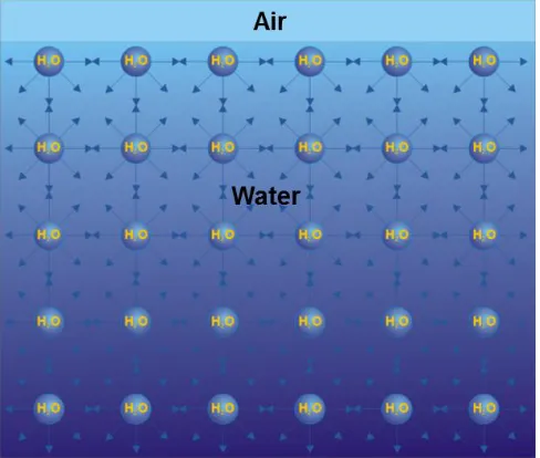
a force parallel to the interface caused by the intermolecular forces between solvent
molecules (Figure 1.1). Surface tension has the unit of Newton per meter (N/m), and
tends to shrink the surface area of liquid in order to decrease the surface energy. The
AWI has the highest surface tension in nature because of strong hydrogen bonding. When
amphiphiles adsorb at the interface, the hydrogen bonds between water molecules are
disrupted, therefore causing a decrease of γ. The dependency of γ on the concentration of
surfactants is described by the Gibbs isotherm:
,
1 =
-ln T P
RT C
(Eq 1.1)
R is the gas constant, T is the temperature, Γ is the surface excess (or surface
concentration), and has the unit as mol/m2 or mg/m2, while C is the bulk concentration,
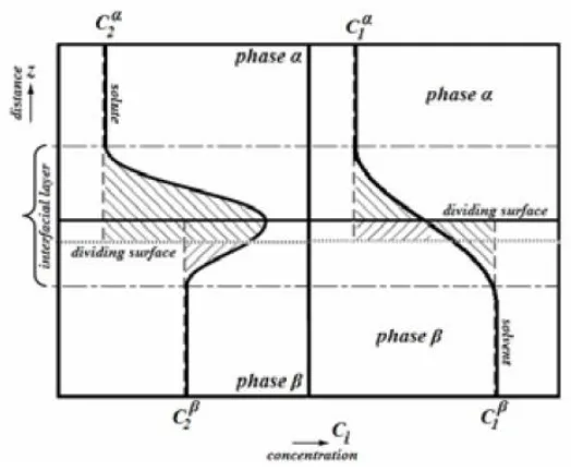
and has the unit as mol/m3 or mg/m3. The idea of surface excess was explained explicitly
in Figure 1.2. Clearly, the surface concentration and bulk concentration are different due
5
the surface excess when surface tension γ and bulk concentration C of amphiphile is
known using Eq 1.1. However, in the case of proteins, the surface excess and surface
tension usually do not follow the Gibbs isotherm. The reasons are discussed in section
IIIa.
Another term frequently used in tensiometry is surface pressure (π), which is the
difference between the surface tension of solvent (γ0, clean surface) and the surface
tension of the solution (γ, the surface that has solute molecules adsorbed). The
relationship between γ and π is defined by Eq 1.2. In the context of aqueous solution, γ0 is
usually used as the surface tension of pure water at 20 oC (72.8 mN/m).19 Thus, the
adsorption of amphiphiles leads to the increase in π.
0
=
-
(Eq 1.2)There are several types of tensiometers including Du Noüy-Padday probe,20 Wilhelmy
plate,21 bubble pressure tensiometer,22 goniometer23 and Du Noüy ring,24 each measures
surface tension/pressure using slightly different principals. Methods used in this thesis are
described in Chapters 2-4 under the “Material and experimental methods” section.
Moreover, Du Noüy-Padday probe, Du Noüy ring and Wilhelmy plate methods are
often coupled with Langmuir-Blodgett (LB) trough to measure the surface pressure at
different surface concentrations of amphiphiles. In LB trough, the solute molecules
adsorb at the flat AWI and form a thin layer. Under adsorption equilibrium, the surface
concentration of solute molecules could be controlled by changing the surface area of the
6
concentration (Γ) is inversely proportional to surface area (A). By moving the barriers
steadily and measuring the surface pressure by tensiometry at the same time, a surface
pressure-surface area (π-A) or surface pressure-surface concentration (π-Γ) isotherm
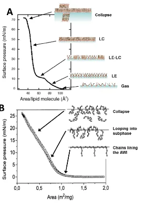
could be measured. The kinks in the isotherm often indicate lateral phase transitions in
lipid monolayers as shown in Figure 1.3 (A),25,26 but the shape changes in the isotherm
are absent in protein systems due to the flexibility of protein chains as indicated in Figure
1.3 (B).27
A prominent advantage of the Langmuir trough is that the interfacial film could be
easily transferred to solid substrate for further characterization by Langmuir-Blodgett
(LB) transfer (solid substrate perpendicular to the interface) or Langmuir-Schaefer (LS)
transfer (solid substrate parallel to the interface), which has greatly expanded the
application of LB trough.28 However, LB trough does have the drawback of being
compound consuming due to the large sample volume required, therefore is not very cost
7
Figure 1.1 Interaction between water molecules at the air-water interface causes surface tension.
On the surface, the interaction between the neighboring water molecules is limited compared to the bulk,
which leads to the increase in free energy and decrease in hydrogen bonding between water molecules.
8
Figure 1.2 Definition of surface excess.
X-axis is the concentration, and Y-axis is the distance normal to the phase boundary. Bold curved lines are
the concentration profiles of the solute (left) and the solvent (right), vertical broken lines are the
concentrations in the reference system. Chain dotted lines indicate the boundaries of the interfacial layer.
Bold horizontal line is the dividing surface and dotted horizontal line is another choice for the location of
the dividing surface. The surface excess is the sum of the shaded areas above and under the dividing
surface. By choosing the upper dividing surface (a suitable one) the resulting surface excess is zero,
whereas by choosing the lower one, surface excess is not zero.
9
Figure 1.3 Comparison of π-A isotherms of lipid and protein at the AWI.
(A) The isotherm of dipalmitoylphosphatidylcholine (DPPC) monolayer and schematic diagrams showing
the lateral phase transitions in the DPPC film at different surface pressures. Gas: gaseous phase, LE:
liquid-expanded phase, LC: liquid-condensed phase, LE–LC: liquid-condensed–liquid-liquid-expanded coexistence.
Collapse: collapse of interfacial film. (B) The isotherm of β-casein and schematic diagrams showing the structural changes in the protein film.
10 b. Fluorescence microscopy
Fluorescence microscopy was first applied to study molecules at the AWI in lipid
monolayer systems in the 1980s,29,30 and was soon applied to study the interaction
between protein and lipids.31,32 Thus, the application of fluorescence microscopy in
studying protein films at the AWI is a natural extension of these works.33,34 Fluorescence
microscopy has sub-micron spatial resolution and high signal-to-noise ratio, hence is
ideal for imaging the film morphology. Besides, the millisecond to a few seconds
temporal resolution of fluorescence microscopy could be utilized to study dynamic
processes occurring at the interface. Fluorescence microscopy could be used
independently or coupled with other methods such as Langmuir trough for in situ
measurement.
With the development of fluorescence microscopy itself, its application in studying
interfacial phenomenon is further expanded. Confocal laser scanning microscopy (CLSM)
greatly enhanced the spatial resolution of widefield fluorescence microscopy with the
added optical sectioning capability. The optical aperture in CLSM eliminates out of focus
light, hence reducing image degradation. Besides, the sectioning ability allows
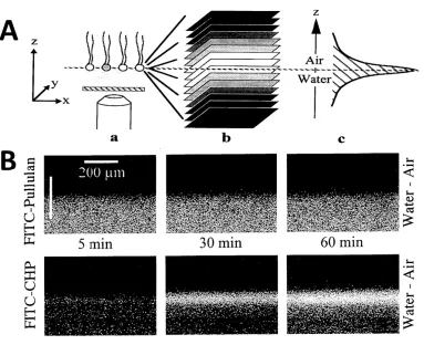
researchers to image the plane perpendicular to the AWI as shown in Figure 1.4. From
the change of fluorescence intensity, one can obtain semi-quantitative information about
11
Figure 1.4 Vertical imaging of molecules at the AWI by CLSM.
(A) Procedures for obtaining vertical fluorescence intensity profile. a) scanning the xy plan with CLSM; b)
stacking 2D images c) processing the image stack to get a fluorescence intensity profile along z-axis. (B)
Fluorescence image stacks of FITC-labeled pullulan (not surface-active) and FITC-labeled
cholesterol-bearing-pullulan (surface-active) at the AWI.
12 c. Atomic force microscopy
Atomic force microscopy (AFM) is a powerful tool to directly image the topography of
thin films with nanometer resolution. It has been adapted to study protein layers formed
at the AWI first by Gunning et. al.12,36,37 They imaged the layer of β-casein at the AWI
and bovine serum albumin (BSA) at the hexadecane/water interface and demonstrated
that the AFM could resolve the detailed structure as small as a few protein molecules.36
However, AFM also has obvious drawbacks: to measure the thickness or image the
morphology, an extra step is required to transfer the film onto a solid substrate from the
AWI. The type of solid substrate and change of hydration state may alter the film
structure after transfer.38 As a result, the transfer quality is crucial for the correct
interpretation of the film structure by AFM measurements. Complementary methods,
which could confirm the transfer quality and compare the film before and after the
transferring step, are desired.33
d. Other methods
Besides the above mentioned methods, several other techniques have been developed
and applied to study molecules at the AWI. Although these approaches were not directly
used in the research projects covered in this thesis, results here are often compared to
those reported in the literature using different techniques. A brief introduction of the
other methods is hence included in this section to facilitate data interpretation.
Interfacial sheer rheology (ISR) is a technique to study the flow properties of interfacial
layers. It measures the storage moduli (G’) and the loss moduli (G’’) of the 2D layer
13
elasticity measures the strength of intermolecular forces, while viscosity measures the
diffusion of molecules in the layer, this technique could provide useful information about
the organization of molecules at the AWI.40 Depending on the size of the probe, ISR
could be divided into macroscopic and microscopic. Macroscopic rheology measures the
average elasticity and viscosity,39 while the microscopic rheology could resolve the
heterogeneity in the 2D layer in the micrometer scale, which was demonstrated to be
especially useful in the case of protein films.41
Reflectometry and ellipsometry are useful techniques in measuring the thickness and
density of thin films. Reflectometry encompasses neutron reflectivity and X-ray
reflectivity, which differ in the type of incident beams used but share similar principles.
In reflectometry and ellipsometry, the signal is generated by the reflection and/or
refraction of the electromagnetic wave by the substances in the interfacial film,42,43 and
these methods do not require any exogenous probes such as the dyes used in fluorescence
microscopy or the rheometer sensor used in ISR. However, the thickness and the surface
concentration have to be obtained by regression analysis using hypothesized models, thus,
prior knowledge of the interfacial film is required in order to make precise
measurements.43
Brewster angle microscopy (BAM) is a technique to image 2D layers at the AWI.
When the p-polarized light beam is directed at the air-liquid interface at the Brewster
angle, all light is refracted. If there are any additional materials at the AWI, a small
amount of light will be reflected, creating contrast for BAM imaging.44 BAM is also a
14
Sum Frequency Generation (SFG) is a non-linear spectroscopic technique suited to
study the conformation of molecules at interfaces. Because the second-order nonlinear
process is forbidden in centrosymmetric media, signals only come from the molecules at
the interface, where the symmetry is broken.47 SFG could scan wavelengths across the
vibrational spectrum, giving information on specific chemical bonds such as the C-H
bond, to deduce how the proteins are oriented at the interface.48
III. Protein behaviors at the air-water interface
a. Adsorption, conformation and structure of protein assembly at the AWI
Different from small-molecule surfactants, during protein adsorption, the increase in
surface pressure and surface excess is often not simultaneous and their correlation does
not follow the Gibbs isotherm.49 This is caused by two distinct characteristics of protein:
1) The proteins in the surface layer could slowly change conformation at the AWI,
leading to continuous change in surface pressure.49 2) Protein could form multilayers at
the interface at high bulk concentrations;50 because surface pressure is dominated by the
surface layer, the surface excess could increase without an obvious change in surface
pressure.51 As a result, the surface pressure and surface excess are not correlated, and the
two parameters need to be measured independently. Due to the large structural and
compositional variance in proteins, the adsorption kinetics for different proteins needs to
be studied on a case-by-case basis.
It is still controversial whether protein adsorption at the AWI is reversible.52,53 But a
slow desorption rate of protein from the interface has been commonly observed, which is
sub-15
layers where the proteins are surrounded by water molecules, the folded structure of the
protein will tend to bury hydrophobic regions within the interior of the molecule and
expose hydrophilic regions at the protein surface, which reduces the solvation energy.55
When protein adsorbs at the interface, it will partially unfold and expose its hydrophobic
regions in order to reduce surface energy, therefore may undergo some structural
distortion. This process could last for hours or even days, demonstrated by the continuous
increase of surface pressure after surface excess reached equilibrium due to the change of
conformation.49 Conformational change may lead to denaturation depending on the
stability of the protein.56
Intermolecular forces, which include the chemical, electrostatic and hydrodynamic
interactions between protein molecules, could further drive the proteins into assemblies
and networks. Rheological studies showed that the shear viscosity of 8 different protein
films kept increasing over 30 hours aging time, which indicates a continuous increase in
intermolecular cohesion or covalent bonding.40 Structural differences of the films formed
by different proteins were reported as a result of the different conformation and stability:
Previous AFM studies showed that the globular shape of BSA was kept in the interfacial
film while a more disordered network was observed in β-casein film shown in Figure
1.5.36 Heterogeneity in the protein film formed by a single component was also reported
by imaging studies. Powers et al. reported microdomains with various geometry in a
synthetic amphiphilic peptide at the AWI using fluorescent imaging.57 BAM imaging also
16
heterogeneity in the micrometer scale was also observed in β-lactoglobulin film at the
17
Figure 1.5 AFM images of interfacial film of BSA or β-casein transferred onto mica.
(A, B) BSA prepared at the hexadecane/water interface, (C) β-casein prepared at the AWI. Height in gray scale (black to white) (A) 0-7.6 nm, (B) 0-7.0 nm, (C) 0-2.7 nm. Scale bar: 100 nm
18
A representative plasma protein, serum albumin has a heart-shaped structure resolved
by crystallography (Table 1.1), but an ellipsoid shape has been historically used in
hydrodynamic studies.59 It was thought to undergo structural elongation after absorption
at the AWI,60 but is not fully denatured due to the stability of the intramolecular
scaffold.56 The degree of conformational change is dependent on serum albumin bulk
concentrations. It was found by SFG that the higher the surface coverage of BSA, the less
change in structure that was observed.61 Recent microrheology study showed that in
albumin films, surface elasticity did not increase accordingly with viscosity, indicating an
annealing mechanism instead of covalent bonding between albumin molecules.62
The shape of fibrinogen could be simplified as three balls connected linearly by two
sticks (Table 1.1), so the packing density of fibrinogen at the interface is limited
geometrically.63 Fibrinogen aqueous solution was shown to have moderate surface
activity.63 In accordance with the geometric estimation, ellipsometry of fibrinogen layers
at the AWI indicate high water content.64 Studies using BAM and SEM showed fibrils are
formed during organization at the AWI.65
IgG is a Y-shaped antibody (Table 1.1). It was found to form a monolayer at the AWI
even at high bulk concentration, and the orientation of the molecule changed from
side-on to end-side-on when the surface area was reduced or the bulk cside-oncentratiside-on was
increased.66 Study using fluorescent antigen binding showed that after adsorption at the
19
Table 1.1 Crystal structures and geometric dimensions of serum albumin, fibrinogen and IgG.
20 b. Interactions between proteins
In biological systems, there is usually more than one type of protein present in solution,
and the same is also true at biological interfaces. Signaling proteins interact with each
other through specific recognition at the water-lipid interface.68 For plasma proteins, the
competition for the available interfacial area is the dominant interaction. The competition
process could be separated into two steps: the diffusion rate, which decides how fast the
protein reaches the interface from the bulk, and the affinity of the protein to the interface,
which decides the most stable protein interfacial assembly.15,69 Because the size of
molecules decides the diffusion rate to the interface, smaller proteins arrive first at the
interface but can be later replaced by larger proteins with higher affinity to the interface.
This is the so-called Vroman effect, which was first discovered by Vroman et al. in blood
protein adsorption on solid surfaces.70 Among the few reported studies on protein-protein
competition at the AWI, Krishnan et al. measured the surface tensions of blood plasma
and serum proteins, and found that different samples exhibited nearly identical
concentration dependence.15 Therefore just from tensiometry, it is hard to resolve the
composition of different protein species at the AWI. Moreover, when different proteins
adsorb at the interface, the mechanical properties of the interfacial film are strongly
affected by the interaction between different species. Rheological study found that
mixing serum albumin into IgG greatly reduced the elasticity of the interfacial film,
indicating that serum albumin intercalated into IgG and weakened the intermolecular
interactions in the film.39 Overall, the competition between proteins at the AWI is
21 c. Interaction with polymers
Due to the wide application of synthetic polymers in biomaterials, biosensors, and drug
delivery,71 understanding and controlling the interaction between synthetic polymers and
proteins at interfaces has become an important topic. New methods have been reported
for incorporating antibody into polymeric thin films for immunosensing,72-74 demanding
deeper understanding of protein-polymer interactions at biological interfaces.
Many polymers—particularly copolymers—are surfactants,75 and tend to adsorb at
interfaces, thus able to compete with protein adsorption. Surfactants are known to interact
with proteins at the AWI to reduce significantly the surface tension and change the
rheological behavior of the surface layer.76 It is proposed that surfactants displace
proteins heterogeneously from the AWI, as described by an “orogenic” model from
previous AFM studies by Morris et al.12 However, direct proof from in situ measurements
has only been provided by BAM studies,77 and aspects of dynamic transitions remain
under-explored.
On the other hand, if polymer layers were pre-spread at the AWI, the polymer film
could modulate the interfacial energy and encourage protein adsorption to the interface.
Enhanced protein adsorption on polymer films has been accomplished by making
superhydrophobic polymer surfaces or incorporating specific protein ligands into the
polymer.4 More recently, tunable adsorption of proteins on polymer films has been
achieved by preferential adsorption on spatially separated chemical components that
differ in hydrophobicity,78,79 or by changing the surface charge of polymer thin films.80,81
22
assembly could further expand the chemical toolbox for controlling protein adsorption
and assembly.
IV. Research scope
Work in this thesis utilized fluorescence microscopy alone or in combination with
tensiometry and AFM to study plasma proteins and their interaction with polymers at the
AWI, contributing to methodological development as well as understanding interfacial
behavior of these plasma proteins. The morphological study of human serum albumin
(HSA) self-assembly controlled by solution conditions will be presented in Chapter 2.
Semi-quantitative study of the adsorption and competition between plasma proteins and a
block copolymer Pluronic F-127 at the AWI will be presented in Chapter 3. The
interaction between Polydimethylsiloxane film and serum proteins will be investigated in
Chapter 4. Finally, conclusions and future directions are summarized in Chapter 5.
V. References
(1) Nozaki, Y.; Tanford, C.: Solubility of amino acids and 2 glycine peptides
in aqueous ethanol and dioxane solutions-establishment of a hydrophobicity scale. J. Biol.
Chem.1971, 246, 2211-2217.
(2) Hessa, T.; Kim, H.; Bihlmaier, K.; Lundin, C.; Boekel, J.; Andersson, H.;
Nilsson, I.; White, S. H.; von Heijne, G.: Recognition of transmembrane helices by the
endoplasmic reticulum translocon. Nature2005, 433, 377-381.
(3) Conner, S. D.; Schmid, S. L.: Regulated portals of entry into the cell.
23
(4) Elbert, D. L.; Hubbell, J. A.: Surface treatments of polymers for
biocompatibility. Annu. Rev. Mater. Sci.1996, 26, 365-394.
(5) Roach, P.; Farrar, D.; Perry, C. C.: Interpretation of protein adsorption:
Surface-induced conformational changes. J. Am. Chem. Soc.2005, 127, 8168-8173.
(6) Cooper, A.; Kennedy, M. W.: Biofoams and natural protein surfactants.
Biophys. Chem.2010, 151, 96-104.
(7) Choi, H. J.; Ebersbacher, C. F.; Quan, F. S.; Montemagno, C. D.: pH
stability and comparative evaluation of ranaspumin-2 foam for application in biochemical
reactors. Nanotechnology2013, 24, 055603.
(8) Yang, Y.; Dicko, C.; Bain, C. D.; Gong, Z.; Jacobs, R. M. J.; Shao, Z.;
Terry, A. E.; Vollrath, F.: Behavior of silk protein at the air–water interface. Soft Matter
2012, 8, 9705-9712.
(9) Krishnaji, S. T.; Huang, W.; Rabotyagova, O.; Kharlampieva, E.; Choi, I.;
Tsukruk, V. V.; Naik, R.; Cebe, P.; Kaplan, D. L.: Thin film assembly of spider silk-like
block copolymers. Langmuir2011, 27, 1000-1008.
(10) Zampieri, F.; Wösten, H. A. B.; Scholtmeijer, K.: Creating surface
properties using a palette of hydrophobins. Materials2010, 3, 4607-4625.
(11) Russev, S. C.; Arguirov, T. V.; Gurkov, T. D.: Beta-casein adsorption
kinetics on air-water and oil-water interfaces studied by ellipsometry. Colloid Surf.
24
(12) Mackie, A. R.; Gunning, A. P.; Ridout, M. J.; Wilde, P. J.; Morris, V. J.:
Orogenic displacement in mixed beta-lactoglobulin/beta-casein films at the air/water
interface. Langmuir2001, 17, 6593-6598.
(13) Zuo, Y. Y.; Veldhuizen, R. A. W.; Neumann, A. W.; Petersen, N. O.;
Possmayer, F.: Current perspectives in pulmonary surfactant - Inhibition, enhancement
and evaluation. Biochimica et Biophysica Acta - Biomembranes2008, 1778, 1947-1977.
(14) Anderson, N. L.: The human pplasma proteome: history, character, and
diagnostic prospects. Molecular & Cellular Proteomics 2002, 1, 845-867.
(15) Krishnan, A.; Wilson, A.; Sturgeon, J.; Siedlecki, C. A.; Vogler, E. A.:
Liquid-vapor interfacial tension of blood plasma, serum and purified protein constituents
thereof. Biomaterials2005, 26, 3445-3453.
(16) Mukundakrishnan, K.; Ayyaswamy, P. S.; Eckmann, D. M.: Bubble
motion in a blood vessel: shear stress induced endothelial cell injury. J. Biomech. Eng.
2009, 131, 074516.
(17) Sobolewski, P.; Kandel, J.; Eckmann, D. M.: Air bubble contact with
endothelial cells causes a calcium-independent loss in mitochondrial membrane potential.
PLoS One2012, 7, e47254.
(18) Deng, Y.; Zhu, X. Y.; Kienlen, T.; Guo, A.: Transport at the air/water
interface is the reason for rings in protein microarrays. J. Am. Chem. Soc. 2006, 128,
25
(19) Vargaftik, N. B.; Volkov, B. N.; Voljak, L. D.: International tables of the
surface-tension of water. J. Phys. Chem. Ref. Data1983, 12, 817-820.
(20) Padday, J. F.; Pitt, A. R.; Pashley, R. M.: Menisci at a free liquid surface:
surface tension from the maximum pull on a rod. J. Chem. Soc., Faraday Trans. I 1975,
71, 1919-1931.
(21) Holmberg, K.: Handbook of Applied Surface and Colloid Chemistry,
Volumes 1-2. John Wiley & Sons; pp 219.
(22) Holmberg, K.: Handbook of Applied Surface and Colloid Chemistry,
Volumes 1-2. John Wiley & Sons; pp 227.
(23) Kwok, D. Y.; Gietzelt, T.; Grundke, K.; Jacobasch, H. J.; Neumann, A. W.:
Contact angle measurements and contact angle interpretation .1. Contact angle
measurements by axisymmetric drop shape analysis and a goniometer sessile drop
technique. Langmuir1997, 13, 2880-2894.
(24) du Nouy, P. L.: An interfacial tensiometer for universal use. J. Gen.
Physiol.1925, 7, 625-631.
(25) Kaganer, V. M.; Mohwald, H.; Dutta, P.: Structure and phase transitions in
Langmuir monolayers. Rev. Mod. Phys.1999, 71, 779-819.
(26) Wüstneck, R.; Perez-Gil, J.; Wüstneck, N.; Cruz, A.; Fainerman, V. B.;
Pison, U.: Interfacial properties of pulmonary surfactant layers. Adv. Colloid Interface Sci.
26
(27) Nino, M. R. R.; Sanchez, C. C.; Patino, J. M. R.: Interfacial characteristics
of beta-casein spread films at the air-water interface. Colloid Surf. B-Biointerfaces 1999,
12, 161-173.
(28) Petty, M. C.: Langmuir-Blodgett Films - An Introduction. Cambridge
University Press; pp 39-63.
(29) McConnell, H. M.; Tamm, L. K.; Weis, R. M.: Periodic structures in lipid
monolayer phase-transitions. Proc. Natl. Acad. Sci. U. S. A.1984, 81, 3249-3253.
(30) Lösche, H.; Möhwald, H.: Fluorescence microscopy on monomolecular
films at an air/water interface. Colloids and Surfaces1984, 10, 217-224.
(31) Heckl, W. M.; Zaba, B. N.; Mohwald, H.: Interactions of cytochromes b5
and c with phospholipid monolayers. Biochim. Biophys. Acta1987, 903, 166-176.
(32) Möhwald, H.: Phospholipid and phospholipid-protein monolayers at the
air/water interface. Annu. Rev. Phys. Chem.1990, 41, 441-476.
(33) Morris, V. J.; Gunning, A. P.: Microscopy, microstructure and
displacement of proteins from interfaces: implications for food quality and digestion. Soft
Matter2008, 4, 943-951.
(34) Liu, W.; Johnson, S.; Micic, M.; Orbulescu, J.; Whyte, J.; Garcia, A. R.;
Leblanc, R. M.: Study of the aggregation of human insulin Langmuir monolayer.
27
(35) Gluck, G.; Ringsdorf, H.; Okumura, Y.; Sunamoto, J.: Vertical sectioning
of molecular assemblies at air water interface using laser scanning confocal fluorescence
microscopy. Chem. Lett.1996, 209-210.
(36) Gunning, A. P.; Wilde, P. J.; Clark, D. C.; Morris, V. J.; Parker, M. L.;
Gunning, P. A.: Atomic force microscopy of interfacial protein films. J. Colloid Interface
Sci.1996, 183, 600-602.
(37) Mackie, A. R.; Gunning, A. P.; Pugnaloni, L. A.; Dickinson, E.; Wilde, P.
J.; Morris, V. J.: Growth of surfactant domains in protein films. Langmuir 2003, 19,
6032-6038.
(38) Viswanathan, R.; Schwartz, D. K.; Garnaes, J.; Zasadzinski, J. A. N.:
Atomic force microscopy imaging of substrate and pH effects on Langmuir-Blodgett
monolayers. Langmuir1992, 8, 1603-1607.
(39) Ariola, F. S.; Krishnan, A.; Vogler, E. A.: Interfacial rheology of blood
proteins adsorbed to the aqueous-buffer/air interface. Biomaterials2006, 27, 3404-3412.
(40) Bos, M. A.; van Vliet, T.: Interfacial rheological properties of adsorbed
protein layers and surfactants: a review. Adv. Colloid Interface Sci.2001, 91, 437-471.
(41) Lee, M. H.; Reich, D. H.; Stebe, K. J.; Leheny, R. L.: Combined passive
and active microrheology study of protein-layer formation at an air-water interface.
28
(42) De Feijter, J. A.; Benjamins, J.; Veer, F. A.: Ellipsometry as a tool to
study the adsorption behavior of synthetic and biopolymers at the air-water interface.
Biopolymers1978, 17, 1759-1772.
(43) Zhou, X. L.; Chen, S. H.: Theoretical foundation of X-ray and neutron
reflectometry. Phys. Rep.-Rev. Sec. Phys. Lett.1995, 257, 223-348.
(44) Honig, D.; Mobius, D.: Direct visualization of monolayers at the
air-water-interface by brewster angle microscopy. J. Phys. Chem.1991, 95, 4590-4592.
(45) Ruiz-Garcia, J.; Moreno, A.; Brezesinski, G.; Mohwald, H.; Mas-Oliva, J.;
Castillo, R.: Phase transitions and conformational changes in monolayers of human
apolipoproteins CI and AII. J. Phys. Chem. B2003, 107, 11117-11124.
(46) Murray, B. S.; Xu, R.; Dickinson, E.: Brewster angle microscopy of
adsorbed protein films at air-water and oil-water interfaces after compression, expansion
and heat processing. Food Hydrocolloids2009, 23, 1190-1197.
(47) Shen, Y. R.: Surface properties probed by 2nd harmonic and sum
frequency generation. Nature1989, 337, 519-525.
(48) Wang, J.; Buck, S. M.; Chen, Z.: Sum frequency generation vibrational
spectroscopy studies on protein adsorption. J. Phys. Chem. B2002, 106, 11666-11672.
(49) Graham, D. E.; Phillips, M. C.: Proteins at liquid interfaces .1. kinetics of
29
(50) Lu, J. R.; Su, T. J.; Penfold, J.: Adsorption of serum albumins at the
air/water interface. Langmuir1999, 15, 6975-6983.
(51) Graham, D. E.; Phillips, M. C.: Proteins at liquid interfaces
.2.adsorption-isotherms. J. Colloid Interface Sci.1979, 70, 415-426.
(52) VanTassel, P. R.; Viot, P.; Tarjus, G.: A kinetic model of partially
reversible protein adsorption. J. Chem. Phys.1997, 106, 761-770.
(53) Fainerman, V. B.; Miller, R.; Ferri, J. K.; Watzke, H.; Leser, M. E.;
Michel, M.: Reversibility and irreversibility of adsorption of surfactants and proteins at
liquid interfaces. Adv. Colloid Interface Sci.2006, 123-126, 163-171.
(54) Mosesson, M. W.: Fibrinogen and fibrin structure and functions. J.
Thromb. Haemost.2005, 3, 1894-1904.
(55) Chandler, D.: Interfaces and the driving force of hydrophobic assembly.
Nature2005, 437, 640-647.
(56) Lad, M. D.; Birembaut, F.; Matthew, J. M.; Frazier, R. A.; Green, R. J.:
The adsorbed conformation of globular proteins at the air/water interface. Phys. Chem.
Chem. Phys.2006, 8, 2179-2186.
(57) Powers, E. T.; Kelly, J. W.: Medium-dependent self-assembly of an
amphiphilic peptide: Direct observation of peptide phase domains at the air-water
30
(58) Garcia-Gonzalez, A.; Flores-Vazquez, A. L.; Maldonado, E.; de la Rosa,
A. P. B.; Ruiz-Garcia, J.: Globulin 11S and its mixture with
L-dipalmitoylphosphatidylcholine at the air/liquid interface. J. Phys. Chem. B 2009, 113,
16547-16556.
(59) Ferrer, M. L.; Duchowicz, R.; Carrasco, B.; de la Torre, J. G.; Acuna, A.
U.: The conformation of serum albumin in solution: A combined phosphorescence
depolarization-hydrodynamic modeling study. Biophys. J.2001, 80, 2422-2430.
(60) Lu, J. R.; Su, T. J.; Thomas, R. K.: Structural conformation of bovine
serum albumin layers at the air-water interface studied by neutron reflection. J. Colloid
Interface Sci.1999, 213, 426-437.
(61) Wang, J.; Buck, S. M.; Chen, Z.: The effect of surface coverage on
conformation changes of bovine serum albumin molecules at the air-solution interface
detected by sum frequency generation vibrational spectroscopy. Analyst 2003, 128,
773-778.
(62) Dhar, P.; Cao, Y. Y.; Fischer, T. M.; Zasadzinski, J. A.: Active interfacial
shear microrheology of aging protein films. Phys. Rev. Lett.2010, 104, 016001.
(63) Hernandez, E. M.; Phang, T. L.; Wen, X. Y.; Franses, E. I.: Adsorption
and direct probing of fibrinogen and sodium myristate at the air/water interface. J.
31
(64) Hernandez, E. M.; Franses, E. I.: Adsorption and surface tension of
fibrinogen at the air/water interface. Colloid Surf. A-Physicochem. Eng. Asp.2003, 214,
249-262.
(65) Sankaranarayanan, K.; Dhathathreyan, A.; Miller, R.: Assembling
fibrinogen at air/water and solid/liquid interfaces using Langmuir and Langmuir-Blodgett
films. J. Phys. Chem. B2010, 114, 8067-8075.
(66) Tronin, A.; Dubrovsky, T.; Nicolini, C.: Comparative study of Langmuir
monolayers of immunoglobulins-G formed at the air-water interface and covalently
immobillized on solid supports. Langmuir1995, 11, 385-389.
(67) Tronin, A.; Dubrovsky, T.; Dubrovskaya, S.; Radicchi, G.; Nicolini, C.:
Role of protein unfolding in monolayer formation on air-water interface. Langmuir1996,
12, 3272-3275.
(68) Li, P.; Banjade, S.; Cheng, H. C.; Kim, S.; Chen, B.; Guo, L.; Llaguno, M.;
Hollingsworth, J. V.; King, D. S.; Banani, S. F.; Russo, P. S.; Jiang, Q. X.; Nixon, B. T.;
Rosen, M. K.: Phase transitions in the assembly of multivalent signalling proteins. Nature
2012, 483, 336-340.
(69) Krishnan, A.; Siedlecki, C. A.; Vogler, E. A.: Mixology of protein
solutions and the Vroman effect. Langmuir2004, 20, 5071-5078.
(70) Vroman, L.; Adams, A. L.; Fischer, G. C.; Munoz, P. C.: Interaction of
high molecular-weight kininogen, factor-XII, and fibrinogen in plasma at interfaces.
32
(71) Stuart, M. A. C.; Huck, W. T. S.; Genzer, J.; Muller, M.; Ober, C.; Stamm,
M.; Sukhorukov, G. B.; Szleifer, I.; Tsukruk, V. V.; Urban, M.; Winnik, F.; Zauscher, S.;
Luzinov, I.; Minko, S.: Emerging applications of stimuli-responsive polymer materials.
Nat. Mater.2010, 9, 101-113.
(72) Caruso, F.; Niikura, K.; Furlong, D. N.; Okahata, Y.: Assembly of
alternating polyelectrolyte and protein multilayer films for immunosensing .2. Langmuir
1997, 13, 3427-3433.
(73) Janmanee, R.; Baba, A.; Phanichphant, S.; Sriwichai, S.; Shinbo, K.; Kato,
K.; Kaneko, F.: Detection of human IgG on poly(pyrrole-3-carboxylic acid) thin film by
electrochemical-surface plasmon resonance spectroscopy. Jpn. J. Appl. Phys. 2011, 50,
01BK02.
(74) Valsesia, A.; Mannelli, I.; Colpo, P.; Bretagnol, F.; Rossi, F.: Protein
nanopatterns for improved immunodetection sensitivity. Anal. Chem. 2008, 80,
7336-7340.
(75) Torchilin, V. P.: Structure and design of polymeric surfactant-based drug
delivery systems. J. Controlled Release2001, 73, 137-172.
(76) Kotsmar, C.; Pradines, V.; Alahverdjieva, V. S.; Aksenenko, E. V.;
Fainerman, V. B.; Kovalchuk, V. I.; Krägel, J.; Leser, M. E.; Noskov, B. A.; Miller, R.:
Thermodynamics, adsorption kinetics and rheology of mixed protein-surfactant
33
(77) Mackie, A. R.; Gunning, A. P.; Ridout, M. J.; Wilde, P. J.; Patino, J. R.: In
situ measurement of the displacement of protein films from the air/water interface by
surfactant. Biomacromolecules2001, 2, 1001-1006.
(78) Lau, K. H. A.; Bang, J.; Kim, D. H.; Knoll, W.: Self-assembly of protein
nanoarrays on block copolymer templates. Adv. Funct. Mater.2008, 18, 3148-3157.
(79) Kidambi, S.; Chan, C.; Lee, I.: Tunable resistive m-dPEG acid patterns on
polyelectrolyte, multilayers at physiological conditions: Template for directed deposition
of biomacromolecules. Langmuir2008, 24, 224-230.
(80) Eloi, J.-C.; Jones, S. E. W.; Poór, V.; Okuda, M.; Gwyther, J.;
Schwarzacher, W.: Electrochemically triggered selective adsorption of biotemplated
nanoparticles on self-assembled organometallic diblock copolymer thin films. Adv. Funct.
Mater.2012, 22, 3273-3278.
(81) Pernites, R. B.; Santos, C. M.; Maldonado, M.; Ponnapati, R. R.;
Rodrigues, D. F.; Advincula, R. C.: Tunable protein and bacterial cell adsorption on
colloidally templated superhydrophobic polythiophene films. Chem. Mater. 2012, 24,
34
Chapter 2
Morphology of protein self-assembly at
the air-water interface controlled by solution
conditions—a study by confocal laser scanning
microscopy
*I. Introduction
The AWI is a unique tool to guide proteins as small building units into larger,
self-assembled structures. Proteins are naturally occurring biopolymers with unsymmetrical
distribution of buried and solvent-exposed active sites, electrostatic charges and
hydrophobic/hydrophilic residues. These features, combined with geometric constraints,
dictate the structures of folded proteins and their larger assemblies. A strategy in
assembling complex biological or synthetic systems is to promote interactions in just one
or two dimensions. Liquid interfaces are particularly useful in this regard, based on the
anisotropic forces residing therein and two-dimensional spatial confinement.1-3 Besides
the special characteristics of liquid interfaces, factors that commonly control
self-assembly are the chemical and structural complementarity of the building blocks.4 For
macromolecules such as proteins, the active site, geometric structure, electrostatic and
hydrophobic interactions are greatly affected by the surrounding solvent molecules and
*
35
other solutes.5,6 Thus, solution conditions provide a convenient handle for controlling
protein assembly at the AWI.
Previous tensiometry, reflectometry and ellipsometry studies explored the change in
surface excess and structural conformation of proteins at the AWI in relation to solution
conditions such as subphase concentration, pH and ionic strength.7-10 These techniques
provide ensemble-averaged properties of adsorbed proteins over a macroscopic scale. To
study assembled structures at the AWI on the nanometer-to-micron scale, imaging
techniques such as atomic force microscopy (AFM), Brewster angle microscopy (BAM)
and fluorescence microscopy have been applied.11-13 AFM affords nanometer spatial
resolution, but involves transferring interfacial films onto a solid surface, which leads to
potential loss of structural fidelity and temporal resolution.11 BAM coupled with
Langmuir trough is a widely used in situ method, but offers lower spatial resolution (2
μm) than many diffraction-limited optical imaging techniques, and offers little kinetic
information.12,14 Earlier studies by Gluck et al. on polysaccharide adsorption at the AWI
showed the potential of fluorescence microscopy to serve as a facile in situ imaging tool
for studying macromolecular self-assembly at the AWI.15 By this technique, Powers et al.
discovered medium-dependent micron-size phase domains of an amphiphilic peptide
self-assembled at the AWI.16 The observations made in this landmark study have not yet been
generalized to protein systems. Here, we demonstrate for the first time the ability to
control the microstructure of protein self-assembly at the AWI through systematic