R E S E A R C H A R T I C L E
Open Access
Clinical features and characteristics of
uveitis associated with juvenile idiopathic
arthritis in Japan: first report of the
pediatric rheumatology association of
Japan (PRAJ)
Junko Yasumura
1*, Masato Yashiro
2, Nami Okamoto
3, Kosuke Shabana
3, Hiroaki Umebayashi
4, Naomi Iwata
5,
Yuka Okura
6, Tomohiro Kubota
7, Masaki Shimizu
8, Minako Tomiita
9, Yasuo Nakagishi
10, Kenichi Nishimura
11,
Ryoki Hara
11, Mao Mizuta
8, Takahiro Yasumi
12, Fumiya Yamaide
13, Hiroyuki Wakiguchi
14, Masao Kobayashi
1and
Masaaki Mori
15Abstract
Background:Although there are many reports on Juvenile Idiopathic arthritis-associated uveitis (JIA-U) from various countries, especially from Europe and North America, there are few reports from Asia. Our aim was to investigate the epidemiology, characteristics and predictors of JIA-U in Japan.
Methods:Data were retrospectively collected on 726 patients with JIA from medical records as of April 2016 at 15 medical centers specialized in pediatric rheumatic diseases. Of these, patients with uveitis were further investigated for the specific characteristics of this manifestation.
Results:The prevalence of uveitis was 6.1% in the 726 JIA patients examined. Incidence of uveitis was significantly higher in patients with an earlier arthritis onset (2.6-vs.-5.8 years,P< 0.0001), oligoarthritis (16.1%-vs.-1.6%,P< 0.001), or anti-nuclear antibodies. On the contrary, it was significantly less common in patients with rheumatoid factor or anti-cyclic citrullinated peptide antibodies. A history of using methotrexate (MTX), infliximab or adalimumab was also associated with uveitis occurrence. The median age at uveitis diagnosis was 5 years, and the median time from arthritis onset to uveitis diagnosis was 2 years. The occurrence of anterior and bilateral uveitis was 79.3 and 53.7%, respectively. There were no symptoms at uveitis diagnosis in 58.5% of cases. Complications arising between the time of uveitis diagnosis and the last observation increased from 31.7 to 56.1%; in particular, cataract was increased 3-fold. While no patients lost their vision, 61.9% did not recover normal vision (≥1.0), and in many cases active uveitis persisted, especially in males. In addition to steroid eye drops (97.6%) and MTX (15.4%), biological agents were used for treating the uveitis in 41.5% of patients.
Conclusions:The epidemiology, characteristics and predictors of JIA-U in Japan are described here for the first time. Although the prevalence of JIA-U in Japan is lower than in predominantly Caucasian cohorts, as reported from North America and Europe, the epidemiology, characteristics and predictors were found to be similar.
Keywords:Juvenile idiopathic arthritis, Uveitis, Epidemiology, Asian
© The Author(s). 2019Open AccessThis article is distributed under the terms of the Creative Commons Attribution 4.0 International License (http://creativecommons.org/licenses/by/4.0/), which permits unrestricted use, distribution, and reproduction in any medium, provided you give appropriate credit to the original author(s) and the source, provide a link to the Creative Commons license, and indicate if changes were made. The Creative Commons Public Domain Dedication waiver (http://creativecommons.org/publicdomain/zero/1.0/) applies to the data made available in this article, unless otherwise stated. * Correspondence:junko-ma562@hiroshima-u.ac.jp
1Department of Pediatrics, Hiroshima University Graduate School of
Biomedical and Health Sciences, 1-2-3 Kasumi, Minami-ku, Hiroshima 734-8551, Japan
Background
Uveitis is the most common extra-articular manifest-ation of juvenile idiopathic arthritis (JIA); it is a ser-ious manifestation carrying the risk of blindness if treatment is delayed or inadequate. The prevalence of JIA-associated uveitis (JIA-U) has been reported as ranging from 4.7 to 20.5% [1–6] with local differences noted. Numerous reports have identified risk factors for JIA-U such as female sex, oligoarthritis, earlier arthritis onset, ANA-positivity, and RF-negativity in predominantly Cauca-sian cohorts [2,7,8]. Do these characteristics also apply to JIA-U in East Asia? To date, there are very few reports from East Asia and the epidemiology, characteristics and risk factors for JIA-U in Japan are unclear.
Although we, the members of the Pediatric Rheumatol-ogy Association of Japan (PRAJ), issued recommendations for ophthalmologic screening intervals for JIA patients in Japan [9,10] (Table1), these were based on other countries’ guidelines [2, 11]. In any event, these recommendations were issued only one year before this study of patients in Japan, and therefore such recommendations did not apply to most of the patients included here. Given that the preva-lence and characteristics of JIA-U in Japan were unclear, and pediatricians and ophthalmologists had little know-ledge of the management of JIA-U, investigating these is-sues specifically in Japanese patients was warranted. Accordingly, in the present study, we aimed to establish the epidemiology, clinical characteristics, and risk factors for JIA-U in Japan and to compare these with data reported from other countries. Thus, we reviewed the charts of out-patients making regular hospital visits as of April 2016.
Methods
Study setting
This is a retrospective study, approved by the Ethics Re-view Board of Tokyo Medical and Dental University
(No. M2015–537), the main study center, on March 4th, 2016, after which approval was obtained from each of the 15 other participating medical centers. Guardians and patients were provided information by means of an opt-out form.
Study design and patients
First, we sent questionnaires to pediatricians who are members of the PRAJ belonging to 15 medical centers to gather data on the characteristics of JIA in Japan. We analyzed data from outpatients with JIA who had regu-larly visited the hospital as of April 2016. All patients were classified according to the International League of Associations for Rheumatology (ILAR) criteria [12]. Sec-ond, we undertook a further questionnaire survey for pa-tients identified as having uveitis in the first questionnaire, and gathered more detailed information about uveitis features from their ophthalmology charts using this second questionnaire. Patients who had already discontinued follow-up of JIA as of April 2016 were not included.
Data collection
All patient data including ophthalmic records were col-lected from medical histories and were recorded by pe-diatricians. The following parameters were evaluated for the primary investigation: sex, age as of April 2016, dur-ation of disease, age at arthritis diagnosis, type of JIA classified by ILAR criteria, laboratory data including anti-nuclear antibody (ANA), rheumatoid factor (RF), anti-cyclic citrullinated peptide (CCP) antibody, serum matrix metalloproteinase-3 (MMP-3), and drugs used for treatment. An ANA titer of ≥1:160 by fluorescent antibody testing was designated positive. Data on ANA, RF and MMP-3 were acquired at the time of arthritis diagnosis. The following parameters were evaluated in
Table 1Recommendations for ophthalmologic screening intervals for JIA patients in Japan [9,10]
JIA category ANA
(titer)a
Ophthalmologic screening intervals
Onset age of≤6 years old Onset age≥7 years old ≤4 years from onset of arthritis
Oligoarthrits, RF-negative polyarthritis, undifferentiated arthritis ≥160 Every 3 months Every 6 months
< 160 Every 6 months Every 6 months
Psoriatic arthritis whose onset age is <4 years old Regardless Every 3 months –
Others Regardless Every 12 months Every 12 months
4 < years,≤7 years from onset of arthritis
Oligoarthrits, RF-negative polyarthritis, undifferentiated arthritis ≥160 Every 6 months Every 12 months
< 160 Every 12 months Every 12 months
Psoriatic arthritis whose onset age is <4 years old Regardless Every 6 months –
Others Regardless Every 12 months Every 12 months
>7 years from onset of arthritis: Every 12 months.
the second investigation: race of parents, family history of uveitis, infection history, possession of HLA-B27, lo-cation of uveitis, age at uveitis diagnosis, time from arth-ritis onset to uveitis diagnosis, timing of uveitis diagnosis, eye manifestations at uveitis diagnosis, eye complications due to uveitis at the first and last observa-tion, activity of arthritis at uveitis diagnosis, current ac-tivity of arthritis, current acac-tivity of uveitis, visual acuity at uveitis diagnosis and at present, drugs used for treat-ment. Visual acuity was recorded as a decimal unit using the Landolt ring method where 1.0 refers to 20/20 of the Snellen fraction, whereas 0.1 refers to 20/200 [13].
Statistical analysis
Statistical analysis was performed using JMP pro® Ver-sion 13 software (SAS Institute Inc., Cary, NC, USA). The Wilcoxon rank sum test or Welch’s t test were used for comparison of continuous variables between two groups, and categorical variables were analyzed using the Chi-Square test or Fisher’s exact test (two-sided). Odds ratios (ORs) are presented with 95% confidence in-tervals (CI). The time from onset of arthritis to diagnosis of uveitis was analyzed by the Kaplan-Meier method. A
p-value < 0.05 was considered significant.
Results
Comparison of characteristics of JIA patients with and without uveitis
We collected data from 730 patients with JIA at 15 med-ical centers. Four patients were excluded due to many missing data. Of the remaining 726 JIA patients, 44 (6.1%) had uveitis. Table 2 shows the characteristics of patients with and without uveitis. There were no signifi-cant differences between the two groups in sex or age at the last visit. Patients with uveitis had been significantly younger at arthritis onset (2.6-vs.-5.8 years, P< 0.0001) and the duration of disease was significantly longer (9.1-vs.-5.1 years, P< 0.0001) than in patients without uveitis. The most frequent disease type in patients with uveitis was oligoarthritis (81.8%), followed by RF-negative polyarthritis, but there were no patients with systemic arthritis or RF-positive polyarthritis. There were very few patients with psoriatic arthritis or non-classified arthritis in either group. The rate of uve-itis in oligoarthruve-itis patients was 16.1% and in all other JIA patients 1.6%; thus, the risk of uveitis in oligoarthri-tis was 10-fold higher (Table 6). In addition, the fre-quency of ANA-positive patients was significantly higher in the group with uveitis (52.3%-vs.-22.2%, P< 0.0001), with a tendency towards higher ANA titers than patients without uveitis (ANA titers ≥1:640; P= 0.018) (data not shown). The frequency of RF-positive cases (2.4%-vs.-24.5%, P= 0.0004) and those with anti-CCP antibodies (0%-vs.-26.2%, P< 0.0001) was significantly
lower in the uveitis group. No significant differences in serum MMP-3 levels at JIA diagnosis were noted be-tween the two groups. The proportion of patients with a history of using oral Methotrexate (MTX) was signifi-cantly higher in patients with uveitis (95.5%-vs.-77.0%,P = 0.0022). Similarly, significant differences were noted for infliximab (IFX), adalimumab (ADA), and toci-lizumab (TCZ) use: IFX and ADA were more frequently used in the uveitis group, whereas TCZ was more fre-quently used by patients without uveitis.
Clinical characteristics of patients with uveitis
We obtained data for a further analysis of 41 of the 44 patients with uveitis. Three patients were excluded due to lack of data from their physicians. Of the 41 patients, 38 had Japanese parents, one had Chinese parents, one had a Chinese and a Japanese parent, and the other had a Chinese and an Iranian parent. They were found to have no family history of uveitis or history of preceding infections. HLA-B27 had been tested only in 34.1% (14 of 41 patients) because of not being covered by insur-ance. Two patients were positive, and 12 patients were negative.
We were able to obtain data on the site of uveitis from only 29 of the 41 patients because of poor record keep-ing by ophthalmologists in some centers. Uveitis in 29 patients was anterior in 79.3% (N= 23), panuveitis in 13.8% (N= 4), posterior in 3.5% (N= 1), and both anter-ior and posteranter-ior in 3.5% (N = 1). Uveitis was bilateral in 53.7% of these 41 patients.
exhibited only persistent uveitis at the final observation, and not arthritis.
Ocular symptoms were present at uveitis diagnosis in 36.6% of patients, whereby ocular hyperemia accounted for 46.7% and decreased vision accounted for 46.7% (Table 3). Ocular complications were noted in 31.7% of patients at diagnosis (three were unknown) and in 56.1% at the final observation. The major complications at diagnosis were posterior synechia of the iris (46.2%),
followed by cataract (30.8%), band-keratopathy (30.8%), and glaucoma (23.1%). At the time of the last observa-tion, the number of patients with cataract had increased 3-fold since uveitis diagnosis (Table 4). There were no significant differences in oral GC use between patients with or without cataracts at the final observation. There were also no significant differences between patients with or without complications at diagnosis in the pro-portions of patients with active uveitis at the final
Table 2Comparison of characteristics in 682 non-uveitis patients vs. 44 JIA-U patients
JIA total without uveitis with uveitis P- valuec
Number of Patients, N (%) 726 682 (93.9%) 44 (6.1%)
Variables
Female, N (%) 492 (67.8%) 461 (67.6%) 31 (70.5%) 0.743
Age at the last visit, N 724 680 44
mediana(yrs.) 12.9 (8.9–17.2) 12.8 (8.8–17.2) 13.6 (10.1–18.3) 0.191
Duration of disease at the last visit, N 718 674 44
mediana(yrs.) 5.4 (2.8–9.2) 5.1 (2.7–8.8) 9.1 (6.5–14.3) < 0.0001*
Age at arthritis onset, N 720 676 44
mediana(yrs.) 5.5 (2.7–10.3) 5.8 (2.8–10.4) 2.6 (1.6–5.1) < 0.0001* Subtypes of JIA
Oligo persistent, N (%) 184 (25.3%) 155 (22.7%) 29 (65.9%) < 0.001*
Oligo extended 40 (5.5%) 33 (4.8%) 7 (15.9%) 0.0076*
Poly RF (−) 95 (13.1%) 90 (13.2%) 5 (11.4%) 1.000
Poly RF (+) 152 (20.9%) 152 (22.3%) 0 (0%) < 0.001*
Systemic 204 (28.1%) 204 (29.9%) 0 (0%) < 0.001*
Psoriatic 4 (0.6%) 4 (0.6%) 0 (0%) 1.000
Enthesitis related 37 (5.1%) 35 (5.1%) 2 (4.5%) 1.000
Undifferentiated 10 (1.4%) 9 (1.3%) 1 (2.3%) 0.467
Blood test
ANA tested 615 571 44
Positiveb, N(%) 150 (24.4%) 127 (22.2%) 23 (52.3%) < 0.0001*
RF tested 592 551 41
Positive, N (%) 136 (23.0%) 135 (24.5%) 1 (2.4%) 0.0004*
Anti-CCP antibody tested 445 404 41
Positive, N (%) 106 (23.8%) 106 (26.2%) 0 (0%) < 0.0001*
MMP-3 tested 552 521 31
MMP-3 (ng/ml), mean ± SD 169.0 ± 301.3 170.6 ± 308.7 142.0 ± 123.4 0.27
Medication Use
Methotrexate, N (%) 567 (78.1%) 525 (77.0%) 42 (95.5%) 0.0022*
Infliximab 30 (4.1%) 18 (2.6%) 12 (27.3%) < 0.0001*
Etanercept 81 (11.2%) 78 (11.4%) 3 (6.8%) 0.4625
Adalimumab 124 (17.1%) 109 (16.0%) 15 (34.1%) 0.0058*
Tocilizumab 297 (40.9%) 289 (42.4%) 8 (18.2%) 0.0014*
Abatacept 24 (3.3%) 24 (3.5%) 0 (0%) 0.3904*
a
interquartile ranges: 25th–75th percentile,b
fluorescent ANA≥1:160,c
observation (P= 0.09, OR: 3.6, 95% CI: 0.9–14.4). How-ever, patients with complications at diagnosis had signifi-cantly more complications at the final observation (P= 0.014, OR: 5.57, 95% CI: 1.51–20.51). No significant dif-ferences were noted between the presence or absence of complications at uveitis diagnosis and the age of the pa-tients at the time of uveitis diagnosis, or time from arth-ritis onset to uveitis diagnosis.
We obtained visual acuity data on 63 affected eyes (30 right eyes and 33 left eyes) in 39 patients. Two were ex-cluded because of lack of data of the side of uveitis and visual acuity. We did not identify any patient who lost vision during the observation period. However, visual acuity of the affected eye less than 0.1 was noted in 6.7% at diagnosis (4 of 60 eyes: 3 data points were missing), and in 6.4% at the final observation (4 of 63 eyes). Visual acuity less than 1.0 at the final observation was noted in 61.9% (39 of 63 eyes) and less than 0.5 was noted in 22.2% (14 of 63 eyes). No significant differences were seen between complications at diagnosis and at the final observation, and prognosis of visual capacity.
Uveitis treatment
Steroid eye drops were mainly used for uveitis treatment (97.6%). Only two patients received steroid injections into the eye. MTX was used in 34.1% (14/41 patients) at diagnosis of uveitis, and in 95.1% (39/41 patients) after diagnosis. Physicians used MTX for treating the uveitis in only 15.4% (6/39 patients) after diagnosis of uveitis. On the other hand, biological agents were used in 70.7% of patients and for treating uveitis in 41.5%. IFX (29.3%) and ADA (36.6%) were the most frequently-used agents. A history of some type of surgery was noted in 36.6% of the patients. Of these, 86.7% (13/15 patients) underwent cataract surgery, most of which were phacoemulsifica-tion and intraocular lens implantaphacoemulsifica-tion. There was no sig-nificant association between the presence of cataract at the time of final observation and a history of surgery (P = 0.0834). Other surgery included vitrectomy, trabecu-lectomy, perioperative surgery and phototherapeutic keratectomy.
Sex differences
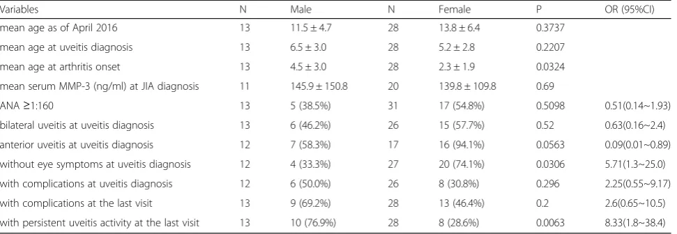
Risk factors were compared between males and females in the group with uveitis (Table 5). No significant sex differences were seen for age at uveitis diagnosis. Al-though there was also no significant difference in age at onset of arthritis in either males or females without uve-itis (6.9 ± 4.4-vs.-6.6 ± 4.3 years, P= 0.37, data not shown), females in the uveitis group had a significantly earlier arthritis onset (2.3 ± 1.9 vs. 4.5 ± 3.0 years old, P = 0.032). While the percentage of ANA-positive females at diagnosis of JIA was significantly higher in patients without uveitis (27.8%-vs.-10.1%, P< 0.0001, data not shown), there were no significant sex differences in the uveitis group. There was a tendency for more female than male patients to have anterior uveitis at diagnosis, but this difference was not significant (94.1%-vs.-58.3%,
P= 0.0563). The proportion of patients without ocular symptoms at diagnosis was higher in females than males (74.1%-vs.-33.3%, P= 0.031). Although there was no sig-nificant sex difference in the proportion of patients with Fig. 1Cumulative uveitis onset rate (vertical axis) in 41 JIA-U
patients, including 2 children developing uveitis before onset of arthritis (time 0). The horizontal axis indicates years from the onset of arthritis to diagnosis of uveitis
Table 3Ocular symptoms at uveitis diagnosis (N= 41a)
N (%)
Ocular symptom (+) 15 (36.6%)
Decreased vision 7 (46.7%)
Ocular hyperemia 7 (46.7%)
Photophobia 2 (13.3%)
Ocular pain 1 (6.7%)
Blurred vision 1 (6.7%)
Myodesopsia (floaters) 1 (6.7%)
White pupil 1 (6.7%)
a
2 data were unknown. There were patients who had multiple symptoms
Table 4Ocular complications in JIA-U patientsa
at uveitis diagnosis (N= 41b)
N (%)
at the last visit (N= 41) N (%)
Ocular complication (+) 13 (31.7%) 23 (56.1%)
Cataract 4 (30.8%) 12 (52.2%)
Glaucoma 3 (23.1%) 5 (21.7%)
Posterior synechia 6 (46.2%) 4 (17.4%)
Band-keratopathy 4 (30.8%) 7 (30.4%)
Other complications 1 (7.7%)c 2 (8.7%)d
a
There were patients who had multiple complications.b
3 data were unknown.
c
papilledema.d
any type of complication, the proportion of males with persistent active uveitis at the final observation was sig-nificantly higher than in females (76.9%-vs.-28.6%, P= 0.006). In addition, there were no significant sex differ-ences in visual acuity or decreased visual acuity at diag-nosis or at final observation. Furthermore, no significant sex difference was observed in time from arthritis onset to uveitis diagnosis.
Discussion
This is the first report surveying the epidemiology and characteristics of JIA-U in Japan. Our results are com-pared with previous reports in Table 6. It is apparent that the prevalence of JIA-U in Japan is lower than re-ported earlier from North America and Europe. These previous reports revealed regional differences in the prevalence of JIA-U: 11.6–20.5% in North America and Europe [1–5] and 4.7% in Taiwan [6]. Another study re-ported that the prevalence of JIA-U in Asia is lower than in Scandinavia as well as the United States [14]. Al-though these differences may be due to differences in study design, there may well also be an influence of race. Because almost all of the patients assessed in our survey were Asians, this population may have a lower incidence than predominantly Caucasian populations.
Our study confirmed that oligoarthritis, earlier arth-ritis onset, ANA-positivity, RF-negativity and anti-CCP antibody-negativity could be risk factors for JIA-U in Japanese as well as in predominantly Caucasian popula-tions. Several reports showed that oligoarthritis was the most common subtype of JIA with uveitis [2, 15, 16]. Likewise, in our study most patients (81.8%) with uveitis had oligoarthritis. While oligoarthritis accounts for ap-proximately 50% of all JIA patients in North America and Europe [2, 4, 5, 17], the proportion is smaller in Japan and many patients have systemic and RF-positive polyarthritis. The reason for the low prevalence of JIA-U
in Japan may be related to the lower proportion of oli-goarthritis in Japan than in North America and Europe. We compared the rate of uveitis in oligoarthritis patients and other arthritis patients between Japan and other countries (Table 6). The risk of uveitis in oligoarthritis patients in Japan is 10-fold, which is much higher than in oligoarthiritis patients in other countries. None of the patients with systemic arthritis and RF-positive polyar-thritis developed uveitis - consistent with previous re-ports [2,4,5,7,18]. Because patients with these types of arthritis have little risk of developing uveitis, in such cases, autoinflammatory diseases such as Blau syndrome, or infections should be considered rather than JIA. In the present study, it was difficult to determine the fre-quency of the HLA-B27 allele because too few patients were tested, but Japanese JIA is characterized by a low incidence of enthesitis-related arthritis, as shown in Table 2. In addition, psoriatic arthritis is also very rare. Previous reports [19,20] indicated that mean ANA titers tend to be high, with a significantly higher prevalence of ANA ≥1:320 in JIA patients with uveitis; our study also indicated a significant difference at≥1:640.
The mean age of onset of JIA-U has been variably re-ported to be between 3.2–10.9 years [1,2,5,18]; almost all cases are diagnosed before arthritis onset or within 4 years of onset [2, 4, 5, 15], and particularly within one year of the first ophthalmologic examination [2, 5] (not shown in Table 6). Furthermore, 10–24% patients de-velop uveitis before arthritis onset [1, 2,18]. More than 80% of the locations affected by uveitis are anterior and 60.6 to 72% are bilateral [1, 2,18]. Ocular complications including glaucoma, cataract, band-keratopathy, and posterior synechia at uveitis diagnosis were seen in be-tween 37.3 and 56% of uveitis patients [1, 2]. Because the presence of complications will influence the visual prognosis, early diagnosis and treatment of uveitis is crucial. However, failure to have regular ocular
Table 5Sex differences of characteristics in JIA-U patients
Variables N Male N Female P OR (95%CI)
mean age as of April 2016 13 11.5 ± 4.7 28 13.8 ± 6.4 0.3737
mean age at uveitis diagnosis 13 6.5 ± 3.0 28 5.2 ± 2.8 0.2207
mean age at arthritis onset 13 4.5 ± 3.0 28 2.3 ± 1.9 0.0324
mean serum MMP-3 (ng/ml) at JIA diagnosis 11 145.9 ± 150.8 20 139.8 ± 109.8 0.69
ANA≥1:160 13 5 (38.5%) 31 17 (54.8%) 0.5098 0.51(0.14~1.93)
bilateral uveitis at uveitis diagnosis 13 6 (46.2%) 26 15 (57.7%) 0.52 0.63(0.16~2.4)
anterior uveitis at uveitis diagnosis 12 7 (58.3%) 17 16 (94.1%) 0.0563 0.09(0.01~0.89)
without eye symptoms at uveitis diagnosis 12 4 (33.3%) 27 20 (74.1%) 0.0306 5.71(1.3~25.0)
with complications at uveitis diagnosis 12 6 (50.0%) 26 8 (30.8%) 0.296 2.25(0.55~9.17)
with complications at the last visit 13 9 (69.2%) 28 13 (46.4%) 0.2 2.6(0.65~10.5)
with persistent uveitis activity at the last visit 13 10 (76.9%) 28 8 (28.6%) 0.0063 8.33(1.8~38.4)
examinations may lead to delayed diagnosis because JIA-U is asymptomatic and insidious in many cases [2]. In the present study, 36.6% of patients had ocular symp-tom at uveitis diagnosis; one of the reasons for the large number of patients with ocular symptoms may be the lack of regular routine ophthalmologic examinations in Japan. Similar to previous reports, the most commonly affected location of the uveitis was anterior, whereas fewer cases were bilateral. In our study, the median age at diagnosis of uveitis was 5 years, 80.5% of the patients developed uveitis before they reached 8 years of age, and 95.1% developed it under 7 years of arthritis onset. Thus, the risk of onset of uveitis is high for patients under 8 years of age and under 7 years from arthritis onset. These results suggest that recommendations for stand-ard ophthalmological follow-up of Japanese JIA patients should consider patient age, and time from arthritis onset.
Except for two patients, all were treated with MTX, a medication previously shown to be effective for uveitis
[21–23]. However, we could not determine whether treatment with MTX was effective in the present study.
Heiligenhaus et al. stated that ocular complications at the first visit to an ophthalmologist could be predictive of ocular complications at final observation [2]. In addition, Woreta et al. concluded that a short interval between arthritis and uveitis onset, ANA-positivity and the degree of ocular inflammation at the initial diagno-sis, were all risk factors for complications [24]. Previous reports showed that cataract is associated with systemic steroid therapy, the amount of steroid eye drops and de-gree of ocular inflammation [25–28]. We found that the proportion of patients with complications increased from 31.7% at the first visit to an ophthalmologist to 56.1% at the final observation, and it was especially strik-ing that the number of patients with cataract increased 3-fold over this period. Patients with complications at the initial diagnosis indeed had significantly more com-plications at the final observation, but there was no asso-ciation between the presence of complications and age
Table 6Comparison of epidemiology and characteristics of JIA-U in our results vs. in previous reports
study our study Saurenmann,
2007 [1]
Heiligenhaus, 2007 [2]
Nordal, 2017 [5] Angeles-Han, 2015 [18]
country Japan Switzerland Germany Norway United States of
America
prevalence 6.1% 13.1% 12% 20.5% 18%
JIA subtype
oligo 81.8% 48% 41% 46.1% 78.9%
RF + poly 0% 0% 0% 0% 0%
Systemic 0% 0.6% 0% 0% 0%
Rate of uveitis in oligo 16.1% 20.9% 17.6% 19.6% 30.8%
Rate of uveitis in all other type 1.6% 8.3% 6.7% 21.3% 7.1%
Risk ratio of uveitis in oligo 10 2.5 2.6 0.9 4.3
femalea 67.6% vs. 70.5%,
p= 0.743
79.6% vs. 63.7%,
p= 0.0009b 74% vs. 62%,p= 0.012 65.6% vs. 66.3% 76.9% vs. 70.2%,p= 0.332
ANA-positive (%)a 52.3% vs. 22.2%,
p< 0.0001
80.9% vs. 51%, p< 0.0001
86% vs. 42%, p< 0.01
42.5% vs. 23.2% 54.9% vs. 35.9%, p= 0.017 RF-positive (%)a 2.4% vs. 24.5%,
p= 0.0004 – –
1.1% vs. 2.7% 0% vs. 11.1%, p= 0.013 the mean age of uveitis
onset (yrs.)
5.6 6.2 5.2 10.8(acute) and
3.2(chronic)
median 4.8
the mean age of arthritis
onset (yrs.)a 3.8 vs. 6.8 4.3 vs. 7.3 3.8 vs. 7.0 – median 2.8 vs. 7.7
time from arthritis onset to uveitis diagnosis
median 2 yrs. mean 1.8 yrs. median 5.5 months – –
uveitis diagnosis before the arthritis diagnosis
4.9% 12.7% 10% – 24%
anterior uveitis 79.3% 100% 83% – 80%
bilateral uveitis 53.7% 60.6% 70% – 72%
asymptomatic uveitis 58.5% 69.7% – – –
a
Comparison of patients with uveitis vs. without uveitis,p-value.b
at diagnosis of uveitis, or the time from arthritis onset to uveitis diagnosis, and ANA-positivity. There were also no significant differences between complications and persisting active uveitis at the final observation. In addition, we found that systemic steroid therapy had no influence on cataract formation, and the presence of cataract at the first visit had no influence on the surgical history. JIA-U carries a risk of decreased visual acuity and blindness [29, 30]. While none of the patients lost their eyesight over the observation period in our study, more than half did not fully recover visual acuity or had decreased visual acuity at the final observation. Steroid eye drops, oral MTX, and biological agents (IFX and ADA) were mainly used for treatment, and especially biological agents had been employed in a high propor-tion of patients. This may be one of the reasons why there was no loss of eyesight and few patients with se-verely decreased vision in our study.
While female sex is reported as a risk factor for JIA-U, severity of the uveitis may be greater in males [31–33]. In comparison, in our study, female sex was not a risk factor for JIA-U, but males with uveitis did have a sig-nificantly higher incidence of active uveitis at the last observation.
This study has some limitations. It included 726 Japa-nese JIA patients, representing approximately 1/4 of all JIA patients in Japan and an estimated half of all patients treated in medical centers specialized in pediatric rheumatic diseases. Although Japan is divided into 47 prefectures, there are only about 80 pediatric rheumatol-ogists and they are unevenly distribution locally, so gen-eral pediatricians treat JIA in areas where there are no pediatric rheumatologists. On the other hand, because severe cases are often referred to a pediatric rheumatolo-gist, many JIA-U will be treated by pediatric rheumatol-ogists. Accordingly, the prevalence of uveitis in Japan as established here may actually be even less than 6% of all JIA patients.
Just as there are few pediatric rheumatologists, so there are also few ophthalmologists who specialize in uveitis in Japan. In addition, although the standardization of uveitis nomenclature (SUN) working group reported criteria to evaluate uveitis [34], few ophthalmologists in Japan use these criteria. Therefore, in the present study, because pedi-atricians extracted the records written by each ophthal-mologist there were some uninterpretable data, especially the location of uveitis. Because of the retrospective nature of this study, there were also some missing data such as data on visual acuity. Although we issued recommendations for ophthalmologic screening intervals for JIA in 2015 [9], because we issued it one year before this study, most pa-tients were not receiving screening according to this rec-ommendation. Hence, only 34% of patients had regular ophthalmologic screening before uveitis diagnosis, and 34%
had ocular symptoms at the time of uveitis diagnosis in this study. JIA-U is typically asymptomatic and insidious [2], so if patients have regular ophthalmologic examinations, ocu-lar symptoms may be identified less frequently at the time of JIA diagnosis, but more often beforehand.
We did not collect data on the degree of ocular in-flammation and the dose of steroids, and so we were unable to analyze potential associations between uve-itis and these factors. Also, we did not collect detailed data on dose and timing of drug use for any drugs other than steroids, so we could not analyze the ef-fects of these treatments on uveitis. Thus, because de-tailed analysis of treatment was difficult in this study, we plan an additional study in cooperation with oph-thalmologists in future.
Conclusions
We investigated whether the prevalence and characteris-tics of JIA-U in Japan are different from those reported elsewhere. We conclude that the prevalence of JIA-U in Japan is lower than reported in countries with predom-inantly Caucasian populations. Risk factors for JIA-U were identified as oligoarthritis, early arthritis onset, ANA-positivity, RF-negativity and anti-CCP antibody-negativity. Arthritis onset was significantly earlier in females. Although the most frequent location of uveitis was anterior, males tended to have more af-fected locations in other the parts of the eye, and the uveitis remission rate was lower than in females. Thus, in males, uveitis may be more severe and harder to cure. Once uveitis had developed, many patients did not fully recover their eyesight. In addition, we found that uveitis may develop before arthritis onset, or under treatment for arthritis and even after cessation of arthritis medica-tion. It is important to recognize these characteristics when caring for Japanese patients with JIA. Patients under 8 years of age and under 7 years of arthritis onset require especially careful specialist ophthalmological monitoring.
Abbreviations
ADA:Adalimumab; ANA: Antinuclear antibodies; anti-CCP: anti-Cyclic citrullinated peptides; CI: Confidence interval; GC: Glucocorticoid; HLA-B27: Human leucocyte antigen B27; IFX: Infliximab; ILAR: International League of Associations for Rheumatology; JIA: Juvenile idiopathic arthritis; JIA-U: Juvenile idiopathic arthritis associated uveitis; MMP-3: Matrix Metalloproteinase-3; MTX: Methotrexate; NSAID: Non-steroidal anti-Inflammatory drugs; OR: Odds ratio; PRAJ: Pediatric Rheumatology Association of Japan; SUN: Standardization of Uveitis Nomenclature; TCZ: Tocilizumab
Acknowledgments
Yamaide (Department of Allergy and Rheumatology, Chiba Children’s Hospital, Chiba, Japan), Dr. Naoki Shimojo, Dr. Yuzaburo Inoue, Dr. Hironori Sato (Department of Pediatrics, Chiba University Graduate School of Medicine, Chiba, Japan), Dr. Shunichiro Takezaki (Department of Pediatrics, Hokkaido University Graduate School of Medicine, Sapporo, Japan), Dr. Fumiko Okazaki (Department of Pediatrics, Yamaguchi University Graduate School of Medicine, Ube, Japan), Dr. Tomo Nozawa, Dr. Asami Oohara, Dr. Ayako Murase and Dr. Tetsuya Tsuchida (Department of Pediatrics, Yokohama City University Graduate School of Medicine, Yokohama, Japan) for collecting the medical records. We thank Dr. Syuji Takei (Department of Pediatrics, Kagoshima University Graduate School of Medicine, Kagoshima, Japan) for analyzing data. We thank Dr. Norihiro Nishimoto (Department of Molecular Regulation for Intractable Disease, Institute of Medical Science, Tokyo Medical University, Tokyo, Japan) and Dr. Toshihiro Matsui (Department of Rheumatology, Clinical Research Center for Allergy and Rheumatology, National Hospital Organization Sagamihara National Hospital, Sagamihara, Japan) for research planning.
Funding
This work was supported by Health Labour Sciences Research Grant: 201510096A.
Availability of data and materials
The datasets used and/or analyzed during the current study are not publicly available for ethical reasons, as well as privacy reasons, but are available from the corresponding author on reasonable request.
Authors’contributions
All authors were involved in the conception, design of the study and revising it critically for important intellectual content. JY and MY were involved in the acquisition of data, analysis, interpretation of data and drafting of the manuscript. All authors read and approved the final manuscript.
Ethics approval and consent to participate
This is a retrospective study, approved by the Ethics Review Board of Tokyo Medical and Dental University (No.M2015–537), the main study center, on March 4th, 2016, and then approval was obtained from the 15 additional medical centers. Guardians and patients were provided information by means of an opt-out form.
Consent for publication
Not applicable.
Competing interests
Tokyo Medical and Dental University (TMDU) received unrestricted research grants for Department of Lifetime Clinical Immunology from AbbVie GK, Ayumi Pharmaceutical, Chugai Pharmaceutical, CSL Behring, Japan Blood Products Organization, Mitsubishi Tanabe Pharma, Nippon Kayaku, Ono Pharmaceutical, Towa Pharmaceutical, and UCB Japan. TMDU paid the salary of Masaaki Mori.
The authors declare that they have no competing interests.
Publisher’s Note
Springer Nature remains neutral with regard to jurisdictional claims in published maps and institutional affiliations.
Author details
1Department of Pediatrics, Hiroshima University Graduate School of
Biomedical and Health Sciences, 1-2-3 Kasumi, Minami-ku, Hiroshima 734-8551, Japan.2Department of Pediatrics, Okayama University Hospital,
2-5-1 Shikata-cho, Kita-ku, Okayama 700-8558, Japan.3Department of
Pediatrics, Osaka Medical College, 2-7 Daigaku-machi, Takatsuki 569-8686, Japan.4Department of General Pediatrics, Miyagi Children’s Hospital, 4-3-17 Ochiai, Aoba-ku, Sendai 989-3126, Japan.5Department of Immunology and
Infectious Diseases, Aichi Children’s Health and Medical Center, 7-426 Morioka-cho, Obu, Aichi 474-8710, Japan.6Department of Pediatrics, KKR
Sapporo Medical Center, 6-3-40 Hiragishi 1-jo, Toyohira-ku, Sapporo 062-0931, Japan.7Department of Pediatrics, Kagoshima University Hospital,
8-35-1 Sakuragaoka, Kagoshima 890-0075, Japan.8Department of Pediatrics,
Graduate School of Medical Sciences, Kanazawa University, 13-1
Takara-machi, Kanazawa 920-8641, Japan.9Department of Allergy and
Rheumatology, Chiba Children’s Hospital, 579-1 Heta-cho, Midori-ku, Chiba 266-0007, Japan.10Department of Pediatric Rheumatology, Hyogo Prefectural
Kobe Children’s Hospital, 1-6-7 Minatojimaminami-machi, Chuo-ku, Kobe 650-0047, Japan.11Department of Pediatrics, Yokohama City University
Graduate School of Medicine, 3-9 Fukuura, Kanazawa-ku, Yokohama 236-0004, Japan.12Department of Pediatrics, Kyoto University Graduate
School of Medicine, 54 Shogoin Kawahara-cho, Sakyo-ku, Kyoto 606-8507, Japan.13Department of Pediatrics, Chiba University Graduate School of
Medicine, 1-8-1 Inohana, Chuo-ku, Chiba 260-8670, Japan.14Department of
Pediatrics, Yamaguchi University Graduate School of Medicine, 1-1-1 Minamikogushi, Ube 755-8505, Japan.15Department of Lifetime Clinical Immunology, Graduate School of Medical and Dental Sciences, Tokyo Medical and Dental University, 1-5-45 Yushima, Bunkyo-ku, Tokyo 113-8510, Japan.
Received: 7 January 2019 Accepted: 1 April 2019
References
1. Saurenmann RK, Levin AV, Feldman BM, Rose JB, Laxer RM, Schneider R, et al. Prevalence, risk factors, and outcome of uveitis in juvenile idiopathic arthritis: a long-term followup study. Arthritis Rheum. 2007;56:647–57. 2. Heiligenhaus A, Niewerth M, Ganser G, Heinz C, Minden K. German uveitis in
childhood study group. Prevalence and complications of uveitis in juvenile idiopathic arthritis in a population-based nation-wide study in Germany: suggested modification of the current screening guidelines. Rheumatology. 2007;46:1015–9.
3. Kotaniemi K, Sihto-Kauppi K, Salomaa P, Säilä H, Ristolainen L, Kauppi M. The frequency and outcome of uveitis in patients with newly diagnosed juvenile idiopathic arthritis in two 4-year cohorts from 1990-1993 and 2000-2003. Clin Exp Rheumatol. 2014;32:143–7.
4. Angeles-Han ST, Pelajo CF, Vogler LB, Rouster-Stevens K, Kennedy C, Ponder L, et al. Risk markers of juvenile idiopathic arthritis-associated uveitis in the childhood arthritis and rheumatology research Alliance (CARRA) registry. J Rheumatol. 2013;40:2088–96.
5. Nordal E, Rypdal V, Christoffersen T, Aalto K, Berntson L, Fasth A, et al. Incidence and predictors of uveitis in juvenile idiopathic arthritis in a Nordic long-term cohort study. Pediatr Rheumatol. 2017;15:66.
6. Yu HH, Chen PC, Wang LC, Lee JH, Lin YT, Yang YH, et al. Juvenile idiopathic arthritis-associated uveitis: a Nationwide population-based study in Taiwan. PLoS One. 2013;8:e70625.
7. Kotaniemi K, Savolainen A, Karma A, Aho K. Recent advances in uveitis of juvenile idiopathic arthritis. Surv Ophthalmol. 2003;48:489–502. 8. Berk AT, Koçak N, Ünsal E. Uveitis in juvenile idiopathic arthritis. Ocul
Immunol Inflamm. 2001;9:243–51.
9. Okamoto N, Iwata N, Umebayashi H, Okura Y, Kinjo N, Kunishima T, et al. Guidance for early treatment of juvenile idiopathic arthritis. Tokyo: Medical Review Corporation; 2015.
10. Okamoto N, Yokota S, Takei S, Okura Y, Kubota T, Shimizu M, et al. Clinical practice guidance for juvenile idiopathic arthritis (JIA) 2018. Mod Rheumatol. 2018;29:1–19.https://doi.org/10.1080/14397595.2018.1514724[Epub ahead of print].
11. American Academy of Pediatrics Section on Rheumatology and Section on Ophthalmology: Guidelines for ophthalmologic examinations in children with juvenile rheumatoid arthritis. Pediatrics. 1993;92:295–6.
12. Petty PE, Southwood TR, Manners P, Baum J, Glass DN, Goldenberg J, et al. International league of associations for rheumatology classification of juvenile idiopathic arthritis: second revision, Edmonton, 2001. J Rheumatol. 2004;31:390–2. 13. Ferris FL 3rd, Kassoff A, Bresnick GH, Bailey I. New visual acuity charts for
clinical research. Am J Ophthalmol. 1982;94:91–6.
14. Carvounis PE, Herman DC, Cha S, Burke JP. Incidence and outcomes of uveitis in juvenile rheumatoid arthritis, a synthesis of the literature. Graefes Arch Clin Exp Ophthalmol. 2006;244:281–90.
15. Kotaniemi K, Kautiainen H, Karma A, Aho K. Occurrence of uveitis in recently diagnosed juvenile chronic arthritis: a prospective study. Ophthalmology. 2001;108:2071–5.
17. Petty R, Laxer R, Lindsley C, Wedderburn L. Textbook of pediatric rheumatology, vol. 119. 7th ed. Philadelphia: Elsevier; 2015.
18. Angeles-Han ST, McCracken C, Yeh S, Jenkins K, Stryker D, Rouster-Stevens K, et al. Characteristics of a cohort of children with Juvenile Idiopathic Arthritis and JIA-associated Uveitis. Pediatric Rheumatol. Online J. 2015;13:–19.
https://doi.org/10.1186/s12969-015-0018-8.
19. Chen CS, Roberton D, Hammerton ME. Juvenile arthritis-associated uveitis: visual outcomes and prognosis. Can J Ophthalmol. 2004;39:614–20. 20. Nordal EB, Songstad NT, Berntson L, Moen T, Straume B, Rygg M. Biomarkers
of chronic uveitis in juvenile idiopathic arthritis: predictive value of antihistone antibodies and antinuclear antibodies. J Rheumatol. 2009;36: 1737–43.
21. Yu EN, Meniconi ME, Tufail F, Baltatzis S, Foster CS. Outcomes of treatment with immunomodulatory therapy in patients with corticosteroid-resistant juvenile idiopathic arthritis-associated chronic iridocyclitis. Ocul Immunol Inflamm. 2005;13:353–60.
22. Heiligenhaus A, Mingels A, Heinz C, Ganser G. Methotrexate for uveitis associated with juvenile idiopathic arthritis: value and requirement for additional anti-inflammatory medication. Eur J Ophthalmol. 2007;17:743–8. 23. Foeldvari I, Wierk A. Methotrexate is an effective treatment for chronic
uveitis associated with juvenile idiopathic arthritis. J Rheumatol. 2005;32: 362–5.
24. Woreta F, Thorne JE, Jabs DA, Kedhar SR, Dunn JP. Risk factors for ocular complications and poor visual acuity at presentation among patients with uveitis associated with juvenile idiopathic arthritis. Am J Ophthalmol. 2007; 143:647–55.
25. Wolf MD, Lichter PR, Ragsdale CG. Prognostic factors in the uveitis of juvenile rheumatoid arthritis. Ophthalmology. 1987;94:1242–8.
26. Kump LI, Castañeda RA, Androudi SN, Reed GF, Foster CS. Visual outcomes in children with juvenile idiopathic arthritis-associated uveitis.
Ophthalmology. 2006;113:1874–7.
27. Angeles-Han S, Yeh S. Prevention and management of cataracts in children with juvenile idiopathic arthritis-associated uveitis. Curr Rheumatol Rep. 2012;14:142–9.
28. Thorne JE, Woreta FA, Dunn JP, Jabs DA. Risk of cataract development among children with juvenile idiopathic arthritis-related uveitis treated with topical corticosteroids. Ophthalmology. 2010;117:1436–41.
29. Thorne JE, Woreta F, Kedhar SR, Dunn JP, Jabs DA. Juvenile idiopathic arthritis-associated uveitis: incidence of ocular complications and visual acuity loss. Am J Ophthalmol. 2007;143:840–6.
30. de Boer J, Wulffraat N, Rothova A. Visual loss in uveitis of childhood. Br J Ophthalmol. 2003;87:879–84.
31. Holland GN, Denove CS, Yu F. Chronic anterior uveitis in children: clinical characteristics and complications. Am J Ophthalmol. 2009;147:667–78. 32. Chia A, Lee V, Graham EM, Edelsten C. Factors related to severe uveitis at
diagnosis in children with juvenile idiopathic arthritis in a screening program. Am J Ophthalmol. 2003;135:757–62.
33. Kalinina Ayuso V, Ten Cate HA, van der Does P, Rothova A, de Boer JH. Male gender and poor visual outcome in uveitis associated with juvenile idiopathic arthritis. Am J Ophthalmol. 2010;149:987–93.
![Table 1 Recommendations for ophthalmologic screening intervals for JIA patients in Japan [9, 10]](https://thumb-us.123doks.com/thumbv2/123dok_us/9525665.1481780/2.595.58.540.542.723/table-recommendations-ophthalmologic-screening-intervals-jia-patients-japan.webp)