R E V I E W
Acquired auditory-visual synesthesia: A window
to early cross-modal sensory interactions
Pegah Afra Michael Funke Fumisuke Matsuo
Department of Neurology, University of Utah, Salt Lake City, UT, USA
Correspondence: Pegah Afra Department of Neurology, University of Utah, 175 North Medical Drive East, Salt Lake City, UT 84132, USA Tel +1 608 213 1458 Email pegah.afra@hsc.utah.edu
Abstract: Synesthesia is experienced when sensory stimulation of one sensory modality elicits an involuntary sensation in another sensory modality. Auditory-visual synesthesia occurs when auditory stimuli elicit visual sensations. It has developmental, induced and acquired varieties. The acquired variety has been reported in association with deafferentation of the visual system as well as temporal lobe pathology with intact visual pathways. The induced variety has been reported in experimental and post-surgical blindfolding, as well as intake of hallucinogenic or psychedelics. Although in humans there is no known anatomical pathway connecting auditory areas to primary and/or early visual association areas, there is imaging and neurophysiologic evidence to the presence of early cross modal interactions between the auditory and visual sensory pathways. Synesthesia may be a window of opportunity to study these cross modal interactions. Here we review the existing literature in the acquired and induced auditory-visual synesthesias and discuss the possible neural mechanisms.
Keywords: synesthesia, auditory-visual, cross modal
The term synesthesia is composed of two parts, syn= together and aesthesia= sensation. It denotes that sensory stimulation of one sensory modality (the inducer) will elicit an involuntary or automatic sensation in another sensory modality (the concurrent).1
For the purpose of clear communication in this paper synesthesia will be addressed by two consecutive words of inducer-concurrent. For example, if auditory stimuli evoke visual sensations, it is called auditory-visual synesthesia. Although there is no standard terminology, this has been the most common trend in the past decade.
Synesthesia has been scientifi cally reported as early as 1883 by Galton2 in an
assay in which he referred to published earlier cases including the ones by Bleuler and Lehman in 1881. As a result there is centenarian literature on developmental as well as drug-induced synesthesias.
The developmental synesthesias are not a neurologic disorder but a different way of experiencing one’s environment. They are life-long (ie, the subjects remember them back to their childhood), idiosyncratic, automatic, involuntary and consistent experiences across ones, lifespan.3 The acquired variety is different in that it is
emer-gent and usually happens subsequent to neuropathologic insult to the brain. There is also induced synesthesia that can happen with sensory deprivation as well as intake of hallucinogenic or psychedelics.4–7
In 1977, Bender brought this topic to neurologic attention by describing a case of acquired auditory-visual synesthesia.8 Since, there have been several case reports
and case series in the acquired variety, but the topic stays reported and under-appreciated in the fi eld of neurology. In this paper we review the existing literature in the most common acquired variety, auditory-visual synesthesia.8–15 We also mention
briefl y less common reported acquired varieties as well as the induced synesthesias.
Psychology Research and Behavior Management downloaded from https://www.dovepress.com/ by 118.70.13.36 on 27-Aug-2020
Afra et al
Developmental synesthesias are not discussed here and the interested reader is encouraged to learn about this by referring to the already existing and extensive literature on the topic by cognitive neuropsychologist colleagues.
Acquired auditory-visual
synesthesia
The majority of the acquired synesthesias have been reported in the setting of sensory deafferentation, when sensory stimuli to another sensory system is experienced ectopically in the deafferented system. There are also few case reports of acquired synesthesia with intact sensory pathways. Here the acquired auditory-visual variety is divided into two groups, depending on the presence or absence of visual deafferentation.
Auditory-visual synesthesia associated
with visual deafferentation
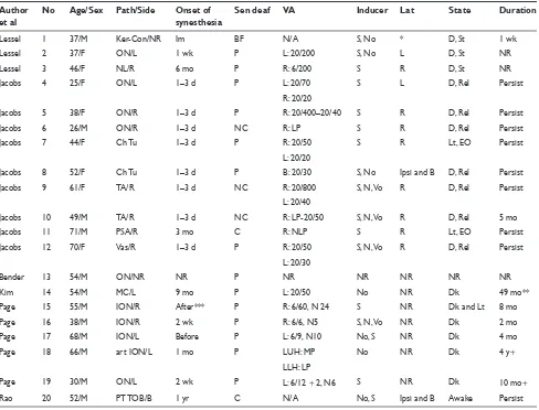
In all the reported cases with sensory deafferentation, the neu-ropathologic abnormality involves the anterior optic pathways (optic nerve, optic chiasm or both). Table 1 summarizes the clinical details of twenty existing cases of acquired auditory visual synesthesia with visual deafferentation across different case series and reports.8–13 In brief the underlying
neuropathol-ogy involves the optic nerve and/or chiasm and consists of demyelination, ischemia, or tumors as well as post-traumatic total ocular blindness. The reported visual sensation is not well-formed and rather simple, like light fl ashes, kaleido-scope, color, etc. The very fi rst case in Table 1 (patient #1) is a case of induced synesthesia due to surgical blindfolding of the external eye after surgery for keratoconus. This case is discussed separately under induced synesthesias. The other 19 cases (#2–20) are discussed below depending on degree of deafferentation, onset of synesthesia, laterality of the con-current in relation to the inducer, duration, vigilance during synesthesia experience and neurophysiologic testing.
Degree of deafferentation
Synesthesia was associated with varying degrees of deaf-ferentation. In two cases complete blindness ie, no light perception was reported by the patient. One (#20) had absent visual evoked potentials (VEPs) and in the other (#11) VEP showed incomplete deafferentation. The rest of the cases (#2–10 and #12–19) had mild to severe partial deafferentaion as determined by visual acuity (see Table 1).
Onset of synesthesia
The reported onset of synesthesia in relation to the clinical onset of the visual symptoms was very variable, ranging
from synestheisa preceding any visual symptoms to cases that synesthesia started months after. We divided the reported onset of synethesia in relation to clinical onset of visual symptoms into four groups of: preceding, acute (one to three days), sub-acute (one to four weeks) and chronic (in months). These are summarized in Table 2. With the exception of one case, synesthesia followed onset of visual symptoms and hence a variable period of deafferentation. There seems to be no relationship between the onset of synesthesia and the underlying neuropathology.
Laterality
Table 1 provides information about the laterality in each patient. Laterality refers to the side of the auditory stimuli (inducer) in relation to the side of the visual perception (concurrent). In 12 of the cases where laterality was reported, the induced visual perception was always ipsilateral to the side of the perceived auditory stimuli. In fi ve of these 12 cases synesthesia was experienced in the area of scotoma itself (#4, #6, #7, #9, and #11). Regardless of laterality of the inducer in relation to the concurrent, the induced visual phenomenon was perceived in the deafferented eye with auditory stimuli in 16 of the 19 cases. In patients #13–14, laterality was not reported. Patient #15 was able to lateralize the auditory stimulus, but the induced phosphenes always happened in front of his eyes.
Vigilance
Most patients experienced their synesthesia when in dark and in a relaxed state. Four of the patients (#7, #11, #15, and #20) experienced their synesthesia in light. These included the two patients who reported no light perception (one with absent VEP and one with VEP showing incomplete deafferentation), and two patients with partial deafferentation.
Duration
In 16 cases the duration of synesthesia was reported. In fi ve of the 16 cases synesthesia was a transient phenomenon of variable duration associated with partial deafferentation. In 11 cases synesthesia was persistent. Interestingly, synesthesia persisted 16 months after enucleation of the left eye in patient #14.
Neurophysiologic evidence
Table 3 summarizes the neurophysiologic studies done across all patients.
a. Visual evoked potentials (VEPs). Thirteen of the patients had VEPs and these showed abnormalities of anterior optic pathways as expected.
Psychology Research and Behavior Management downloaded from https://www.dovepress.com/ by 118.70.13.36 on 27-Aug-2020
Acquired auditory-visual synesthesia
Table 1 Clinical patient summary of reported cases of acquired auditory-visual synesthesia in the literature Author
et al
No Age/Sex Path/Side Onset of synesthesia
Sen deaf VA Inducer Lat State Duration
Lessel 1 37/M Ker-Con/NR Im BF N/A S, No * D, St 1 wk
Lessel 2 37/F ON/L 1 wk P L: 20/200 S, No L D, St NR
Lessel 3 46/F NL/R 6 mo P R: 6/200 S R D, St NR
Jacobs 4 25/F ON/L 1–3 d P L: 20/70 S L D, Rel Persist
R: 20/20
Jacobs 5 38/F ON/R 1–3 d P R: 20/400–20/40 S R D, Rel Persist
Jacobs 6 26/M ON/R 1–3 d NC R: LP S R D, Rel Persist
Jacobs 7 44/F Ch Tu 1–3 d P R: 20/50 S R Lt, EO Persist
L: 20/20
Jacobs 8 52/F Ch Tu 1–3 d P B: 20/30 S, No Ipsi and B D, Rel Persist
Jacobs 9 61/F TA/R 1–3 d NC R: 20/800 S, N, Vo R D, Rel Persist
L: 20/40
Jacobs 10 49/M TA/R 1–3 d NC R: LP-20/50 S, N, Vo R D, Rel 5 mo
Jacobs 11 71/M PSA/R 3 mo C R: NLP S R Lt, EO Persist
Jacobs 12 70/F Vas/R 1–3 d P R: 20/50 S, N, Vo R D, Rel Persist
L: 20/30
Bender 13 54/M ON/NR NR P NR NR NR NR NR
Kim 14 54/M MC/L 9 mo P L: 20/50 No NR Dk 49 mo**
Page 15 55/M ION/R After*** P R: 6/60, N 24 S NR Dk and Lt 8 mo
Page 16 38/M ION/R 2 wk P R: 6/6, N5 S, N, Vo NR Dk 2 mo
Page 17 68/M ION/L Before P L: 6/9, N10 No, S NR Dk 4 mo
Page 18 66/M art ION/L 1 mo P LUH: MP No NR Dk 4 y+
LLH: LP
Page 19 30/M ON/L 2 wk P L: 6/12 + 2, N6 S NR Dk 10 mo+
Rao 20 52/M PT TOB/B 1 yr C N/A No, S Ipsi and B Awake Persist
Abbreviations: No, number of patients as referenced in the text; F, female; M, male; Path, pathology; NR, not reported; Ker-Con, keraotconus; ON, optic neuritis; NL, neurilemma; Ch Tu, chiasmal tumor; TA, temporal arteritis; PSA, post-surgical amauresis; Vas, vascular occlusive disease; MC, melanocytoma; ION, ischemic optic neuropathy; art, arteritic; PT TOB, post-traumatic total ocular blindness; Im, Immediate; wk, week; d, day; mo, month; yr, year; Sens deaf, degree of sensory deafferentation by patient report; BF, blindfolded; P, partial; C, complete; NC, near complete; VA, visual acuity of the abnormal eye; NR, not reported; LP, light perception; NLP, no light perception; MP, movement perception; LUH, left upper half; LLH, left lower half; R, right; L, left; Ind, inducer; S, sound; No, Noise; Vo, voice; Lat, laterality of inducer-concurrent (see text); Ipsi, ipsilateral; B, bilateral; D, drowsy; St, still; Rel, relaxed; Dk, dark; Lt, light; +, persistent after the stated time.
Notes: *Flash of light localized approximately to the site at which the sound originated; **Total of 49 months including 16 months post-enucleation of the left eye; ***not reported in days, weeks or months.
b. Electroretinogram (ERG). Two of the patients (#15 and #16) had this test done and there was no ERG activation after sound stimuli.
c. Auditory evoked potentials (AEPs). In nine of the cases AEPs were performed showing intact auditory path-ways. There was no mention of presence or absence of any occipital activation with the exception of #20, a case report by Rao and colleagues.13 In this case of
post-traumatic total blindness, there was occipital activa-tion with auditory evoked potentials.
Other (not included in Table 1)
In all patients with the exception of three (#13, #14, and #20), the presence of positive visual phenomenon were
addressed. Four of these 16 patients (#4, #5, #7, and #10) had concomitant positive visual phenomenon that was not sound induced. One patient (#16) had phosphenes provoked by eye movements in his initial presentation, but not concomitant with synesthesia. The remaining 11 patients did not experi-ence positive visual phenomenon.
Auditory-visual synesthesia associated
with intact visual pathways
but neuropathologic affection of CNS
The reported cases are confi ned to synesthesia experienced with pathologic involvement of temporal lobe.14,15Vike and colleagues14 reported a case of synesthesia
with intact visual pathways. This patient had a gliotic mass
Psychology Research and Behavior Management downloaded from https://www.dovepress.com/ by 118.70.13.36 on 27-Aug-2020
Afra et al
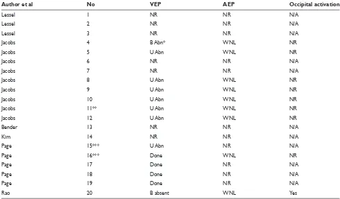
Table 3 Neurophysiological studies in patients with auditory-visual synesthesia with visual deafferentation
Author et al No VEP AEP Occipital activation
Lessel 1 NR NR N/A
Lessel 2 NR NR N/A
Lessel 3 NR NR N/A
Jacobs 4 B Abn* WNL NR
Jacobs 5 U Abn WNL NR
Jacobs 6 NR NR N/A
Jacobs 7 NR NR N/A
Jacobs 8 U Abn WNL NR
Jacobs 9 U Abn WNL NR
Jacobs 10 U Abn WNL NR
Jacobs 11** U Abn WNL NR
Jacobs 12 U Abn WNL NR
Bender 13 NR NR N/A
Kim 14 NR NR N/A
Page 15*** U Abn NR N/A
Page 16*** Done WNL NR
Page 17 Done NR N/A
Page 18 Done NR N/A
Page 19 Done NR N/A
Rao 20 B absent WNL Yes
Abbreviations: No, number of patients as referenced in the text; VEP, visual evoked potential; AEP, auditory evoked potential; NR, not reported; B, bilateral; U, unilateral; Abn, abnormality; Done, exact results not reported; WNL, normal; N/A, not applicable (because AEPs were not done).
Notes: *Due to presence of left optic neuritis and past history of right retrobulbar neuritis (four years prior); **VEP showed incomplete deafferentation despite amauresis reported by patient; ***No electroretinogram (ERG) activation by sound.
Table 2 Summary of onset of synesthesia in relation to visual deafferentation
Onset of synesthesia No Pathology: number of cases
Before 17 ION: 1
Acute (1–3 d) 4, 5, 6, 7, 8, 9, 10, 12 ON: 3, ChTu: 2, TA: 1
Sub-acute (1–4 wk) 2,16,18,19 ION: 1, ON: 2, arteritic ION: 1
Chronic (months) 3, 11, 14, 20 NL: 1, MC: 1, PT TOB: 1
Abbreviations: No, number of patients as referenced in the text; ION, ischemic optic neuritis; ON, optic neuritis; ChTu, chiasmal tumor; TA, temporal arteritis; NL, neurilemoma; MC, melanocytoma; PT TOB, post-traumatic total ocular blindness.
involving his left mesial temporal area and the adjacent midbrain. Synesthesia in this case was reported to be bilateral but more on the left (ipsilateral side). Patient had normal AEP and VEPs and no comment was made about occipital activation. Synesthesia was aborted after the removal of tumor.
Synesthesia-like symptoms was reported by Jacome and colleagues15 in a case of left temporal lobe seizures. During
his aura the patient experienced what the authors called “internal synesthesia”. After sudden onset of the pain on the right side of his face, he simultaneously heard the word
“fi ve” in his both ears and saw the number “5” or “fi ve” on a grey background before his eyes.
Other acquired synesthesias
Acquired auditory-tactile synesthesia
Ro and colleagues16 reported a case of auditory-tactilesynesthesia in a 36-year-old right-handed female with a lacunar infarct involving the ventrolateral (VL) nucleus of thalamus. One year after the stroke, patient exhibited multisensory neglect for vision and touch and tactile and visual antiextinction. Three years after her stroke, the patient
Psychology Research and Behavior Management downloaded from https://www.dovepress.com/ by 118.70.13.36 on 27-Aug-2020
Acquired auditory-visual synesthesia
was reported to experience auditory-tactile synesthesia contralateral to the thalamic lesion. Magnetic resonance imaging (MRI) and diffusion tensor imaging (DTI) done in 1.3, three, and six years showed the stroke in right thalamus. White matter asymmetries between right and left hemisphere were found in MRI/DTI done three and six years after the stroke. Authors suggested that abnormal connectivity from right VL to the cortex in the form of disorganized and fewer fi ber bundles originating from right thalamus may be the neural basis synesthesia.
Acquired tactile-visual synesthesia
Armel and colleagues reported a case of acquired tactile-visual synesthesia17 in a patient with retinitis pigmentosa
and progressive visual decline, until complete blindness at age 40. Synesthesia in from of tactile stimuli evoking vivid visual sensations started at age 42. Interestingly the touch threshold was reported to be signifi cantly less when the hand was held behind the patient as opposed to front of him. The phenomenon occurred ipsilateral to the stimulation.
Induced auditory-visual synesthesia
Induced auditory-visual synesthesia
with deafferentation (by transient
blindfolding)
The first of the three patients reported by Lessell and colleagues9 had post-surgical blindfolding due to
kerato-conus surgery. There was no neuropathologic involvement of the anterior optic pathways. This patient had immediate onset of his synesthesia that lasted for one week despite the continued blindfolding. No laterality or neurophysiologic testing was reported.
In a visual deprivation experiment with blindfolding by Merabet and colleagues,18 two of the 13 subjects experienced
their visual hallucinations acutely and in response to auditory stimuli (one to TV and the other to Mozart’s Requiem).
Induced auditory-visual synesthesia without
deafferentation (use of hallucinogens
and psychedelics)
Synesthesia has been reported with LSD5 as
accompani-ment to other visual hallucinations. These sound-induced visual perceptions and the visual hallucinations can be well formed and complex. The cause of synesthesia is thought to be chemical deafferentation of lateral geniculate body, which will deprive the occipital cortex from visual stimuli.5
LSD is known to selectively activate serotonin 2A receptor.
Brang and Ramachandran19 hypothesized that serotonin 2A
receptors are the “synesthesia receptor”. Auditory visual synesthesia also has been reported with other serotonin 2A agonists including mescaline,4 and ayahuasca.20 Ayahuasca
contains serotonergic psychledic N,N-dimethyltryptamine (DMT) and beta-carboline, a monoamine oxidase inhibitor.21
DMT is a partial agonist of serotonin 2A receptors and beta-carboline inhibits the visceral metabolism of DMT, increas-ing its systemic levels.
Salvia divinorum is a psychotropic mint and unscheduled dietary supplement that also produces visual hallucinations and synesthesia.6 Its active ingredient, salvinorin A, is a
selective kappa-opioid receptor agonist, and hence points to a different mechanism of action compare to serotonin 2A receptor agonists.
Synesthesia has also been reported with hashish.7 The
active component, delta-9-tetrahydrocannabinol exerts its central effects through the CB1 cannabinoid receptor.22
Other induced synesthesias
Ramachandrian and Rogers-Ramachandrian23 induced
visual-tactile synesthesia in patients with phantom limbs (ie, in an already deafferented system). They resurrected the phantom limb visually by using a virtual reality box that contained a mirror. The refl ection of patient’s intact hand was superimposed on the place of the phantom limb. When the normal hand was moved, the phantom limb was perceived to move by looking at the mirror. Six of the ten patients had kinesthetic sensation of the involved limb. Interestingly, in four of the fi ve patients this technique relieved the painful spasms of the phantom limb.
Shannon and colleagues20 reported that with ayahuasca,
synesthesias are mainly from a nonvisual modality to a visual modality. Although the most common experience is auditory-visual but other synesthesias including olfactory-auditory-visual and tactile-visual synesthesias occur. Tactile-visual synesthesias have been reported in mescaline-treated subjects.24
Discussion
The pathophysiologic basis of auditory-visual synesthesia is not understood. It is known that the human neonate is an auditory-visual synesthete.25 During the fi rst month of life evoked
responses to spoken language were recorded not only over the temporal cortex but also over the occipital areas. The extension of auditory evoked responses over the occipital areas continues until six months of age. After six months of age the auditory evoked responses over the visual areas gradually decrease until they disappear at 36 months.26 There is evidence of persistence
Psychology Research and Behavior Management downloaded from https://www.dovepress.com/ by 118.70.13.36 on 27-Aug-2020
Afra et al
of these auditory-visual connections in the early blinds, when auditory evoked potentials are recorded over the occipital areas or when there is functional evidence of auditory27,28 or
somatosensory processing (Braille reading) by functional MRI (fMRI)29 in the occipital cortex.
In case of acquired auditory-visual synesthesia there is an emergent auditory evoked visual awareness, in which the quality of the reported visual perception is rather simple like light fl ashes, kaleidoscope, color, etc. There is no known anatomical substrate for low level auditory-visual sensory interactions in humans. As a matter of fact in the classical functional neuroanatomy, the primary visual, auditory and sensorimotor koniocortices and the early association areas are considered unimodal.30 In such model the cross modal
interactions happens only in higher heteromodal asso-ciation cortices. Many recent studies have challenged this classical view. For example in the macaque monkeys, it is well known that lesioning superior temporal polysensory (STP) area will cause auditory and somatosensory neglect. Using tracer injections, both Falchier and colleagues31 and
Rockland and Ojima32 demonstrated connections between
auditory and primary visual areas (V1). The former study was done in macaque monkeys and demonstrated projections from parabelt areas of auditory cortex and STP area to the region of primary visual cortex (area 17) that sub-serves the 10–20 degrees of the peripheral visual fi eld. In the latter study there were direct projections from auditory association areas of monkeys to areas V1 and V2. It is not known if the same connections exist in humans. Martuzzi and colleagues33
evaluated the potential for existence of such an anatomical substrate in humans by an f MRI study. They demonstrated the presence of bilateral BOLD responses in the primary visual cortices subsequent to auditory stimuli.
Neurophysiologic studies in normal human adult by Shams and colleagues34 introduced the sound-induced
illusory fl ash effect. That is when a single visual fl ash is accompanied by multiple auditory beeps, the single fl ash is perceived as multiple fl ashes. The brain evoked potentials recorded from occipital electrodes (O1, O2, and Oz) were similar for a physical fl ash and illusory fl ash. Bhattacharya and colleagues35 compared illusory and nonillusory trials in
this experiment and found early gamma band responses for the illusory trials.
In humans, acute visual deprivation (by blindfolding) causes cortical excitability. This is evidenced by decreased phosphene threshold to TMS of occipital regions and enhanced activation of visual cortex to incoming input, as measured by f MRI.36 In synesthesia secondary to blindfolding
(surgical9 or experimental18) when there is complete and
sudden deafferentation but no neuronal injury, the onset of synesthesia is rather acute. The acuteness of the onset points to the presence of some existing auditory-visual pathways that could be unmasked due to hyperexcitability of the cortex deprived of visual stimuli.
When synesthesia occurs as a result of neuropathologic insult to the anterior optic pathways,8–13 the region of the
primary visual cortex corresponding to the affected axons will be deafferented. The imbalance between the excitation and inhibition will result in increased excitability in and surrounding the deafferented area.37 There will be
reorga-nization and change in weight of synaptic neurons before recovery. It has been shown that recovery from optic neuritis to normal or near normal vision is associated with a change in the distribution of cerebral response to visual stimulation in f MRI.38 There is extensive extra-striate activation that may
be evidence of long-term changes in networks involved in neuronal processing of visual stimuli.
It is puzzling why some patients experience synesthesia with mild degrees of deafferentation and some with severe degrees of deafferentation, and some never experience it at all. Also it is strange that some patients develop it preceding or acutely after visual symptoms (in the subclinical or clinical denervation supersensitivity period) while others develop it subacute to chronically during or after recovery (ie, during or after any cortical re-organization or plasticity). This may be due to inter-individual differences between patients and their central sensory synapses and the answer is subject to future research.
The reported acquired auditory-visual synesthesias not associated with deafferentation of the visual systems were all associated with temporal lobe pathology. Temporal lobe harbors the primary auditory cortex and auditory association areas in the Heschl’s and superior temporal gyri as well as higher order association areas in the lateral temporal lobe. These areas could be easily recruited by seizures of temporal lobe origin. Alternatively any temporal lobe space occupy-ing lesion can exert mass effect on them. In the case of the mass lesion, the synesthesia disappeared after the removal of the lesion.
The mechanism of drug induced synesthesia in case of LSD has been attributed to chemical deafferentation of lateral geniculate body,5 which will deprive the occipital cortex from
visual stimuli. Mechanism involving serotonin 2A receptor activation has been hypothesized by Brang and Ramachandran. Other receptors including kappa opioid receptor and canna-binoid receptors may also be involved in the mechanism of
Psychology Research and Behavior Management downloaded from https://www.dovepress.com/ by 118.70.13.36 on 27-Aug-2020
Acquired auditory-visual synesthesia
synesthesia. The quality of auditory evoked visual symptoms in drug induced cases can be more complex and well formed. This may point to involvement of higher order association areas in the genesis of the visual symptoms as opposed to lower level (primary or early association) audiovisual connections. Although synesthesia is a symptom of neurologic disease, currently there is no specifi c ICD diagnosis for it. The purpose of this paper is to bring this symptom to neurologic attention and suggest that auditory-visual synesthesia as well as other forms of synestheisa may be much more common than encountered in the neurologic literature and they provide a window into understanding the basis of human cognitive sensory function. Their presence as a neurologic symptom warrant neurophysiologic testing as well as functional imaging (EP, EEG, MEG, and f MRI) to investigate the spatio-temporal sequence of human cognitive sensory functions that are otherwise not accessible for testing in normal conditions.
Acknowledgment
We would like to thank Dr Mark Hallett for the review of this manuscript and his helpful comments. The authors report no confl icts of interest.
References
1. Grossenbacher PG, Lovelace CT. Mechanisms of synesthesia: cognitive and physiological constraints. Trends Cogn Sci. 2001;5:36–41. 2. Galton F. Inquiries into human faculty and its development. London:
Macmillan and Co; 1883.
3. Hubbard EM. Neurophysiology of synesthesia. Curr Psychiatry Rep. 2007;9:193–199.
4. Simpson L, McKellar P. Types of synaesthesia. J Ment Sci. 1955;101:141–147.
5. Hollister LE. Chemical psychoses: LSD and related drugs. Springfi eld: Charles C Thomas Publisher; 1968.
6. Babu KM, McCurdy CR, Boyer EW. Opioid receptors and legal highs: Salvia divinorum and Kratom. Clinical Toxicol (Phila). 2008;46:146–152.
7. Marks LE. On colored-hearing synesthesia: a cross-modal translations of sensory dimensions. Psychol Bull. 1975;82:303–331.
8. Bender MB. Neuro-ophtalmology. In: Baker AB, Baker LH (editors).
Clinical Neurology. Hagerstown, MD: Harper and Row Publishers Inc; 1977. vol 1, p. 37.
9. Lessell S, Cohen MM. Phosphenes induced by sound. Neurology.
1979;29:1524–1526.
10. Jacobs L, Karpik A, Bozian D, et al. Auditory-visual synesthesia: sound-induced photisms. Arch Neurol. 1981;38:211–216.
11. Page NG, Bolger JP, Sanders MD. Auditory evoked phosphenes in optic nerve disease. J Neurol Neurosurg Psychiatry. 1982;45:7–12. 12. Kim IK, Dryja TP, Lessell S, Gragoudas ES. Melanocytoma of the optic
nerve associated with sound-induced phosphenes. Arch Ophthalmol. 2006;124:273–277.
13. Rao A, Nobre AC, Alexander I, Cowey A. Auditory evoked visual awareness following sudden ocular blindness: an EEG and TMS investigation. Exp Brain Res. 2007;176:288–298.
14. Vike J, Jabbari B, Maitland CG. Auditory-visual synesthesia. Report of a case with intact visual pathways. Arch Neurol. 1984;41:680–681.
15. Jacome DE, Gumnit RJ. Audioalgesic and audiovisuoalgesic synesthesias: epileptic manifestation. Neurology. 1979;29:1050–1053. 16. Ro T, Farne A, Johnson RM, et al. Feeling sounds after a thalamic
lesion. Ann Neurol. 2007;62:433–441.
17. Armel KC, Ramachandran VS. Acquired synesthesia in retinitis pigmentosa. Neurocase. 1999;5:293–296.
18. Merabet LB, Maquire D, Warde A, et al. Visual hallucinations during prolonged blindfolding in sighted subjects. J Neuroophthalmol. 2004;24:109–113.
19. Brang D, Ramachandran VS. Psychopharmacology of synesthesia; the role of serotonin S2a receptor activation. Med Hypotheses. 2008;70:903–904.
20. Shanon B. The Antipodes of the Mind: Charting the phenomenology of Ayahuasca experience. Oxford: Oxford University Press; 2002. 21. Riba J, Valle M, Urbano G, et al. Human pharmacology of Ayahuasca:
subjective and cardiovascular effects, monoamine metabolite excretion, and pharmacokinetics. J Pharmacol Exp Ther. 2003;306:73–83. 22. Iversen L. Cannabis and the brain. Brain. 2003;126:1252–1270. 23. Ramachandran VS, Rogers-Ramachandran D. Synaesthesia in phantom
limbs induced with mirrors. Proc R Soc Lond B. 1996;263:377–386. 24. Sigel RK, Jarvik ME. Drug induced hallucinations in animals and man.
In: Siegel RK, West LJ (editors). Hallucinations: Behavior, experience and theory. New York: John Wiley and Sons; 1975. p. 81–161. 25. Maurer D. Neonatal synesthesia: implications for the processing
of speech and faces. In: Boysson-Bardies B de, Schonen S de, Jusczyk P, et al. (editors). Developmental Neurocognition: Speech and face processing in the fi rst year of life. Dordrecht: Kluwer Academic Publishers; 1993. p. 109–124.
26. Neville HJ. Developmental specifi city in neurocognitive development on humans. In: Gazzaniga M (editor). The Cognitive Neurosciences.
Cambridge: MIT Press; 1995. p. 219–231.
27. Roder B, Teder-Salejarvi W, Sterr A, et al. Improved auditory spatial tuning in blind humans. Nature. 1999;400:162–166.
28. Gougoux F, Zatorre RJ, Lassonde M, et al. A functional neuroimaging study of sound localization: visual cortex activity predicts performance in early-blind individuals. PLoS Biol. 2005;3:e27.
29. Sadato N, Pascual-Leone A, Grafman J, et al. Activation of the primary visual cortex by Braille reading in blind subjects. Nature.
1996;380:526–528.
30. Mesulam MM. Patterns in behavioral neuroanatomy: Association areas, the limbic system and hemispheric specialization. In: Mesulam MM (editor). Principles of Behavioral Neurology. Philadelphia, PA: F A Davis Company; 1985. p. 1–70.
31. Falchier A, Clavagnier S, Barone P, Kennedy H. Anatomical evi-dence of multimodal integration in primate striate cortex. J Neurosci.
2002;22:5749–5759.
32. Rockland KS, Ojima H. Multisensory convergence in calcarine visual cortex. Int J Psychophysiol. 2003;50:19–26.
33. Martuzzi R, Murray MM, Michel CM, et al. Multisensory interactions within human primary cortices revealed by BOLD dynamics. Cereb Cortex. 2007;17:1672–1679.
34. Shams L, Kamitani Y, Thompson S, Shimojo S. Sound alters visual evoked potentials in humans. Neuroreport. 2001;12:3849–3852. 35. Bhattacharya J, Shams L, Shimojo S. Sound-induced illusory
flash perception: role of gamma band responses. Neuroreport.
2002;13:1727–1730.
36. Boroojerdi B, Bushara KO, Corwell B, et al. Enhanced excitability of the human visual cortex induced by short-term light deprivation. Cereb Cortex. 2000;10:529–534.
37. Eysel UT, Schweigart G, Mittmann T, et al. Reorganization in the visual cortex after retinal and cortical damage. Restor Neurol Neurosci.
1999;15:153–164.
38. Werring DJ, Bullmore ET, Toosy AT, et al. Recovery from optic neuritis is associated with a change in the distribution of cerebral response to visual stimulation: a functional magnetic resonance imaging study. J Neurol Neurosurg Psychiatry. 2000;68:441–449.
Psychology Research and Behavior Management downloaded from https://www.dovepress.com/ by 118.70.13.36 on 27-Aug-2020
Psychology Research and Behavior Management downloaded from https://www.dovepress.com/ by 118.70.13.36 on 27-Aug-2020