R E S E A R C H A R T I C L E
Open Access
Expression and functional analysis of the
Wnt/beta-catenin induced
mir-135a-2
locus
in embryonic forebrain development
Giuliana Caronia-Brown
*, Angela Anderegg and Rajeshwar Awatramani
Abstract
Background:Brain size and patterning are dependent on dosage-sensitive morphogen signaling pathways–yet
how these pathways are calibrated remains enigmatic. Recent studies point to a new role for microRNAs in tempering the spatio-temporal range of morphogen functions during development. Here, we investigated the role ofmiR-135a, derived from themir-135a-2locus,in embryonic forebrain development.
Method:1. We characterized the expression ofmiR-135a,and its host geneRmst, by in situ hybridization (ish). 2. We conditionally ablated, or activated, beta-catenin in the dorsal forebrain to determine if this pathway was necessary and/or sufficient forRmst/miR-135aexpression. 3. We performed bioinformatics analysis to unveil the most predicted pathways targeted bymiR-135a.4. We performed gain and loss of function experiments onmir-135a-2and analyzed by ishthe expression of key markers of cortical hem, choroid plexus, neocortex and hippocampus.
Results:1.miR-135a, embedded in the host long non-coding transcriptRmst, is robustly expressed, and functional, in the medial wall of the embryonic dorsal forebrain, a Wnt and TGFβ/BMP-rich domain. 2. Canonical Wnt/beta-catenin signaling is critical for the expression ofRmstandmiR-135a, and the cortical hem determinantLmx1a. 3. Bioinformatics analyses reveal that the Wnt and TGFβ/BMP cascades are among the top predicted pathways targeted bymiR-135a. 4. Analysis ofmir-135a-2null embryos showed that dorsal forebrain development appeared normal. In contrast, modest mir-135a-2overexpression, in the early dorsal forebrain, resulted in a phenotype resembling that of mutants with Wnt and TGFβ/BMP deficits - a smaller cortical hem and hippocampus primordium associated with a shorter neocortex as well as a less convoluted choroid plexus. Interestingly, late overexpression ofmir-135a-2revealed no change.
Conclusions:All together, our data suggests the existence of aWnt/miR-135aauto-regulatory loop, which could serve to limit the extent, the duration and/or intensity of the Wnt and, possibly, the TGFβ/BMP pathways.
Keywords:Wnts,Rmst,miR-135a,Forebrain,Lmx1a,beta-catenin
Background
MicroRNAs are micro-modulators of gene expression, eliciting small changes in the expression of a wide array of targets [1]. In the last ten years, their role in almost every facet of nervous system development and function has been considered including neuronal and glial differentiation, syn-aptogenesis, and neuro-degeneration [2–10]. Yet only re-cently, some studies have focused on their role in modulating the dosage or duration of the most fundamental
developmental molecules – morphogens [11, 12]. Given the exquisite dosage sensitivity of morphogens, an argu-ment has been proposed that these pathways are prime substrates for microRNA micro-management [12].
Wnt and TGFβ/BMP morphogens expressed by the
roof plate, or adjacent neuroepithelium, act through sig-naling cascades implicated in various facets of dorsal neural tube development [13]. Throughout the Central Nervous System (CNS), various studies have revealed a role for Wnt signaling in the expansion of the brain via increases in cell proliferation and survival [14–18]. * Correspondence:[email protected]
Department of Neurology and Center for Genetic Medicine, Northwestern University Feinberg School of Medicine, 7-113 Lurie Bldg., 303 E. Superior Street, Chicago, IL 60611, USA
Other studies have revealed roles in specification of key neuronal progenitor types, as well as in the timing of neurogenesis [16, 19–22]. Additionally, several studies have suggested that the dosage of the Wnt pathway is critical for normal specification, neurogenesis and differ-entiation [15, 19, 23, 24]. TGFβ/BMP signaling has also been implicated in proliferation, specification, neurogen-esis and gliogenneurogen-esis [25–31]. Akin to the Wnt pathway, several studies have suggested that the TGFβ/BMP path-way is exquisitely dosage sensitive [32, 33]. Despite an emerging literature on the cross talk between these two key pathways [31, 34], the potential nodes of intersection and their net molecular outputs remain to be fully elucidated. It is likely that the molecular synchronization of these pathways is required for dorsal neural tube development.
The cortical hem, positioned adjacent to the hippocam-pus, between the choroid plexus on the medial side and the cortical neuroepithelium on the lateral side, is a Wnts and TGFβ/BMPs-rich embryonic structure [29, 35, 36]. The hem has been demonstrated to specify the hippo-campus primordium [37–42], to serve as a source of Cajal-Retzius cells [43], to be required for choroid plexus formation [16, 29, 36] and to play a role in regulating the size and patterning of the neocortex [44].
Previously, we identified a microRNA, miR-135a,
whose expression was correlated to the long non-coding transcript Rhabdomyosarcoma 2 associated transcript
(Rmst). We deduced that miR-135a was derived from
mir-135a-2 locus embedded in Rmst and we demon-strated that Rmstand miR-135aare co-expressed in the ventral midbrain, isthmus, as well as dorsal regions of the neural tube [11]. At least in the midbrain, modest and early overexpression of this microRNA yields phe-notypes consistent with a reduction of Wnt signaling [11]. Given the potential importance of this microRNA, we have explored its expression, activity and induction in the dorsal forebrain as well as generated mir-135a-2
knockout and overexpressor mice. We reveal that
miR-135a is strongly expressed, and is functional, in the medial wall of the telencephalon including the cortical hem and hippocampus primordium, but more weakly expressed in the choroid plexus and in the neocortex. We show that canonical Wnt/beta-catenin signaling is critical for Rmst and miR-135a expression, and also for Lmx1a
expression, a key cortical hem determinant. While
mir-135a-2 loss of function did not result in appreciable changes in the cortical hem and neocortex sizes or in choroid plexus complexity, its modest over-expression re-sulted in smaller cortical hem and neocortical domains and also in a less convoluted choroid plexus. All together, our data lead us to conclude that this Wnt induced micro-RNA is a potential modulator of the Wnt and TGFβ/BMP signaling pathways during dorsal forebrain development.
Methods Nomenclature
miRbase uses a 3 or 4 letter prefix to designate the microRNA species, such that‘mmu’refers to the mouse. The un-capitalized ‘mir’ refers to the pre-microRNA (mmu-mir-135). In this manuscript, we have only inves-tigated the murine mir-135 therefore we have omitted the prefix. Distinct genomic loci that belong to the same family (mir-135) are typically indicated with an additional letter and number such as mir-135a-1,
mir-135a-2 andmir-135b. The capitalized‘miR’refers to the mature form (miR-135). mir-135a-1 and mir-135a-2
give rise to only one mature form called mmu-miR-135a-5p. For simplicity, we will refer to the mature
form as miR-135a. However, our experiments on
mir-135a-2 knockout mice imply that the predominant mature form ofmiR-135a in the dorsal forebrain is pro-duced from themir-135a-2locus.
Mouse lines
Animals were maintained in compliance with National Institutes of Health guidelines. The Northwestern University IACUC approved the protocols for this study. E0.5 designates the morning of the day when a vaginal plug was detected. For beta-catenin gain and loss of function experiments,Ctnnb1lox(ex3)[45] or beta-catenin floxed mice (Ctnnb1flox/flox) [46], were crossed with
Emx1::IRES-Cre [47] and embryos were used for in situ hybridization (ish) or RT-qPCR experiments. The miR-135a“sensor”construct was previously described [11]. To evaluate miR-135b expression, we used mir-135bflox/flox
mice (Jackson lab) [48], which harbor a LacZ cassette and crossed them to wild type females. E12.5 embryos of the
mir-135bflox/+ genotype were stained for Xgal as previ-ously described [49]. To generate the mir-135a-2 knock-out mice, we utilized ZFN technology (Sigma). One advantage of this approach is that no selection cassette or residual FRT, or loxP sites, will remain in the intron, and the resultant deletion will be clean. Sixteen different ZFNs were custom designed to bind and cleave themir-135a-2
locus, within 100 bp upstream or downstream of the stem-loop precursor. The ZFN (GCCATCAGGATAGC
nAACTATAGCCTGTGGAC) that demonstrated the
deletion, which removed the entiremir-135a-2precursor, and was used for the experiments here described. RT-qPCR with TaqMan primers was used to confirm drastic reduction of the mature formmiR-135a.
Sensor transgenic embryos (n= 4), andmir-135a-2 knock-out (mir-135a-2KO) mice were generated at the Northwestern Transgenic and Targeted Mutagenesis Laboratory.
Generation and genotyping of mir-135a-2
over-expressor transgenic mouse line have been previously described in [11]. To generate conditional mir-135a-2OE
embryos, mir-135a-2OE mouse line was crossed with
Emx1::IRES-Cre(henceforthEmx1::Cre) [47] orNestin::Cre
(henceforth Nes::Cre) [50]. As controls for these ex-periments, we used littermates negative for Cre. To generate Emx1::Cre;mir-135a-2OE;mir-135a-2+/-embryos,
Emx1::Cre;mir-135a-2OE adult mice were crossed with
mir-135a-2knockout mice.
Tissue processing
Brains were fixed with 4 % PFA and either embedded in 30 % sucrose-10 % gelatin-PBS and sectioned with a Leica SM2010R sliding microtome, or in OCT and sectioned with a Leica cryostat. Sections (20–40μm) were processed for ish with Digoxigenin (Dig)-labeled riboprobes [43]. Bound Dig was detected with anti-Dig antibody (1:5000, Roche). To detectRmst, we used two probes as described in [11]. To detectmiR-135a, we performed locked-nucleic acid (LNA)ishwith Exigon probe # 39037-01 and followed recommended protocol for non-radioactive hybridization by Dr. Wigard Kloosterman, the Plasterk Group, Hubrecht Laboratory, Utrecht, The Netherlands, with the following modifications: no de-paraffinization step; PK treatment 5’– 10’at 37C (20-40μm sections); T hyb 53C; probe [25nM]; blocking solution 10 % lamb serum-TBST; anti-Dig-AP was diluted 1:5000 in 1 % lamb serum-TBST. eGFP expres-sion, in double transgenic embryos, was detected by im-munofluorescence with anti-GFP rabbit polyclonal (1:1500, Invitrogen) without antigen retrieval on 20 μm cryostat sections. Secondary antibody was donkey anti-rabbit 488 (Invitrogen). No immunostaining was necessary to detect tdTomato expression. For Xgal staining, brains were lightly fixed in 2 % PFA-PIPES solution, washed in PBS and Xgal stained from few hours to overnight at 37C. To generate coronal sections, after Xgal staining, brains were fixed in 4 % PFA overnight and processed for cryostat sectioning (50 μm). For immunohistochemistry (IHC) assay, brains were also fixed in 4 % PFA overnight and sectioned at 20μm with a Leica cryostat. After citrate antigen retrieval, sections were incubated with anti-phospho-Smad 1/5/8 rabbit polyclonal (1:50, Cell signaling). Secondary antibody was biotinylated anti-rabbit polyclonal from ABC KIT (1:200, Vectastain). In this study, gene expression compari-sons between control and mutant mice were based on at least 3 brains per group per age for each gene.
Quantification of cortical tissue and cortical hem area At E12.5, the length of the neocortex was measured from the pallium-subpallium boundary (PSB), chosen as a land-mark, to the cortical hem. Quantification was performed at three levels of the brain, 80μm apart, along the rostro-caudal axis. At the same stage, we additionally quantified the cortical hem area (Lmx1a+). For all of the measure-ments, we used ImageJ software (series 1.4, NIH, public domain). Data are expressed as a mean ± the standard error (SEM) (n= 3).
RT-qPCR
For RT-qPCR experiments, dorsal or ventral forebrain tissue was dissected from controls (wild types) and mu-tants (Emx1::Cre;Ctnnb1lox(ex3), Emx1::Cre;Ctnnb1 cKO,
mir-135a-2 knockout and overexpressor) (n= 3). Briefly, E12.5 embryos were collected in ice cold PBS, the fore-brain was exposed, and a piece of the dorsal, or ventral, forebrain tissue was snipped with forceps and processed for RNA extraction. Total RNA, including small RNAs, was extracted using themirVana kit (Ambion). To quan-tify miR-135a and miR-135b expression levels, we used the TaqMan PCR Assay (ID 000460 and ID 002261, Ap-plied Biosystems) and normalized our data to microRNA
sno202(ID 001232, Applied Biosystems).
Statistical analysis
To determine statistical significance of our quantification experiments, we first determined if data followed the nor-mal distribution by the Anderson-Darling Test for Nornor-mal- Normal-ity. All of our data sets had a p value > 0.05, indicating normality. To assess the statistical significance of changes in the cortical hem area and neocortical domain length, the two experimental groups (control and mutant mice) were compared with two samples equal variance, two tailed, Student’s t-test. To calculate the relative fold changes in
miR-135a and miR-135b expression by RT-qPCR experi-ments, we applied the comparative C(T) method also re-ferred to as the 2 (-DeltaDeltaC(T)) method [51] and normalized our data to microRNAsno202. Unpaired Stu-dent’sttest was applied to determine statistical significance.
Bioinformatics analysis
To determine miR-135a most predicted targeted path-ways, we used the Diana-miRPath, a microRNA pathway analysis web server that combines predicted and vali-dated microRNA targets in CDS or 3’-UTR regions with sophisticated merging and meta-analysis algorithms [52].
Results
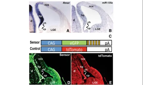
Rmst and miR-135a expression and activity in the embryonic forebrain
ventral midbrain region of the embryonic Central Nervous System (CNS) [11]. We provided evidence thatmiR-135a
expression was correlated with Rmst, and deduced that
miR-135a was derived from mir-135a-2 locus located in the final detected intron of Rmst [11, 53]. Since the em-bryonic dorsal forebrain is known to be a Wnt-rich region and dependent on Wnt signaling [36, 54, 55], we deter-mined if Rmst and miR-135a were also co-expressed in this region of the CNS. We therefore first characterized
Rmstexpression at embryonic and adult stages (Fig. 1 and Additional file 1: Figure S1). In embryonic forebrain sec-tionsRmst is robustly detected in the medial wall of the telencephalon, encompassing a Wnt and TGFβ/BMP-rich signaling center, the cortical hem [35, 56]. At E12.5,Rmst
was strongly expressed in the dorsal telencephalon in a medialHigh to dorso-lateralLow gradient (Fig. 1a). Expres-sion was robust in the cortical hem and the adjacent hippocampus primordium. A very weak hybridization signal was detected in the choroid plexus and along the neocortical domain. Rmst was also strongly expressed in the septum, diencephalon, and eminentia thalami. At later stages,Rmstexpression was localized to various forebrain nuclei, and the fimbria (Additional file 1: Figure S1).
We next performed Locked Nucleic Acid (LNA) ish
experiments to detect mature miR-135a, focusing on the embryonic forebrain. We found that this microRNA spe-cies, like Rmst, was expressed in the medial wall of the telencephalon in a medialHighto dorso-lateralLowgradient (Fig. 1b). Expression was robust in the cortical hem and the adjacent hippocampus primordium. A very weak hybridization signal was detected in the choroid plexus and along the neocortical domain. Some signal was also detected in the LGE, however, sinceRmstis not expressed in this region, this signal could represent cross hybri-dization with a closely related microRNA. Alternatively, in this region miR-135a and Rmst expression could be uncoupled.
To demonstrate thatmiR-135ais functional in these do-mains, we generated double transgenic embryos harboring a“sensor” and a control transgene. A“sensor” construct contains a constitutively expressed reporter gene (eGFP), under control of a CAG promoter, and multiple binding sites formiR-135ain the 3’UTR region (Fig. 1c, cartoon, yellow bars). In cells expressing miR-135a, its perfect complementarity to sequences in the 3’UTR should result in suppression of the eGFP reporter. A control transgene construct contains a tdTomato reporter, but lacks the
miR-135a binding sites and, therefore, should be
constitutively active. In E12.5 double transgenic embryos, we found that the eGFP reporter (Fig. 1d), but not tdTomato (Fig. 1e), was selectively reduced in the medial wall of the telencephalon where miR-135a is strongly expressed, as well as in the choroid plexus and in the dor-sal neocortical domain, where Rmst and miR-135a are more weakly expressed. eGFP was also not detected in the diencephalon (Fig. 1d). Thus, miR-135ais expressed, and displays activity, in the embryonic dorsal forebrain.
beta-catenin signaling induces RmstandmiR-135a
expression
Previously, we reported that in the embryonic midbrain, the transcription factor Lmx1b induces Wnt1/Wnt sig-naling as well as Rmstand mir-135a-2 locus [11]. Since
Lmx1b and Lmx1a are partially redundant, we postu-lated thatLmx1a might also be a regulator ofRmstand
mir-135a-2. However, since, in the dorsal forebrain,
Rmst and miR-135a expressions exceed the Lmx1a do-main, one possibility is that in addition to Lmx genes, Wnt signaling abets the induction of theRmstand mir-135a-2 locus. To address this question, we performed gain and loss of function experiments of beta-catenin, a key effector of Wnt signaling pathway [57]. Emx1::Cre
mouse line was used to drive recombination throughout the dorsal, but not ventral, forebrain [47]. For gain of function experiments, by conditionally deleting beta-catenin exon 3 (Ctnnb1lox(ex3)), which encompasses GSK3βphosphorylation sites, we effectively elevated Wnt signaling [23, 45]. In mutants (Emx1::Cre;Ctnnb1lox(ex3)) (Fig. 2b), but not in controls (Fig. 2a),Rmst was detected throughout the dorsal forebrain. Conversely, for loss of function experiments, we conditionally removed exons 2-6 of beta-catenin to prevent formation of a functional beta-catenin protein thus impairing Wnt signaling [46]. In mutants (Emx1::Cre;Ctnnb1 cKO) (Fig. 2c), but not in
controls (Fig. 2a), we observed drastic loss ofRmst expres-sion throughout the dorsal forebrain and in the remaining cortical hem tissue (Fig. 2c, asterisk).miR-135aexpression levels, quantified by RT-qPCR on E12.5 dissected dorsal telencephalon, were strongly induced inEmx1::Cre; Ctnn-b1lox(ex3)mutants, and reduced inEmx1::Cre;Ctnnb1cKOs (Fig. 2d). Overall, these data demonstrate that beta-catenin signaling is necessary and sufficient forRmst and
miR-135aexpression in the dorsal forebrain.
beta-catenin signaling is necessary for Lmx1a expression We also determined if Lmx1a, a key cortical hem deter-minant [58], is a target of Wnt/beta-catenin signaling in the forebrain, as in other brain regions [11, 22, 23]. While in Emx1::Cre;Ctnnb1lox(ex3) embryos (Fig. 2f), we did not observe a drastic change in the cortical hem size (Lmx1a+, Fig. 2f and Wnt3a+, Additional file 2: Figure S2) or in
Lmx1a expression (Fig. 2f) with respect to controls (Fig. 2e), inEmx1::Cre;Ctnnb1cKOs, we observed a drastic reduction ofLmx1a signal in the remaining cortical hem tissue (Fig. 2g, asterisk).Lmx1awas detected in the chor-oid plexus of both mutants (Fig. 2f and g). These results suggest that Wnt/beta-catenin signaling is necessary, but not sufficient, forLmx1aexpression in the dorsal forebrain. Given thatLmx1ain part functions to repressLhx2[58], a negative regulator of the hem [39], Wnt/beta-catenin in-duction ofLmx1ais likely to be an important event in the cortical hem establishment and/or maintenance.
miR-135a is predicted to target the Wnt and TGFβ/BMP pathways, but loss of function does not affect dorsal forebrain development
To begin to elucidatemiR-135afunctions, we performed bioinformatics analysis to determine the most common pathways targeted by miR-135a. To do that, we took advantage of the Diana–miRPath software [52], which
Fig. 2Rmst, miR-135aandLmx1aare altered in beta-catenin mutants.a-cRmstexpression in E12.5 wild type control (a),Emx1::Cre;Ctnnb1lox(ex3)
(b) andEmx1::Cre;Ctnnb1cKO mutant (c) brains.dQuantification ofmiR-135aexpression levels by RT-qPCR on E12.5 dissected dorsal telencephalon of control (wild type),Emx1::Cre;Ctnnb1lox(ex3)andEmx1::Cre;Ctnnb1cKO mutants (n= 3).miR-135afold induction was normalized to microRNAsno202. e-gLmx1aexpression in E12.5 controls (e) and beta-catenin mutant (fandg) brains. Asterisks incandghighlight remaining cortical hem tissue in
utilizes predicted, and validated, microRNA targets to perform a hierarchical clustering of microRNA and pathways based on their interactions. We found that Wnt and TGFβ/BMP signaling pathways are among the top pathways targeted by miR-135a with an extremely high statistical significance (p value of approximately 2.9E-07 and 3.6E-10, respectively) (Additional file 3: Figure S3A). It is worth noticing that the genes targeted by
miR-135ain the TGFβ/BMP (Additional file 3: Figure S3B)
and Wnt pathways (Additional file 4: Figure S4) include ligands, receptors and downstream transcriptional regula-tors suggesting that this microRNA likely acts through multiple levels of the Wnt and TGFβ/BMP cascades to modulate the outcome of their signaling.
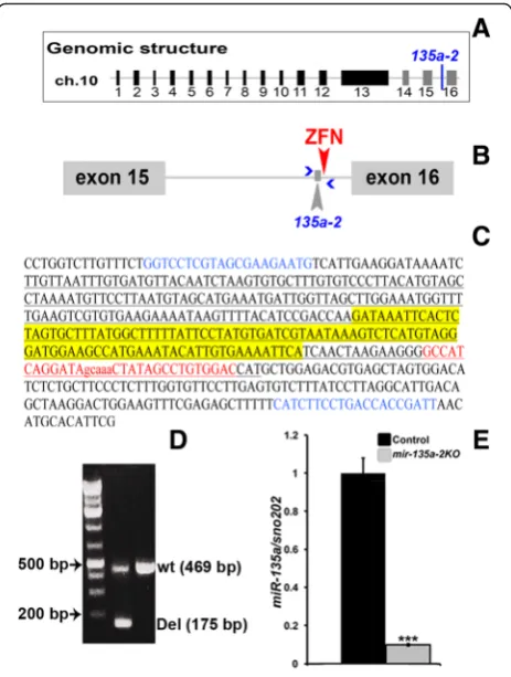
To determine the role of miR-135ain embryonic dor-sal forebrain development, we generated a mouse line in whichmir-135a-2was deleted by pronuclear injection of a specific Zn finger nuclease (Fig. 3a and b), designed to cleave <100 bp from the mature sequence. Of ~60 pups examined, 1 harbored a ~294 bp deletion (Fig. 3c and d) and was used to generate a line for the experiments here described. RT-qPCR on dissected E12.5 dorsal forebrain tissue confirmed a drastic reduction inmiR-135a expres-sion levels (Fig. 3e). We thus deduced that, in the dorsal forebrain akin to the ventral midbrain,miR-135a is pre-dominantly produced frommir-135a-2locus.
Because a growing literature has described interactions
between microRNAs and long non-coding RNAs
(lncRNAs) as a novel mechanism to regulate gene expres-sion as well as microRNA function [59–61], we investi-gated the possibility of an interaction between Rmst and
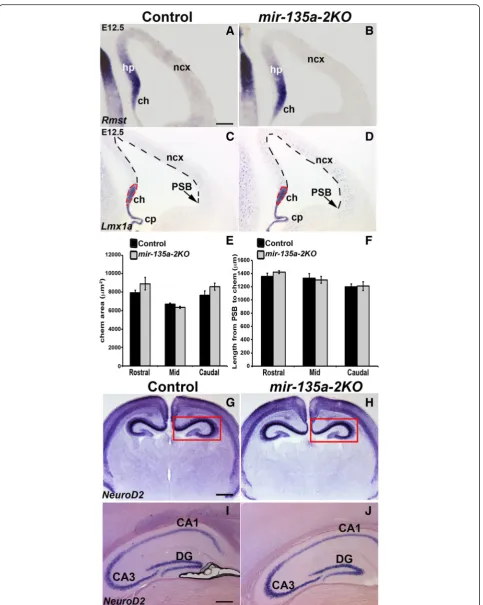
miR-135a. A search on miRcode web site, which repre-sents a comprehensive map of putative microRNA target sites across the GENCODE long non-coding transcrip-tome [62], indicated lack ofmiR-135aresponsive elements in Rmst. To confirm the bioinformatics prediction, we performedishforRmston E12.5 coronal sections of mir-135a-2knockout mice and we did not observe any change in its expression (Fig. 4a and b). These data seem to there-fore exclude the possibility ofRmstbeing a potential target of miR-135a. Next, we performed in situ hybridization withLmx1a(Fig. 4c and d), a marker for the cortical hem and the choroid plexus to investigate any possible change in proper development of these Wnt and TGFβ/BMP responsive domains. We did not, however, observe any apparent change in the cortical hem size (Fig. 4d) as was also confirmed by quantification analysis at three levels of the forebrain, equally spaced along the rostro-caudal axis (referred to as Rostral, Mid and Caudal) (Fig. 4e). No dif-ference was detected in the extent of the cortical domain (Fig. 4d), which was again measured at three levels of the forebrain, equally spaced along the rostro-caudal axis (Rostral, Mid and Caudal) from the inflection that marks the pallium-subpallium boundary (PSB), chosen as a
landmark, to the cortical hem (Fig. 4f). No change in the choroid plexus complexity was observed (Fig. 4d).
The cortical hem is a signaling center known to induce and pattern the adjacent hippocampus [38, 41]. We therefore performed in situ hybridization with neuronal markerNeuroD2to assess any morphological changes in the hippocampal complex at post-natal and adult stages of mir-135a-2 knockouts. No changes were observed (Fig. 4g-j). These findings clearly demonstrate that, at least by these criteria, mir-135a-2 loss of function does not alter forebrain development.
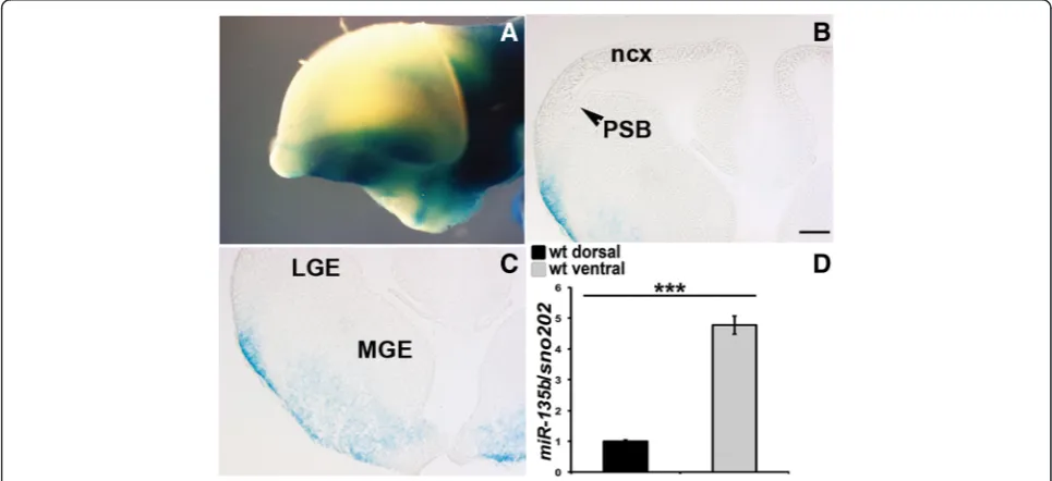
microRNAs often display redundancy with family members [63, 64]. To determine whether miR-135b, a closely related microRNA was expressed in a similar do-main, we obtained mice in which LacZ had been inserted into the mir-135b locus. In E12.5 whole mount
Fig. 3mir-135a-2deletion by ZFN nuclease.aRepresentation ofRmst
genomic structure.mir-135a-2is indicated between exons 15 and 16.
bCartoon of the last intron ofRmstshowing the location ofmir-135a-2
(Fig. 5a) and forebrain sections (Fig. 5b and c), we did not observe any LacZ staining in the dorsal forebrain (Fig. 5a and b) whereas we found LacZ expression in cells emanating from the ganglionic eminences in the ventral forebrain (Fig. 5c). Consistent with this, RT-qPCR ex-periments for miR-135b on wild type dorsal and ventral forebrain dissected tissues, revealed a ventral enrichment of this microRNA (Fig. 5d). These data suggest that
miR-135b does not serve a redundant function in the dorsal forebrain.
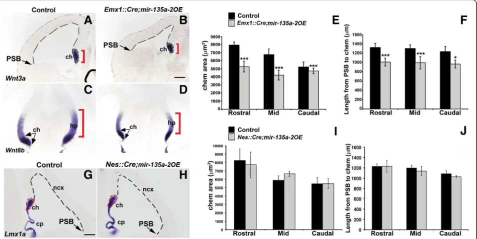
Early mir-135a-2 overexpression affects dorsal forebrain development
To complement our loss of function results, we took advantage of previously generated transgenic mice to con-ditionally expressmir-135a-2for gain of function experi-ments. This time, we reasoned that if the Wnt/miR-135a
circuitry identified in the midbrain [11] is functionally conserved in the forebrain, mir-135a-2 overexpression
might result in Wnts related phenotypes. To test this hy-pothesis, mice in which mir-135a-2precursor expression is under control of a CAG promoter [11] (henceforth mir-135a-2OE), were crossed with Emx1::Cre line [47] to overexpress mir-135a-2throughout the Emx1 domain of the dorsal, but not ventral, forebrain as early as E9.5 [43]. RT-qPCR on dissected E12.5 dorsal forebrain tissue con-firmed thatmiR-135aexpression levels, inEmx1::Cre; mir-135a-2OE mutants, were 1.5 fold more than in controls (Additional file 5: Figure S5). In such mutants we ob-served a clear reduction in the size of the cortical hem (Wnt3a+, Wnt8b+) and the hippocampus primordium (Wnt8b+) (Fig. 6a-d). Additionally, we observed a reduc-tion in size of the neocortical domain (Fig. 6a-b, dashed lines), a phenotype previously shown in mice with genetic ablation of the cortical hem [44, 65], but not reported in
BmpRIA/IB cKOs which display a smaller cortical hem [42]. The choroid plexus, normally specified by TGFβ/ BMPs [29, 42] and demarcated by rTtr1, was overall less
(See figure on previous page.)
Fig. 4mir-135a-2loss of function characterization.a-bE12.5 coronal sections of control (mir-135a-2+/-) andmir-135a-2KO brains (mir-135a-2-/-) processed forishforRmstshowing no change in its expression.c-dishforLmx1ain control (c) andmir-135a-2KO (d) brains. Dashed lines highlight the cortical hem area and the neocortical domain size.eQuantification of the cortical hem area (μm2± SEM) along the rostro-caudal axis (n= 3).fQuantification of the neocortex length (μm ± SEM) from the PSB to the hem at the same levels (n= 3).g-jishforNeuroD2on coronal sections of post-natal P1 (g-h) and adult (i-j) control and mutant brains showing no change in hippocampus morphology (boxed ingand h). ch, cortical hem; cp, choroid plexus; hp, hippocampus; PSB, pallium-subpallium boundary; ncx, neocortex; CA1, CA3, hippocampus fields; DG, dentate gyrus. Scale bar 100μm in panelsa-d, 400μm in panelsgandh; 200μm in panelsiandj
Fig. 5miR-135bexpression in the embryonic forebrain.a-cXgal staining of E12.5 whole mount (lateral view) (a) and E12.5 coronal sections (b-c), showing expression oflacZexclusively in the ventral forebrain ofmir-135bflox/+mice harboring a LacZ reporter gene in the
convoluted (Additional file 6: Figure S6). Altogether,
Emx1::Cre;mir-135a-2OE mutants show characteristics previously described in mice deficient for Wnt or TGFβ/ BMP activity, or in which the cortical hem has been genet-ically ablated [16, 29, 42, 44, 65, 66], although they display a milder phenotype.
Next, we quantified the cortical hem area along the rostro-caudal axis. We estimated a reduction in its size of ~34 % at rostral level, ~38 % at mid level and ~18 % at caudal level (Fig. 6e) in mutants compared to con-trols. Finally, we quantified the extent of the neocortical domain and found a significant reduction in size of the mutant neocortices in comparison to controls (Fig. 6f ). Since the cortical hem and the hippocampus primor-dium are reduced in Emx1::Cre;mir-135a-2OE embryos, these mutants showed a significant reduction of all hippo-campal structures (CA fields and dentate gyrus) from post-natal stage P1 (Additional file 7: Figure S7, A-B and E) to adulthood (Additional file 7: Figure S7C and D) when com-pared to controls.
Finally, we examined the expression of several bioin-formatically predicted miR-135a targets. Of these, only phospho-Smad (1/5/8) and Msx2 revealed consistent changes, showing apparent reduction in their level and
expression domain extent (Additional file 8: Figure S8). Their reduction might be due to direct repression, overall net down regulation of these pathways, or both.
Late mir-135a-2 overexpression does not affect dorsal forebrain development
Conditional transgenes in neural progenitor cells have been associated with non-specific phenotypes [67]. To rule out non-specific effects ofmir-135a-2overexpression, we also overexpressedmir-135a-2by usingNes::Credriver [50], which like Emx1::Cre,is active in the hippocampus primordium, along the neocortical domain, and reported to mediate Cre recombination in the hem at ~ E12.5 [10, 68–71]. Nes::Credriven overexpression of mir-135a-2was additionally useful to determine whether the pheno-type resulting frommir-135a-2overexpression in Emx1::-Cre;mir-135a-2OE mice, was time sensitive. In E12.5 (Fig. 6g and h) and E13.5 (Additional file 9: Figure S9)
Nes::Cre;mir-135a-2OE mutants, we did not observed microcephaly. Both the cortical hem and neocortical sizes were not affected (Fig. 6i and j). Also, no change was observed in the choroid plexus (Fig. 6g and h). These data suggest that late overexpression of this
microRNA in neural progenitors, does not affect forebrain development, and that the transgene does not appear to display significant toxicity in neuronal progenitors.
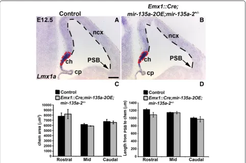
mir-135a-2 overexpression in mir-135a-2+/-mice does not result in forebrain abnormalities
To further demonstrate the specificity of our results, we
removed one copy of endogenous mir-135a-2 from
embryos conditionally overexpressing mir-135a-2. We reasoned that if the phenotype observed in Emx1::Cre
;-mir-135a-2OE mutants is due to mir-135a-2 over-expression, then, removal of one copy of the endogenous
mir-135a-2should alleviate this phenotype. E12.5 embryos of Emx1::Cre;mir-135a-2OE;mir-135a-2+/- (mutants) and
Emx1::Cre;mir-135a-2+/- (controls) genotypes were ana-lyzed for the expression of Lmx1a (Fig. 7a and b) and
Wnt3a (Additional file 10: Figure S10). The cortical hem area and the extent of the cortical domain were quantified as previously described (Fig. 7c and d). No alterations in these domains and in the choroid plexus were observed,
suggesting that normalmiR-135alevels are important for embryonic forebrain development.
Discussion
From the present study we deduced six prominent con-clusions. First, miR-135a, and Rmst, are expressed and functional in the embryonic dorsal forebrain. Second, in the dorsal forebrain, maturemiR-135a is predominantly derived from the mir-135a-2 locus. Third, miR-135a is dispensable for forebrain development. Fourth, modest
mir-135a-2 overexpression, within an early but not late time window, results in a phenotype consistent with Wnt and TGFβ/BMP signaling deficits – a reduced cortical hem, hippocampus primordium and neocortex, and a less convoluted choroid plexus. Fifth, Wnt/beta-catenin pathway is necessary and sufficient to induce Rmst and
miR-135aexpression pointing to the existence of aWnt/ miR-135aauto-regulatory loop, which could serve to limit the extent, the duration and/or intensity of the Wnt path-way. Finally, Wnt/beta-catenin pathway is also necessary
Fig. 7mir-135a-2OEin mice heterozygous for endogenousmir-135a-2.a-bishshowingLmx1aexpression in E12.5 control (Emx1::Cre;mir-135a-2+/-) (a) and mutant (Emx1::Cre;mir-135a-2OE;mir-135a-2+/-) (b) brains. Dashed lines highlight the cortical hem area and the neocortical domain size.
cQuantification of the cortical hem area (μm2± SEM) along the rostro-caudal axis (
forLmx1aexpression, a key cortical hem determinant. A model of these interactions is depicted in Fig. 8.
Wnt and TGFβ/BMP morphogens act through gradients of signaling cascades and have been implicated in various facets of dorsal neural tube development [16, 19–22].
Several studies have suggested that the dosage of the Wnt and TGFβ/BMP pathways is critical and has to be tightly controlled through intricate networks of positive and nega-tive feedback loops [15, 19, 23, 32, 33]. It is therefore chal-lenging to understand how these pathways are modulated in time and space during embryonic development, how cells receive and integrate multiple signals and whether po-tential nodes of intersection exist. Recently, it has been demonstrated that microRNAs contribute to gene net-works that transform the graded activity of a morphogen in robust cell fate decisions by establishing context-dependency, threshold responses and sharpening temporal and spatial expression patterns [4, 12, 72]. miR-135a and its host long non-coding transcript, Rmst,were shown to be expressed and functional in the embryonic Wnt-rich domain of the midbrain [11]. Here, we have shown that
miR-135aandRmstare also co-expressed in the Wnt and TGFβ/BMP-rich domains of the embryonic dorsal fore-brain suggesting a correlation between this microRNA, the Wnts and TGFβ/BMPs-rich domains across the embryonic CNS. Interestingly, akin to several Wnts and TGFβ/BMPs, strong expression of Rmstin the embryonic hippocampal primordium declines over time and, at post-natal stages, become restricted to the fimbria and virtually undetectable in the adult hippocampus.Rmst expression has also been detected in human fetal cortical radial glia, suggesting a conserved role for this locus [73]. Taking together, the
Rmst/miR-135a expression pattern in mice and humans, the finding that Wnt signaling is indeed able to induce their expression and the strong bioinformatics predictions, we postulate that this microRNA might play a role in fine-tuning the Wnt pathway and, possibly, the TGFβ/BMP pathway.
Embryos lacking Wnt3a[16],functional LEF1 [66], or with genetic ablation of the cortical hem [44, 65], display loss of the hippocampus and shrinkage of the neocortex [44, 65], while mice with disrupted TGFβ/BMP signaling fail to develop or properly differentiate the choroid plexus [29]. Modest mir-135a-2 overexpression appears to recapitulate these phenotypes, albeit in a milder fashion. Coupled with the findings that this microRNA is bioinformatically predicted to target both positive and negative regulators in the Wnt and TGFβ/BMP path-ways, the Wnt related phenotypes observed in the mid-brain [11], and proven interactions between miR-135a
and targets like GSK,Tcf7l2,Ccnd1and APCin heterol-ogous systems or cancer cell lines [11, 74–78], we posit that miR-135a modulates the Wnt and TGFβ/BMP
sig-naling cascade in the developing forebrain.
mir-135a-2 loss of function embryos did not display overt forebrain phenotypes, at least by the criteria that we assayed. One plausible explanation comes from the observation that members of a microRNA family are often predicted to target the same or overlapping sets of
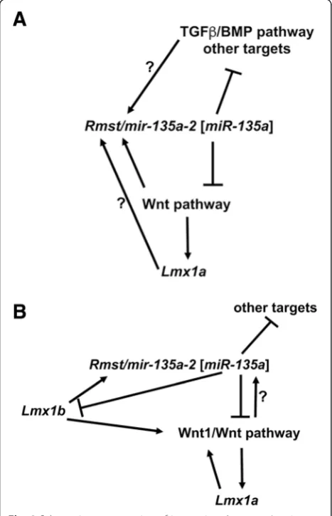
Fig. 8Schematic representation of interactions betweenLmx1
genes, the Wnt pathway andRmst/mir-135a-2 [miR-135a].aIn the embryonic forebrain, canonical Wnt/beta-catenin pathway induces
Rmst/mir-135a-2 [miR-135a], as well as the cortical hem determinant
Lmx1a. miR-135a, in turn, negatively targets Wnt pathway mRNAs establishing an auto-regulatory loop.miR-135ais also predicted to target TGFβ/BMP pathway mRNAs as well as other targets. It is possible that the TGFβ/BMP pathway is also able to induceRmst/mir-135a-2 [miR-135a]and establish amiR-135a/TGFβ/BMP auto-regulatory loop. It is also possible thatLmx1acontributes toRmst/mir-135a-2 [miR-135a]
expression. However, these interactions remain to be demonstrated. Ultimately, this complex network of positive and negative interactions plays a role in determining proper dorsal forebrain size (cortical hem, hippocampus primordium, choroid plexus and neocortex).bA similar scenario was previously identified in the embryonic midbrain [11] whereLmx1bdrives the expression of Wnt1/Wnt pathway and of
genes and therefore to act in a functionally redundant manner [63]. Supporting this idea, single microRNA loss of function or single microRNA silencing, through anti-sense oligonucleotides and sponge techniques, typically results in subtle or no phenotypes [64, 79–81]. Because
mir-135b was expressed in the embryonic ventral fore-brain, we ruled out any compensatory effect due to this microRNA. We considered the possibility that the other member of this family,mir-135a-1might be expressed in a similar and overlapping pattern. Because mir-135a-1
and mir-135a-2 produce an identical mature form, it is not possible to analyze their differential expression by standard techniques. However, RT-qPCR experiments for maturemiR-135aon dorsal forebrain tissue ofmir-135a-2
null embryos revealed greatly reduced expression levels. Therefore, we concluded that the mainmiR-135amature form in the dorsal forebrain could be attributed to mir-135a-2 precursor. Redundancy, on the other hand, can either be attributed to very low levels of miR-135a pro-duced by the mir-135a-1 precursor or to some other microRNA having a similar seed sequence. This latter, more likely, possibility has been raised in the recent litera-ture [82].
Identifying regulators of the Wnt pathway has been highlighted as an important goal [83]. miR-135a is up regulated in several tumor types characterized by high levels of Wnt/beta-catenin signaling, including colorectal tumors as well as certain subtypes of medulloblastomas [74, 76]. It is possible that, in these tumors, Wnt signal-ing, in accordance with our data in the forebrain, in-ducesmiR-135a. If so,miR-135awould serve as a useful marker for aberrant Wnt signaling in these tumors, and possibly others, and in therapeutic protocols designed to circumscribe unrestrained Wnt signaling. In colorectal cancers,miR-135ahas been proposed to act as a positive regulator of Wnt signaling by targeting APC, a key mol-ecule of the beta-catenin destruction complex [74]. Thus, while it is emerging that this microRNA is intim-ately correlated with Wnt signaling in the embryonic CNS ([11]and this work), in tumors [74–76] and in other cell types with high Wnt signaling [77, 78], it is possible that the net effect of miR-135a depends on the physio-logical milieu. Elucidating the targets may reveal how a single microRNA, predicted to target both positive and negative regulators of a signaling pathway, may have dis-tinct outcomes depending on the context. Ultimately, an array of biochemical and genetic approaches will be required to accurately define direct miR-135a targets and the molecular underpinnings of mir-135a-2mutant phenotypes.
Finally, our study has highlighted a role for Wnt/ beta-catenin signaling in the expression of cortical
hem determinant Lmx1a, a LIM-homeodomain
tran-scription factor.
Wnt1/Wnt signaling has been shown to form an auto-regulatory loop with Lmx1a to control dopaminergic neurons differentiation in the embryonic ventral mid-brain [22, 23, 84, 85]. In the dorsal embryonic foremid-brain,
Lmx1a is expressed in the cortical hem, a Wnt-rich region. WhileLmx1ais not required for cortical hem in-duction, it is critical for proper regulation of cell fate decisions [58]. In the absence of Lmx1a, the hippocam-pal selector gene Lhx2 is ectopically expressed in the cortical hem leading to excessive production of hippo-campal cells and decreased production of Cajal-Retzius cells [58]. Here, through gain and loss of function experi-ments of beta-catenin, we show that, in the dorsal forebrain akin to the ventral midbrain [22, 24], Wnt/beta-catenin signaling is necessary, but not sufficient, for Lmx1a ex-pression. Wnt/beta-catenin induction of Lmx1a is likely to be a key event in the proper cortical hem cell fate estab-lishment and/or maintenance.
Additional files
Additional file 1: Figure S1.Rmstexpression during forebrain development. (A-F)Rmst ishon coronal sections of wild type brains from E11.5 to adult stage. (A) At E11.5,Rmstis expressed in the epithalamus, medial wall of the dorsal telencephalon, the eminentia thalami, and the septum. (B) At E12.5 (as in Fig. 1),Rmstwas detected in the medial wall of the telencephalon and in the diandephalon. (C) At E14.5,Rmstis still highly expressed in the cortical hem and hippocampus primordium, thalamus and hypothalamus. (D) At E16.5,Rmstexpression is high in the hippocampus and fimbria but low in scattered cells of the migratory stream. Expression in the thalamus is maintained. (E) At post-natal stages P1,
Rmstis restricted to the fimbria, yet expressed outside of the hippocampal formation to become virtually undetectable in the hippocampus of adult mice (F) (hippocampal staining is not considered specific to theRmstprobe). dm, dorso-medial wall of telencephalon; emT, eminentia thalami; Et, epithalamus; tel, telencephalon; v3, third ventricle; lv, lateral ventricle; Sp, septum; d, diencephalon; hp, hippocampus; ch, cortical hem; cp, choroid plexus; ncx, neocortex; LGE, lateral ganglionic eminences; th, thalamus, hy, hypothalamus; fm, fimbria; ms, migratory stream; CA1 and CA3, hippocampal fieds; DG, dentate gyrus. Scale bar 400μm in panels A, C and D; 100μm in panel B; 200μm in panels E and F. (TIF 1931 kb)
Additional file 2: Figure S2.Wnt3aexpression in mice with elevated beta-catenin. (A-B)Wnt3a in situhybridization on control (A) and mutant (Emx1::Cre;Ctnnb1lox(ex3)) E12.5 coronal sections. Brackets highlight the cortical hem. ch, cortical hem; cp, choroid plexus. Scale bar 100μm. (TIF 2822 kb)
Additional file 3: Figure S3.miR-135abioinformatics analysis. A) Top pathways targeted bymiR-135a. TGFβ/BMP and Wnt signaling pathways rank at position number 2 and 3 with high statistical significance. P values and number of genes targeted in each pathway are indicated. B) Overview of TGFβ/BMP cascade with highlighted, and listed, 16 genes targeted bymiR-135a. It is worth noticing that the number of listed genes reflects only the data available on the Diana web site and the algorithm used for the search, as a higher number of putativemiR-135a
targets have already been reported using multiple search engines [11]. (TIF 1865 kb)
Additional file 5: Figure S5.Quantification ofmiR-135aexpression in
Emx1::Cre;mir-135a-2OEmice. RT-qPCR showing a 1.5 fold change in
miR-135aexpression level in E12.5 dissected dorsal forebrain tissue from mutant mice compared to controls (n= 3). Data are shown as a fold change and have been normalized to microRNAsno202. ***,pvalue <0.001. (TIF 145 kb)
Additional file 6: Figure S6.Earlymir-135a-2overexpression affects choroid plexus development. (A-B) Coronal sections of E12.5 control (mir-135a2-OE) and mutant brains (Emx1::Cre;mir-135a-2OE) showing expression of choroid plexus specific markerrTtr1. The dashed red line is used to highlight the change in choroid plexus complexity. cp, choroid plexus. Scale bar 100μm. (TIF 2429 kb)
Additional file 7: Figure S7.Earlymir-135a-2overexpression affects hippocampus development. (A-D) Coronal sections of post-natal stage P1 and adult brains processed forishforNeuroD2. Panels A, B show overall morphology and size of the hippocampus. Asterisks point towards the pallium-subpallium boundary. Insets show the extent of the hippocampus from the CA1 field to the tip of the dentate gyrus, quantification of which, at mid and caudal level of the brain, is reported in panel E. Length is expressed inμm ± SEM (n= 4). Consistent with a reduced cortical hem size at embryonic stages, the hippocampus was significantly reduced in its extent in mutant brains with respect to controls. ***,p<0.001. Abbreviations: pSub, para subiculum; Sub, subiculum; DG, dentate gyrus; Cng, cingulate cortex. Scale bar 400μm in panels A and B; 200μm in panels C and D. (TIF 2162 kb)
Additional file 8: Figure S8.Expression of bioinformatically predicted
miR-135atargets. (A-D) Coronal sections of E12.5 controls (mir-135a-2OE) and mutant (Emx1::Cre;mir-135a2-OE) brains, processed for immunohistochemistry (IHC) (A-B) or in situ hybridization (C, D). phospho-Smad (1/5/8) IHC on control (A) and mutant (B) brain coronal sections shows reduced signal in the cortical hem and the choroid plexus domains of mutants (arrowheads). Panels C and D show a reducedMsx2expression domain. ch, cortical hem; cp, choroid plexus. Scale bar 25μm in panels A and B; 50μm in panels C and D. (TIF 5061 kb)
Additional file 9: Figure S9.Latemir-135a-2overexpression does not overtly affect forebrain development. Coronal sections of E13.5 control (mir-135a-2OE) and mutant brains (Nes::Cre;mir-135a-2OE) processed forin situhybridization forLmx1a. Little to no change was observed in the cortical hem and neocortical domain (dashed lines) or in the choroid plexus. ch, cortical hem; cp, choroid plexus; ncx, neocortex. Scale bar 400μm. (TIF 2507 kb)
Additional file 10: Figure S10.Little to no change in cortical hem of
Emx1::Cre;mir-135a-2OE;mir-135a-2+/-mutant mice. (A-B)ishshowing cortical hem markerWnt3ain E12.5 control (Emx1::Cre;mir-135a-2+/-) (A) and mutant (Emx1::Cre;mir-135a-2OE;mir-135a-2+/-) (B) brains. ch, cortical hem. Scale bar 100μm. (TIF 542 kb)
Competing interests
The authors declare that they have no competing interests.
Authors’contributions
GCB and RA conceived the experiments and wrote the manuscript; GCB performed the experiments and analyzed the data; AA generated
mir-135a-2knockout,mir-135a-2overexpressor mice lines and the“sensor” construct to determinemiR-135aactivity. All authors read and approved the final manuscript.
Acknowledgements
We thank Makoto Taketo forCtnnb1lox(ex3)mouse line. Northwestern
Transgenic and Targeted Mutagenesis Laboratory for pronuclear injections. We thank Profs. Elizabeth Grove and Shubha Tole for their feedback on the manuscript.
Funding
This work was supported by the Brain Research Foundation, the National Institute of Health (grant number 1R01NS071081-01) and National Institute of Neurological Disorder (grant number 1F31NS065670-01A2). The funders had no role in study design, data collection and analysis, decision to publish or preparation of the manuscript.
Received: 4 January 2016 Accepted: 1 April 2016
References
1. Lewis BP, Burge CB, Bartel DP. Conserved seed pairing, often flanked by adenosines, indicates that thousands of human genes are microRNA targets. Cell. 2005;120(1):15–20. doi:10.1016/j.cell.2004.12.035. 2. Fineberg SK, Kosik KS, Davidson BL. MicroRNAs potentiate neural
development. Neuron. 2009;64(3):303–9. doi:10.1016/j.neuron.2009.10.020. 3. Yun B, Anderegg A, Menichella D, Wrabetz L, Feltri ML, Awatramani R.
MicroRNA-deficient Schwann cells display congenital hypomyelination. J Neurosci. 2010;30(22):7722–8. doi:10.1523/JNEUROSCI.0876-10.2010. PubMed PMID: 20519547, PubMed Central PMCID: PMC2906453. 4. Chen JA, Huang YP, Mazzoni EO, Tan GC, Zavadil J, Wichterle H. Mir-17-3p
controls spinal neural progenitor patterning by regulating Olig2/Irx3 cross-repressive loop. Neuron. 2011;69(4):721–35. doi:10.1016/j.neuron.2011.01.014. PubMed PMID: 21338882, PubMed Central PMCID: PMC3062262.
5. Haramati S, Chapnik E, Sztainberg Y, Eilam R, Zwang R, Gershoni N, et al. miRNA malfunction causes spinal motor neuron disease. Proc Natl Acad Sci U S A. 2010;107(29):13111–6. doi:10.1073/pnas.1006151107. PubMed PMID: 20616011, PubMed Central PMCID: PMC2919953.
6. Li X, Carthew RW. A microRNA mediates EGF receptor signaling and promotes photoreceptor differentiation in the Drosophila eye. Cell. 2005;123(7):1267–77. doi:10.1016/j.cell.2005.10.040.
7. Lin HP, Oksuz I, Hurley E, Wrabetz L, Awatramani R. Microprocessor complex subunit DiGeorge syndrome critical region gene 8 (Dgcr8) is required for schwann cell myelination and myelin maintenance. J Biol Chem. 2015; 290(40):24294–307. doi:10.1074/jbc.M115.636407. PubMed PMID: 26272614; PubMed Central PMCID: PMCPMC4591815.
8. Nowakowski TJ, Mysiak KS, Pratt T, Price DJ. Functional dicer is necessary for appropriate specification of radial glia during early development of mouse telencephalon. PLoS One. 2011;6(8):e23013. doi:10.1371/journal.pone. 0023013. PubMed PMID: 21826226, PubMed Central PMCID: PMC3149632. 9. Kawase-Koga Y, Otaegi G, Sun T. Different timings of Dicer deletion affect neurogenesis and gliogenesis in the developing mouse central nervous system. Dev Dyn. 2009;238(11):2800–12. doi:10.1002/dvdy.22109. PubMed PMID: 19806666, PubMed Central PMCID: PMC2831750.
10. Davis TH, Cuellar TL, Koch SM, Barker AJ, Harfe BD, McManus MT, et al. Conditional loss of Dicer disrupts cellular and tissue morphogenesis in the cortex and hippocampus. J Neurosci. 2008;28(17):4322–30. doi:10.1523/ JNEUROSCI.4815-07.2008.
11. Anderegg A, Lin HP, Chen JA, Caronia-Brown G, Cherepanova N, Yun B, et al. An Lmx1b-miR135a2 Regulatory Circuit Modulates Wnt1/Wnt Signaling and Determines the Size of the Midbrain Dopaminergic Progenitor Pool. PLoS Genet. 2013;9(12):e1003973. doi:10.1371/journal.pgen. 1003973. PubMed PMID: 24348261, PubMed Central PMCID: PMC3861205. 12. Inui M, Montagner M, Piccolo S. miRNAs and morphogen gradients.
Curr Opin Cell Biol. 2012;24(2):194–201. doi:10.1016/j.ceb.2011.11.013. 13. Inestrosa NC, Arenas E. Emerging roles of Wnts in the adult nervous system.
Nat Rev Neurosci. 2010;11(2):77–86. doi:10.1038/nrn2755.
14. Chenn A, Walsh CA. Regulation of cerebral cortical size by control of cell cycle exit in neural precursors. Science. 2002;297(5580):365–9. doi:10.1126/science.1074192.
15. Megason SG, McMahon AP. A mitogen gradient of dorsal midline Wnts organizes growth in the CNS. Development. 2002;129(9):2087–98. 16. Lee SM, Tole S, Grove E, McMahon AP. A local Wnt-3a signal is required for
development of the mammalian hippocampus. Development. 2000;127(3):457–67. Epub 2000/01/13.
17. McMahon AP, Joyner AL, Bradley A, McMahon JA. The midbrain-hindbrain phenotype of Wnt-1-/Wnt-1- mice results from stepwise deletion of engrailed-expressing cells by 9.5 days postcoitum. Cell. 1992;69(4):581–95. 18. Machon O, Backman M, Machonova O, Kozmik Z, Vacik T, Andersen L, et al.
A dynamic gradient of Wnt signaling controls initiation of neurogenesis in the mammalian cortex and cellular specification in the hippocampus. Dev Biol. 2007;311(1):223–37. doi:10.1016/j.ydbio.2007.08.038.
20. Galceran J, Farinas I, Depew MJ, Clevers H, Grosschedl R. Wnt3a-/–like phenotype and limb deficiency in Lef1(-/-)Tcf1(-/-) mice. Genes Dev. 1999;13(6):709–17. PubMed PMID: 10090727, PubMed Central PMCID: PMC316557, Epub 1999/03/25.
21. Woodhead GJ, Mutch CA, Olson EC, Chenn A. Cell-autonomous beta-catenin signaling regulates cortical precursor proliferation. J Neurosci. 2006;26(48): 12620–30. doi:10.1523/JNEUROSCI.3180-06.2006. PubMed PMID: 17135424, PubMed Central PMCID: PMC2867669.
22. Joksimovic M, Yun BA, Kittappa R, Anderegg AM, Chang WW, Taketo MM, et al. Wnt antagonism of Shh facilitates midbrain floor plate neurogenesis. Nat Neurosci. 2009;12(2):125–31. doi:10.1038/nn.2243. Epub 2009/01/06. 23. Joksimovic M, Awatramani R. Wnt/beta-catenin signaling in midbrain
dopaminergic neuron specification and neurogenesis. J Mol Cell Biol. 2014. doi:10.1093/jmcb/mjt043.
24. Nouri N, Patel MJ, Joksimovic M, Poulin JF, Anderegg A, Taketo MM, et al. Excessive Wnt/beta-catenin signaling promotes midbrain floor plate neurogenesis, but results in vacillating dopamine progenitors. Mol Cell Neurosci. 2015;68:131–42. doi:10.1016/j.mcn.2015.07.002. PubMed PMID: 26164566; PubMed Central PMCID: PMCPMC4633300.
25. Furuta Y, Piston DW, Hogan BL. Bone morphogenetic proteins (BMPs) as regulators of dorsal forebrain development. Development. 1997;124(11):2203–12. 26. Liem Jr KF, Tremml G, Jessell TM. A role for the roof plate and its resident
TGFbeta-related proteins in neuronal patterning in the dorsal spinal cord. Cell. 1997;91(1):127–38. Epub 1997/10/23 22:32.
27. Augsburger A, Schuchardt A, Hoskins S, Dodd J, Butler S. BMPs as mediators of roof plate repulsion of commissural neurons. Neuron. 1999;24(1):127–41. Epub 2000/02/17.
28. Samanta J, Burke GM, McGuire T, Pisarek AJ, Mukhopadhyay A, Mishina Y, et al. BMPR1a signaling determines numbers of oligodendrocytes and calbindin-expressing interneurons in the cortex. J Neurosci. 2007;27(28):7397–407. 29. Hebert JM, Mishina Y, McConnell SK. BMP signaling is required locally to
pattern the dorsal telencephalic midline. Neuron. 2002;35(6):1029–41. Epub 2002/10/02.
30. See J, Mamontov P, Ahn K, Wine-Lee L, Crenshaw 3rd EB, Grinspan JB. BMP signaling mutant mice exhibit glial cell maturation defects. Mol Cell Neurosci. 2007;35(1):171–82.
31. Choe Y, Kozlova A, Graf D, Pleasure SJ. Bone morphogenic protein signaling is a major determinant of dentate development. J Neurosci. 2013;33(16):6766–75. doi:10.1523/JNEUROSCI.0128-13.2013. PubMed PMID: 23595735, PubMed Central PMCID: PMC3684166.
32. Doan LT, Javier AL, Furr NM, Nguyen KL, Cho KW, Monuki ES. A Bmp reporter with ultrasensitive characteristics reveals that high Bmp signaling is not required for cortical hem fate. PLoS One. 2012;7(9):e44009. doi:10.1371/ journal.pone.0044009. PubMed PMID: 22984456, PubMed Central PMCID: PMC3439469.
33. Hu JS, Doan LT, Currle DS, Paff M, Rheem JY, Schreyer R, et al. Border formation in a Bmp gradient reduced to single dissociated cells. Proc Natl Acad Sci U S A. 2008;105(9):3398–403. doi:10.1073/pnas.0709100105. PubMed PMID: 18292231, PubMed Central PMCID: PMC2265170.
34. Itasaki N, Hoppler S. Crosstalk between Wnt and bone morphogenic protein signaling: a turbulent relationship. Dev Dyn. 2010;239(1):16–33. doi:10.1002/ dvdy.22009.
35. Subramanian L, Remedios R, Shetty A, Tole S. Signals from the edges: the cortical hem and antihem in telencephalic development. Semin Cell Dev Biol. 2009;20(6):712–8. doi:10.1016/j.semcdb.2009.04.001. Epub 2009/05/19. PubMed PMID: 19446478; PubMed Central PMCID: PMC2791850. 36. Grove EA, Tole S, Limon J, Yip L, Ragsdale CW. The hem of the embryonic
cerebral cortex is defined by the expression of multiple Wnt genes and is compromised in Gli3-deficient mice. Development. 1998;125(12):2315–25. 37. Monuki ES, Porter FD, Walsh CA. Patterning of the dorsal telencephalon and
cerebral cortex by a roof plate-Lhx2 pathway. Neuron. 2001;32(4):591–604. Epub 2001/11/24doi: S0896-6273(01)00504-9 [pii].
38. Tole S, Grove EA. Detailed field pattern is intrinsic to the embryonic mouse hippocampus early in neurogenesis. J Neurosci. 2001;21(5):1580–9. Epub 2001/02/27.
39. Mangale VS, Hirokawa KE, Satyaki PR, Gokulchandran N, Chikbire S, Subramanian L, et al. Lhx2 selector activity specifies cortical identity and suppresses
hippocampal organizer fate. Science. 2008;319(5861):304–9. doi:10.1126/science. 1151695. PubMed PMID: 18202285, PubMed Central PMCID: PMC2494603. 40. Zhou CJ, Zhao C, Pleasure SJ. Wnt signaling mutants have decreased
dentate granule cell production and radial glial scaffolding abnormalities.
J Neurosci. 2004;24(1):121–6. doi:10.1523/JNEUROSCI.4071-03.2004. Epub 2004/01/13. PubMed PMID: 14715945.
41. Grove EA, Tole S. Patterning events and specification signals in the developing hippocampus. Cereb Cortex. 1999;9(6):551–61. Epub 1999/09/25. 42. Caronia G, Wilcoxon J, Feldman P, Grove EA. Bone morphogenetic protein
signaling in the developing telencephalon controls formation of the hippocampal dentate gyrus and modifies fear-related behavior. J Neurosci. 2010;30(18):6291–301. doi:10.1523/JNEUROSCI.0550-10.2010. Epub 2010/05/07. 43. Yoshida M, Assimacopoulos S, Jones KR, Grove EA. Massive loss of
Cajal-Retzius cells does not disrupt neocortical layer order. Development. 2006;133(3):537–45. doi:10.1242/dev.02209.
44. Caronia-Brown G, Yoshida M, Gulden F, Assimacopoulos S, Grove EA. The cortical hem regulates the size and patterning of neocortex. Development. 2014;141(14):2855–65. doi:10.1242/dev.106914.
45. Harada N, Tamai Y, Ishikawa T, Sauer B, Takaku K, Oshima M, et al. Intestinal polyposis in mice with a dominant stable mutation of the beta-catenin gene. EMBO J. 1999;18(21):5931–42. doi:10.1093/emboj/18.21.5931. PubMed PMID: 10545105, PubMed Central PMCID: PMC1171659.
46. Brault V, Moore R, Kutsch S, Ishibashi M, Rowitch DH, McMahon AP, et al. Inactivation of the beta-catenin gene by Wnt1-Cre-mediated deletion results in dramatic brain malformation and failure of craniofacial development. Development. 2001;128(8):1253–64.
47. Gorski JA, Talley T, Qiu M, Puelles L, Rubenstein JL, Jones KR. Cortical excitatory neurons and glia, but not GABAergic neurons, are produced in the Emx1-expressing lineage. J Neurosci. 2002;22(15):6309–14. doi: 20026564.
48. Park CY, Jeker LT, Carver-Moore K, Oh A, Liu HJ, Cameron R, et al. A resource for the conditional ablation of microRNAs in the mouse. Cell Reports. 2012;1(4):385–91. doi:10.1016/j.celrep.2012.02.008. PubMed PMID: 22570807, PubMed Central PMCID: PMC3345170.
49. Awatramani R, Soriano P, Rodriguez C, Mai JJ, Dymecki SM. Cryptic boundaries in roof plate and choroid plexus identified by intersectional gene activation. Nat Genet. 2003;35(1):70–5. doi:10.1038/ng1228. 50. Tronche F, Kellendonk C, Kretz O, Gass P, Anlag K, Orban PC, et al.
Disruption of the glucocorticoid receptor gene in the nervous system results in reduced anxiety. Nat Genet. 1999;23(1):99–103. doi:10.1038/12703. 51. Schmittgen TD, Livak KJ. Analyzing real-time PCR data by the comparative
C(T) method. Nat Protoc. 2008;3(6):1101–8.
52. Vlachos IS, Kostoulas N, Vergoulis T, Georgakilas G, Reczko M, Maragkakis M, et al. DIANA miRPath v.2.0: investigating the combinatorial effect of microRNAs in pathways. Nucleic Acids Res. 2012;40(Web Server issue): W498–504. doi:10.1093/nar/gks494. PubMed PMID: 22649059; PubMed Central PMCID: PMC3394305.
53. Uhde CW, Vives J, Jaeger I, Li M. Rmst is a novel marker for the mouse ventral mesencephalic floor plate and the anterior dorsal midline cells. PLoS One. 2010;5(1):e8641. doi:10.1371/journal.pone.0008641. PubMed PMID: 20062813, PubMed Central PMCID: PMC2799666.
54. Backman M, Machon O, Mygland L, van den Bout CJ, Zhong W, Taketo MM, et al. Effects of canonical Wnt signaling on dorso-ventral specification of the mouse telencephalon. Dev Biol. 2005;279(1):155–68. doi:10.1016/ j.ydbio.2004.12.010. Epub 2005/02/15.
55. Kim AS, Lowenstein DH, Pleasure SJ. Wnt receptors and Wnt inhibitors are expressed in gradients in the developing telencephalon. Mech Dev. 2001;103(1-2):167–72. Epub 2001/05/04.
56. Bulchand S, Grove EA, Porter FD, Tole S. LIM-homeodomain gene Lhx2 regulates the formation of the cortical hem. Mech Dev. 2001;100(2):165–75. 57. Willert J, Epping M, Pollack JR, Brown PO, Nusse R. A transcriptional
response to Wnt protein in human embryonic carcinoma cells. BMC Dev Biol. 2002;2:8. PubMed PMID: 12095419, PubMed Central PMCID: PMC117803.
58. Chizhikov VV, Lindgren AG, Mishima Y, Roberts RW, Aldinger KA, Miesegaes GR, et al. Lmx1a regulates fates and location of cells originating from the cerebellar rhombic lip and telencephalic cortical hem. Proc Natl Acad Sci U S A. 2010;107(23):10725–30. doi:10.1073/pnas.0910786107. Epub 2010/05/26. PubMed PMID: 20498066; PubMed Central PMCID: PMC2890798. 59. Quan M, Chen J, Zhang D. Exploring the secrets of long noncoding RNAs.
Int J Mol Sci. 2015;16(3):5467–96. doi:10.3390/ijms16035467. PubMed PMID: 25764159; PubMed Central PMCID: PMCPMC4394487.
61. Jalali S, Bhartiya D, Lalwani MK, Sivasubbu S, Scaria V. Systematic transcriptome wide analysis of lncRNA-miRNA interactions. PLoS One. 2013;8(2):e53823. doi:10.1371/journal.pone.0053823. PubMed PMID: 23405074; PubMed Central PMCID: PMCPMC3566149.
62. Jeggari A, Marks DS, Larsson E. miRcode: a map of putative microRNA target sites in the long non-coding transcriptome. Bioinformatics. 2012;28(15):2062–3. doi:10.1093/bioinformatics/bts344. PubMed PMID: 22718787; PubMed Central PMCID: PMCPMC3400968.
63. Fischer S, Handrick R, Aschrafi A, Otte K. Unveiling the principle of microRNA-mediated redundancy in cellular pathway regulation. RNA Biol. 2015;12(3):238–47. doi:10.1080/15476286.2015.1017238.
64. Mendell JT, Olson EN. MicroRNAs in stress signaling and human disease. Cell. 2012;148(6):1172–87. doi:10.1016/j.cell.2012.02.005. PubMed PMID: 22424228, PubMed Central PMCID: PMC3308137.
65. Caronia-Brown G, Grove EA. Timing of Cortical Interneuron Migration Is Influenced by the Cortical Hem. Cereb Cortex. 2010. doi:10.1093/cercor/bhq142. Epub 2010/08/18.
66. Galceran J, Miyashita-Lin EM, Devaney E, Rubenstein JL, Grosschedl R. Hippocampus development and generation of dentate gyrus granule cells is regulated by LEF1. Development. 2000;127(3):469–82. Epub 2000/01/13. 67. Qiu L, Rivera-Perez JA, Xu Z. A non-specific effect associated with
conditional transgene expression based on Cre-loxP strategy in mice. PLoS One. 2011;6(5):e18778. doi:10.1371/journal.pone.0018778. PubMed PMID: 21572998, PubMed Central PMCID: PMC3091857.
68. Hodge RD, Garcia III AJ, Elsen GE, Nelson BR, Mussar KE, Reiner SL, et al. Tbr2 expression in Cajal-Retzius cells and intermediate neuronal progenitors is required for morphogenesis of the dentate gyrus. J Neurosci. 2013;33(9): 4165–80. doi:10.1523/JNEUROSCI.4185-12.2013. PubMed PMID: 23447624; PubMed Central PMCID: PMCPMC3623668.
69. Konno D, Iwashita M, Satoh Y, Momiyama A, Abe T, Kiyonari H, et al. The mammalian DM domain transcription factor Dmrta2 is required for early embryonic development of the cerebral cortex. PLoS One. 2012;7(10):e46577. doi:10.1371/journal.pone.0046577. PubMed PMID: 23056351; PubMed Central PMCID: PMCPMC3462758.
70. Li Q, Bian S, Hong J, Kawase-Koga Y, Zhu E, Zheng Y, et al. Timing specific requirement of microRNA function is essential for embryonic and postnatal hippocampal development. PLoS One. 2011;6(10):e26000. doi:10.1371/ journal.pone.0026000. PubMed PMID: 21991391; PubMed Central PMCID: PMCPMC3186801.
71. Lavado A, He Y, Pare J, Neale G, Olson EN, Giovannini M, et al. Tumor suppressor Nf2 limits expansion of the neural progenitor pool by inhibiting Yap/Taz transcriptional coactivators. Development. 2013;140(16):3323–34. doi:10.1242/dev.096537. PubMed PMID: 23863479; PubMed Central PMCID: PMCPMC3737715.
72. Shi Y, Zhao X, Hsieh J, Wichterle H, Impey S, Banerjee S, et al. MicroRNA regulation of neural stem cells and neurogenesis. J Neurosci. 2010;30(45):14931–6. doi:10.1523/JNEUROSCI.4280-10.2010. PubMed PMID: 21068294, PubMed Central PMCID: PMC3071711.
73. Johnson MB, Wang PP, Atabay KD, Murphy EA, Doan RN, Hecht JL, et al. Single-cell analysis reveals transcriptional heterogeneity of neural progenitors in human cortex. Nat Neurosci. 2015;18(5):637–46. doi:10.1038/nn.3980. 74. Nagel R, le Sage C, Diosdado B, van der Waal M, Oude Vrielink JA, Bolijn A,
et al. Regulation of the adenomatous polyposis coli gene by the miR-135 family in colorectal cancer. Cancer Res. 2008;68(14):5795–802. doi:10.1158/ 0008-5472.CAN-08-0951.
75. Leung CO, Deng W, Ye TM, Ngan HY, Tsao SW, Cheung AN, et al. miR-135a leads to cervical cancer cell transformation through regulation of beta-catenin via a SIAH1-dependent ubiquitin proteosomal pathway. Carcinogenesis. 2014;35(9):1931–40. doi:10.1093/carcin/bgu032. 76. Gokhale A, Kunder R, Goel A, Sarin R, Moiyadi A, Shenoy A, et al. Distinctive
microRNA signature of medulloblastomas associated with the WNT signaling pathway. J Cancer Res Ther. 2010;6(4):521–9. doi:10.4103/0973-1482.77072. 77. Chen C, Peng Y, Peng Y, Peng J, Jiang S. miR-135a-5p inhibits 3T3-L1
adipogenesis through activation of canonical Wnt/beta-catenin signaling. J Mol Endocrinol. 2014;52(3):311–20. doi:10.1530/JME-14-0013.
78. Yang X, Wang X, Nie F, Liu T, Yu X, Wang H, et al. miR-135 family members mediate podocyte injury through the activation of Wnt/beta-catenin signaling. Int J Mol Med. 2015;36(3):669–77. doi:10.3892/ijmm.2015.2259. PubMed PMID: 26134897; PubMed Central PMCID: PMCPMC4533775.
79. Krutzfeldt J, Rajewsky N, Braich R, Rajeev KG, Tuschl T, Manoharan M, et al. Silencing of microRNAs in vivo with‘antagomirs’. Nature. 2005;438(7068):685–9. doi:10.1038/nature04303.
80. Ebert MS, Sharp PA. MicroRNA sponges: progress and possibilities. RNA. 2010;16(11):2043–50. doi:10.1261/rna.2414110. PubMed PMID: 20855538, PubMed Central PMCID: PMC2957044.
81. Park CY, Choi YS, McManus MT. Analysis of microRNA knockouts in mice. Hum Mol Genet. 2010;19(R2):R169–75. doi:10.1093/hmg/ddq367. PubMed PMID: 20805106, PubMed Central PMCID: PMC2981466.
82. Bertero T, Grosso S, Robbe-Sermesant K, Lebrigand K, Henaoui IS, Puissegur MP, et al.“Seed-Milarity”confers to hsa-miR-210 and hsa-miR-147b similar functional activity. PLoS One. 2012;7(9):e44919. doi:10.1371/journal.pone.0044919. PubMed PMID: 23028679; PubMed Central PMCID: PMC3441733. 83. Clevers H, Nusse R. Wnt/beta-catenin signaling and disease. Cell. 2012;
149(6):1192–205. doi:10.1016/j.cell.2012.05.012.
84. Chung S, Leung A, Han BS, Chang MY, Moon JI, Kim CH, et al. Wnt1-lmx1a forms a novel autoregulatory loop and controls midbrain dopaminergic differentiation synergistically with the SHH-FoxA2 pathway. Cell Stem Cell. 2009;5(6):646–58. doi:10.1016/j.stem.2009.09.015. PubMed PMID: 19951692, PubMed Central PMCID: PMC2788512.
85. Hoekstra EJ, von Oerthel L, van der Heide LP, Kouwenhoven WM, Veenvliet JV, Wever I, et al. Lmx1a encodes a rostral set of mesodiencephalic dopaminergic neurons marked by the Wnt/B-catenin signaling activator R-spondin 2. PLoS One. 2013;8(9):e74049. doi:10.1371/journal.pone.0074049. PubMed PMID: 24066094, PubMed Central PMCID: PMC3774790.
• We accept pre-submission inquiries
• Our selector tool helps you to find the most relevant journal
• We provide round the clock customer support
• Convenient online submission
• Thorough peer review
• Inclusion in PubMed and all major indexing services
• Maximum visibility for your research
Submit your manuscript at www.biomedcentral.com/submit