R E S E A R C H
Open Access
Chromosomal copy number analysis on
chorionic villus samples from early
spontaneous miscarriages by high
throughput genetic technology
Jiandong Shen, Wei Wu, Chao Gao, Humphrey Ochin, Dianyun Qu, Jiazi Xie, Li Gao, Yadong Zhou, Yugui Cui
and Jiayin Liu
*Abstract
Background:About 10–15 % of all clinically recognized pregnancies result in spontaneous miscarriages, and chromosomal abnormalities are the most common reason. The conventional karyotyping on chorionic villus samples (CVSs) is limited by cell culture and its resolution. This study aimed at evaluating the efficiency of the application of high throughput genetic technology, including array comparative genomic hybridization (array CGH) and next generation sequencing (NGS) on the chromosomal copy number analysis of CVSs from early spontaneous miscarriages.
Results:Four hundred and thirty-six CVSs from early spontaneous abortion were collected. Genomic DNA was extracted using a routine method, and the chromosomal copy number variants (CNVs) were analyzed by array CGH and NGS. Two hundred and twenty-five samples (51.6 %) with abnormal chromosomes were identified among 436 samples, of which 188 samples (41.3 %) were aneuploidy, 23 samples (5.3 %) were segmental deletion and/or duplication cases, and 14 samples (3.2 %) were triploid. Two of the three cases with small segmental deletion and duplication were validated to be transferred from their fathers who were carriers of submicroscopic reciprocal translocation.
Conclusion:A high chromosomal abnormality detection rate on CVSs from early spontaneous miscarriage was achieved by array CGH and NGS. Specifically, the detection of submicroscopic recombination, which is sometimes missed by conventional karyotyping, was important for genetic counseling for the couples that suffered from recurrent miscarriages.
Keywords:Array comparative genomic hybridization, Next generation sequencing, Spontaneous miscarriage, Chorionic villus samples, Chromosome
Background
Miscarriage is the most common complication of preg-nancy. About 10–15 % of all clinically recognized pregnan-cies result in spontaneous miscarriage [1]. Chromosomal abnormalities account for ~45 % of early spontaneous mis-carriages [2]. G-banding karyotyping is a traditional method of chromosomal analysis, and it plays an important role in
investigating the reason for spontaneous abortion. How-ever, G-banding karyotyping is hampered by poor chromo-some preparations, culture failure, and maternal cell contamination. Molecular karyotype approaches, such as multiplex fluorescence in situ hybridization (mFISH), multiplex ligation-dependent probe amplification (MLPA), and quantitative real-time polymerase chain reaction (qPCR), have overcome some disadvantages inherent to conventional cytogenetic techniques. However, they are criticized for their restricted resolutions and/or limited coverage on the whole genome [3–5]. In this study, we
* Correspondence:[email protected]
Department of Reproductive Medicine, The First Affiliated Hospital of Nanjing Medical University, State Key Laboratory of Reproductive Medicine, Nanjing 210029, China
apply array comparative genomic hybridization (array CGH) and next generation sequencing (NGS) technology to detect chromosomal abnormalities on chorionic villus samples (CVSs) from women who had early spontaneous miscarriages.
Methods
Samples collection and DNA extraction
The study was approved by Institutional Review Board of the First Affiliated Hospital of Nanjing Medical University. With informed consent, four hundred and thirty-six CVSs from women who had spontaneous miscarriages were col-lected, and all the miscarriages occurred between 5 to 12 weeks in gestational age. Each sample was rinsed in normal saline solution three times. Then 10 mg of the tissue was submitted to extract genomic DNA using a DNA extraction kit (Tiangen, China).
Chromosomal copy number analysis by array CGH Genomic DNA samples were fluorescently labelled and competitively hybridized to CytoChip Focus Constitu-tional microarrays (Illumina, USA) with a normal male control gDNA in an array CGH experiment format. A laser scanner InnoScan w710AL (Innopsys, France) was used to excite the hybridized fluorophores and read and store the resulting images of the hybridization. Scanned images were then analyzed and quantified by an algorithm with fixed settings in BlueFuse Multi Software (Illumina, USA) (available protocol at www.cytochip.com).
Chromosomal copy number analysis by NGS and validation
Whole genome sequencing by NGS technology was per-formed on an Ion torrent PGM (ThermoFisher, USA) platform according to the standard protocol (protocol available at https://ioncommunity.thermofisher.com/com-munity/protocols-home). Genomic DNA from CVSs was sheared to 250–300 bp fragments using Ion Shear Plus Reagents Kit (ThermoFisher, USA). Ion Torrent barcoded libraries were made using Ion Plus Fragment Library Kit (ThermoFisher, USA). Ion PGM Template OT2 200 Kit (ThermoFisher, USA) was used for template amplification and enrichment of target sequence. Ion Sphere Particles (ISPs) were recovered and template-positive ISPs were enriched using an Ion OneTouch ES (ThermoFisher, USA). Sequencing was performed using an Ion PGM Sequencing 200 Kit v2 (ThermoFisher, USA) on ‘318’ se-quencing chip for a total of 500 nucleotide flows, yielding average read lengths of 220–230 bp. Ten DNA samples were pooled together and labeled with different barcodes on ‘318’ chip. The average whole genomic sequence depth was ~0.02×, and the average read number was ~500 K. The primary sequencing BAM data were submitted to the Celloud cloud server (available at
http://www.celloud.org/), which was offered by a third-party company (JBRH, China), in order to analyze the chromosomal copy number variants (CNVs). The pipeline of the data analysis was done according to the previous report [6]. Before using NGS to detect chromosomal CNVs routinely, valid-ation work was performed. Ten CVSs with different types known of chromosomal abnormalities, which were confirmed by array CGH, were submitted to se-quence blindly. Subsequently the consistency of the results between NGS and array CGH were analyzed.
G-banding karyotyping
Lymphocytes that were isolated from the patients were cultured and harvested after stimulation with phyto-hemagglutinin for 72 h. Metaphase chromosomes were prepared according to standard cytogenetic protocols. Karyotypes were described according to the Inter-national System for Human Cytogenetic Nomenclature 2013 (ISCN 2013).
Parental origin analysis of chromosomal aberration by FISH FISH was performed on metaphase chromosomes of the lymphocytes using telomere probes (Vysis, IL) according to the previous protocol [7]. Lymphocytes were cultured and harvested after stimulation with phytohemagglutinin for 72 h, and metaphase chromosomes were fixed on slides. After degeneration at 78 °C for 5 min, probes with fluorescence labeling were hybridized to the chromo-somes on the slides at 37 °C for 16 h. The slides were washed in 2 × SSC (Sigma, USA) and dyed with DAPI (Vysis, IL). The signals under a fluorescence microscope were observed (Leica, GER).
Results
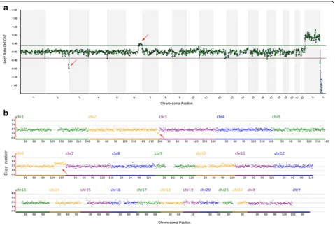
The NGS results of the 10 samples were completely con-sistent with those of array CGH (see Table 1 and Fig. 1); therefore, more samples were submitted for testing using the NGS method. A total of 436 samples were tested, 256 cases of which were tested by array CGH, and 180 cases were tested by NGS. Because detection coverage was theoretically consistent based on array CGH and NGS, we calculated the detection results by using the two methods together. Two hundred and twenty-five cases were found to have abnormal chromosomes, which accounted for 51.6 % of all the cases. There were 188 (43.1 %) cases with aneuploidy, 23 (5.3 %) cases with chromosomal segmental duplication and/or deletion, and 14 (3.2 %) cases with polyploidy (see Table 2). A total of 110 female samples and 101 male samples were found in the normal samples.
Monosomy was mainly found in sex chromosomes except for one case with monosomy 21. Some aneuploidy samples involved two chromosomes. The total chromosomal nu-merical abnormality frequency was 198, for all the chromo-somes except chromosome 1. Trisomy 16 was the most common in 50/198 (25.3 %), followed by chromosomes X, 22, 15, 14, and 21 (see Fig. 2).
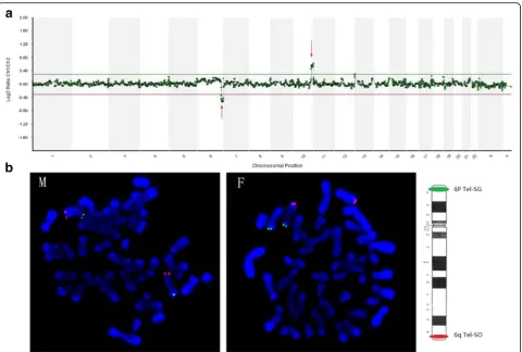
Segmental deletion and/or duplication was found in 23/436 (5.3 %) (see Table 3). Seventeen couples’ kar-yotypes were available, and fifteen couples were re-ported as having normal karyotype, except for 2 reciprocal translocation carriers. Three couples with normal karyotype were submitted for FISH analysis, and two of them were identified to be submicroscopic Table 1Validation of copy number analysis by NGS
NO Results by array CGH Consistency with NGS
C0005 +2;XY Yes
C0003 +16;XX Yes
C0015 +21;XX Yes
C0029 -X Yes
C0142 +15(Mosaic);XY Yes
C0021 69, XXY Yes
C0123 69, XYY Yes
C0012 -(2q37.3-qter)(4.6 M), +(6q23.2-qter)(36.1 M);XX Yes C0146 +(9q34.11-qter)(9.2 M),−(14q32.13-qter)(14.8 M);XX Yes
C0179 Euploid;XX Yes
reciprocal balanced translocation (see Table 4 and Fig. 3).
Discussion
Chromosomal abnormality is the main reason for first-trimester miscarriages. Conventional G-banding karyo-typing is used as a golden standard method to detect chromosomal aneuploidy and imbalances. However, the overall detection failure rate was ~20 % [2, 4], and some-times false negative outcomes resulted from the over-growth of maternal cells in relation to fetal cells [5, 8, 9]. Array CGH is a rapid, automated, reliable, and high-resolution technique used to diagnose unbalanced chromosomal abnormalities in CVSs from miscarriage patients [10, 11]. Recently, NGS was validated as being able to reliably detect the CNVs in CVSs [12]. Array CGH and NGS are both high throughput genetic test platforms that had revolutionary impacts on traditional cytogenetics [13–15]. We validated the efficiency of chromosomal copy number analysis through the NGS method in our lab and summarized the total detection efficiency of array CGH and NGS methods on CVSs. Here, a total number of 436 CVSs from early miscar-riages were analyzed by array CGH and NGS with a
100 % diagnosis rate. We achieved a 51.6 % detection rate.
The occurrence frequency of each aneuploid chromo-some was analyzed in this study, which suggested that errors were involved in all chromosomes besides chromosome 1. However, limited chromosome probe panels were always used to analyze the prenatal samples [16], miscarriage samples [4, 17], and pre-implantation embryos [18, 19]. According to this study, the limited probe panel (Chr13,18,21,X,Y) can only detect 43/198 (21.7 %) aneuploidy in CVSs, and the probe panel (Chr13, 16, 18, 21, 22, X ,Y) can only detect 115/198 (58.1 %) as well. It is obvious that a limited chromosome analysis method for CVSs is not suitable because many positive samples would be ignored. This also reflects why pre-implantation genetic screening using mFISH was proved to have no benefit for improving in vivo fertilization outcomes [20].
It is generally acknowledged that the resolution of rou-tine G-banding karyotyping is 5-10 Mb. However, when the chromosomal segmental imbalances were involved in atypical bands, or poor digestion and dyeing were taken place in the procedure of chromosome prepar-ation, even more than 10 Mb segmental duplications or Table 2Summary of chromosomal copy number analysis of CVSs
Method Maternal age Chromosomal abnormality Euploidy Total Aneuploidy Dup/Del Polyploidya Total
Array CGH 30.8 ± 4.3 115(44.9 %) 7(2.7 %) 9(3.5 %) 131(51.2 %) 125(48.8 %) 256 NGS 31.1 ± 4.8 73(40.6 %) 16b(8.9 %) 5c(2.8 %) 94(52.2 %) 86(47.8 %) 180
Total 30.9 ± 4.5 188(43.1 %) 23(5.3 %) 14(3.2 %) 225(51.6 %) 211(48.4 %) 436
a
array CGH and NGS could identify some polyploidy such as 69 XXY and 69 XYY, but could not find 69 XXX, which was no sex chromosomal segregation.b
two of them combined with aneuploidy;c
one of them combined with segmental duplication and tetrasomy
deletions were sometimes missed, such as samples C0044 and C0146 (see Table 4). Array CGH had been used to identify small-size CNVs on miscarriage samples [2]; however, most of the CNVs found in those studies had no parental origin analysis and no significant clinical value was concluded. In this study, segmental deletion and/or duplication was observed in 5.3 % of the samples by array CGH or NGS. We failed to analyze the origin of the segmental changes in all 15 of the couples with normal karyotypes. However, FISH was performed on three of them to investigate the parental origin, and two
of them were proved to be hereditary from the paternal submicroscopic reciprocal balanced translocations. With exact chromosomal diagnosis, pre-implantation genetic diagnosis (PGD) was recommended to these couples [21]. Although we could not conclude the exact inci-dence of submicroscopic recombination in miscarriage couples with normal karyotypes, we emphasized that a clinician should be aware of submicroscopic reciprocal translocation in couples with recurrent miscarriages.
With consideration of identifying submicroscopic re-ciprocal translocation, high throughput genetic testing is Table 3Parents karyotype analysis of the CVSs with segmental copy number variants
No Copy number variants for CVSs Parents karyotype C0012 -(2q37.3-qter)(4.6 M); +(6q23.2-qter)(36.1 M) Normal C0044 -(6q25.3-qter)(12.5 M); +(10q26.11-qter)(13.8 M) Normal C0146 +(9q34.11-qter)(9.2 M);−(14q32.13-qter)(14.8 M) Normal C0391 -(1p36.21-pter)(12.36 M) Normal C0001 -(18p11-pter)(14.0 M); +(18q11-qter)(59.4 M) Normal C0052 +(17q21.31-qter)(38.9 M) Normal C0227 -(8p12-pter)(28.8 M); +(8q24.3-qter)(4.9 M) Normal C0361 +(5q13.2-qter)(106.42 M);−(15q26.1-qter)(11.35 M) Normal C0376 -(5p15.1-pter)(16.6 M); +(9q21.32-qter)(56.6 M) Normal C0403 -(5p14.1-pter)(25.68 M); +(19q13.33-qter)(8.35 M) Normal C0407 +(2q12.1-q33.1)(93.02 M) Normal C0419 +19(q13.33-qter)(7.56 M) Normal C0420 +(11q23.3-pter)(111.15 M);−(22q11.1-q11.21)(3.92 M) Normal C0432 +(9p21.3-pter)(21.71 M);−3(q28-qter)(8.3 M) Normal C0439 -(1p36.21-pter)(13.69 M); +19 Normal
C0404 +(5p13.33-pter)(31.09 M);−(10q24.32-qter)(29.19 M) Paternal reciprocal translocation carrier C0193 +(2p24.3-pter)(15.11 M);−(13q22.1-qter)(38.47 M) Paternal reciprocal translocation carrier C0063 +(16p11.2-qter)(56.4 M) Loss to Follow-up
C0195 -(8q24.13-qter)(19.83 M); +(11q23.3-qter)(17.22 M) Loss to Follow-up C0294 +(11q22.3-q24.2)(22.08 Mb);−(11q24.2-qter)(7.12 Mb) Loss to Follow-up C0358 -(13q21.31-qter)(48.84 Mb) Loss to Follow-up C0385 +14(q11.2-q12)(8.67 M);−X Loss to Follow-up C0389 -(18p11.21-pter)(14.08 Mb); +(18p11.21-qter)(59.8 Mb); +(19q12-qter)(27.58 Mb) Loss to Follow-up
Table 4Origin analysis of 3 cases with small-size segmental imbalances by FISH
No. Copy number variants FISH test Parental origin Probea Paternal Maternal
C0012 -(2q37.3-qter)(4.6 M); 6p SG; 6q SO Normal Normal De novo +(6q23.2-qter)(36.1 M)
C0044 -(6q25.3-qter)(12.5 M); 6p SG; 6q SO Carrier Normal Paternal +(10q26.11-qter)(13.8 M)
C0146 +(9q34.11-qter)(9.2 M); 14q SO Carrier Normal Paternal -(14q32.13-qter)(14.8 M)
a
recommended for analyzing the chromosomal copy num-ber of CVSs from spontaneous abortions. Array CGH and NGS were robust in the detection of chromosomal CNVs, and 69 XXY and 69 XYY could be detected as well for the special segregation of sex chromosomes. However, the limitations also should be considered. NGS and array CGH cannot detect all polyploidies, such as 69 XXX, 92 XXXX, and 92 XXYY, as well as balanced translocations. In present study, more normal female samples were ob-served than normal male samples (110vs101), which was likely caused by confusion of 69 XXX and 46 XX. In the future, single nucleotide polymorphism analysis could be adopted in order to identify the polyploidies by NGS.
Conclusions
In conclusion, a high chromosomal abnormality detection rate on CVSs from patients who had spontaneous miscar-riages was achieved by array CGH and NGS. Particularly, submicroscopic recombination could be detected, which was important to genetic counseling. Array CGH and NGS are comprehensive, rapid, and high-resolution chromosomal copy number analysis methods.
Competing interests
The authors declare that they have no competing interests.
Authors’contributions
JS carried out the molecular cytogenetic studies, participated in the design of the study and data analysis, and drafted the manuscript. WW carried out the genetic counseling and sample collecting. HO participated in genetic analysis and revising the manuscript. CG, DQ and JX participated in the array CGH and NGS and FISH essay. LG participated in sample collecting and preparing. YZ participated in G-banding karyotyping. YC participated in the design of the study. JL conceived of the study, and participated in its design and coordination and helped to draft the manuscript. All authors read and approved the final manuscript.
Acknowledgement
We would like to thank Dr. Tao Feng for helping to analyze the NGS data. This work was supported by China National 973 program (grant number 2012CB944902), Jiangsu Provincial Science and Technology Project (grant number BL20122009 and BE2011798), and the Priority Academic Program Development of Jiangsu Higher Education Institutions (PAPD).
Funding information
National Fund for Natural Science (81200444).
Received: 11 August 2015 Accepted: 21 December 2015
References
1. Rai R, Regan L. Recurrent miscarriage. Lancet. 2006;368:601–11.
2. van den Berg MM, van Maarle MC, van Wely M, Goddijn M. Genetics of early miscarriage. Biochim Biophys Acta. 1822;2012:1951–9.
3. Deshpande M, Harper J, Holloway M, Palmer R, Wang R. Evaluation of array comparative genomic hybridization for genetic analysis of chorionic villus sampling from pregnancy loss in comparison to karyotyping and multiplex ligation-dependent probe amplification. Genet Test Mol Biomarkers. 2010; 14:421–4.
4. Shearer BM, Thorland EC, Carlson AW, Jalal SM, Ketterling RP. Reflex fluorescent in situ hybridization testing for unsuccessful product of conception cultures: a retrospective analysis of 5555 samples attempted by conventional cytogenetics and fluorescent in situ hybridization. Genet Med. 2011;13:545–52.
5. Diego-Alvarez D, Garcia-Hoyos M, Trujillo MJ, Gonzalez-Gonzalez C, Rodriguez de Alba M, Ayuso C, et al. Application of quantitative fluorescent PCR with short tandem repeat markers to the study of aneuploidies in spontaneous miscarriages. Hum Reprod. 2005;20:1235–43.
6. Hou Y, Fan W, Yan L, Li R, Lian Y, Huang J, et al. Genome analyses of single human oocytes. Cell. 2013;155:1492–506.
7. Shen JD, Liang DS, Zhou ZM, Xia Y, Long ZG, Wu LQ. Pallister-Killian syndrome: meiosis II non-disjunction may be the first step in the formation of isochromosome 12p. Chin Med J (Engl). 2010;123:3482–5.
8. Robberecht C, Schuddinck V, Fryns JP, Vermeesch JR. Diagnosis of miscarriages by molecular karyotyping: benefits and pitfalls. Genet Med. 2009;11:646–54.
9. Karaoguz MY, Nas T, Konac E, Ince D, Pala E, Menevse S. Is cytogenetic diagnosis of 46, XX karyotype spontaneous abortion specimens erroneous? Fluorescence in situ hybridization as a confirmatory technique. J Obstet Gynaecol Res. 2005;31:508–13.
10. Menten B, Swerts K, Delle Chiaie B, Janssens S, Buysse K, Philippe J, et al. Array comparative genomic hybridization and flow cytometry analysis of spontaneous abortions and mors in utero samples. BMC Med Genet. 2009;10:89.
11. Schaeffer AJ, Chung J, Heretis K, Wong A, Ledbetter DH, Lese Martin C. Comparative genomic hybridization-array analysis enhances the detection of aneuploidies and submicroscopic imbalances in spontaneous miscarriages. Am J Hum Genet. 2004;74:1168–74.
12. Xie W, Tan Y, Li X, Lin G, Jiang H, Chen F, et al. Rapid detection of aneuploidies on a benchtop sequencing platform. Prenat Diagn. 2013;33:232–7.
13. Miller DT, Adam MP, Aradhya S, Biesecker LG, Brothman AR, Carter NP, et al. Consensus statement: chromosomal microarray is a first-tier clinical diagnostic test for individuals with developmental disabilities or congenital anomalies. Am J Hum Genet. 2010;86:749–64.
14. Biesecker LG, Green RC. Diagnostic clinical genome and exome sequencing. N Engl J Med. 2014;370:2418–25.
15. Koboldt DC, Steinberg KM, Larson DE, Wilson RK, Mardis ER. The next-generation sequencing revolution and its impact on genomics. Cell. 2013; 155:27–38.
16. Tepperberg J, Pettenati MJ, Rao PN, Lese CM, Rita D, Wyandt H, et al. Prenatal diagnosis using interphase fluorescence in situ hybridization (FISH): 2-year multi-center retrospective study and review of the literature. Prenat Diagn. 2001;21:293–301.
17. Jobanputra V, Esteves C, Sobrino A, Brown S, Kline J, Warburton D. Using FISH to increase the yield and accuracy of karyotypes from spontaneous abortion specimens. Prenat Diagn. 2011;31:755–9.
18. Staessen C, Verpoest W, Donoso P, Haentjens P, Van der Elst J, Liebaers I, et al. Preimplantation genetic screening does not improve delivery rate in women under the age of 36 following single-embryo transfer. Hum Reprod. 2008;23:2818–25.
19. Debrock S, Melotte C, Spiessens C, Peeraer K, Vanneste E, Meeuwis L, et al. Preimplantation genetic screening for aneuploidy of embryos after in vitro fertilization in women aged at least 35 years: a prospective randomized trial. Fertil Steril. 2010;93:364–73.
20. Mastenbroek S, Twisk M, van der Veen F, Repping S. Preimplantation genetic screening: a systematic review and meta-analysis of RCTs. Hum Reprod Update. 2011;17:454–66.
21. Fiorentino F, Spizzichino L, Bono S, Biricik A, Kokkali G, Rienzi L, et al. PGD for reciprocal and Robertsonian translocations using array comparative genomic hybridization. Hum Reprod. 2011;26:1925–35.
• We accept pre-submission inquiries
• Our selector tool helps you to find the most relevant journal
• We provide round the clock customer support
• Convenient online submission
• Thorough peer review
• Inclusion in PubMed and all major indexing services
• Maximum visibility for your research
Submit your manuscript at www.biomedcentral.com/submit