R E S E A R C H
Open Access
In vitro exposure of a 3D-tetraculture
representative for the alveolar barrier at the
air-liquid interface to silver particles and
nanowires
Ionel Fize
ș
an
1, Sébastien Cambier
2, Elisa Moschini
2, Aline Chary
2, Inge Nelissen
3, Johanna Ziebel
2,
Jean-Nicolas Audinot
4, Tom Wirtz
4, Marcin Kruszewski
5,6, Anca Pop
1, Béla Kiss
1, Tommaso Serchi
2,
Felicia Loghin
1and Arno C. Gutleb
2*Abstract
Background:The present study aimed to evaluate the potential differences in the biological effects of two types of spherical silver particles of 20 and 200 nm (Ag20 and Ag200), and of PVP-coated silver nanowires (AgNWs) with a diameter of 50 nm and length up to 50μm, using a complex 3D model representative for the alveolar barrier cultured at air-liquid interface (ALI). The alveolar model was exposed to 0.05, 0.5 and 5μg/cm2of test compounds at ALI using a state-of-the-art exposure system (Vitrocell™Cloud System). Endpoints related to the oxidative stress induction, anti-oxidant defence mechanisms, pro-inflammatory responses and cellular death were selected to evaluate the biocompatibility of silver particles and nanowires (AgNMs) and to further ascribe particular biological effects to the different morphologic properties between the three types of AgNMs evaluated.
Results: Significant cytotoxic effect was observed for all three types of AgNMs at the highest tested doses. The increased mRNA levels of the pro-apoptotic geneCASP7suggests that apoptosis may occur after exposure to AgNWs. All three types of AgNMs increased the mRNA level of the anti-oxidant enzymeHMOX-1and of the metal-binding anti-oxidantmetallothioneins(MTs), with AgNWs being the most potent inducer. Even though all types of AgNMs induced the nuclear translocation of NF-kB, only AgNWs increased the mRNA level of pro-inflammatory mediators. The pro-inflammatory response elicited by AgNWs was further confirmed by the increased secretion of the 10 evaluated interleukins.
Conclusion: In the current study, we demonstrated that the direct exposure of a complex tetra-culture alveolar model to different types of AgNMs at ALI induces shape- and size-specific biological responses. From the three AgNMs tested, AgNWs were the most potent in inducing biological alterations. Starting from 50 ng/cm2, a dose representative for an acute exposure in a high exposure occupational setting, AgNWs induced prominent changes indicative for a pro-inflammatory response. Even though the acute responses towards a dose representative for a full-lifetime exposure were also evaluated, chronic exposure scenarios at low dose are still unquestionably needed to reveal the human health impact of AgNMs during realistic conditions.
Keywords:Air-liquid interface, Tetra-culture, Silver nanoparticles, Silver nanowires, Inflammation;
© The Author(s). 2019Open AccessThis article is distributed under the terms of the Creative Commons Attribution 4.0
International License (http://creativecommons.org/licenses/by/4.0/), which permits unrestricted use, distribution, and reproduction in any medium, provided you give appropriate credit to the original author(s) and the source, provide a link to the Creative Commons license, and indicate if changes were made. The Creative Commons Public Domain Dedication waiver (http://creativecommons.org/publicdomain/zero/1.0/) applies to the data made available in this article, unless otherwise stated.
* Correspondence:arno.gutleb@list.lu
2Environmental Research and Innovation (ERIN) Department, Luxembourg Institute of Science and Technology, Belvaux, Luxembourg
Full list of author information is available at the end of the article
Fizeșanet al. Particle and Fibre Toxicology (2019) 16:14
Background
The rapid advancements in the field of nanotechnology produced a vast number of engineered nanomaterials (ENMs) with unique physical and chemical properties. Exhibiting a broad utility and versatility, ENMs are cur-rently incorporated in a large number of consumer products, offering also unique advantages as diagnostic and therapeutic tools in the emerging field of nanomedi-cine [1]. Silver nanomaterials (AgNMs) are one of the most widely used ENMs, with more than 300 commer-cially available products in Europe [2], due to their anti-microbial activity [3, 4]. Silver nanowires (AgNWs) are incorporated in flexible thin films with high conductiv-ity, and further used in the production of liquid crystal displays, touch screens and solar cells [5,6].
Respiratory exposure is of primary concern due to re-lease of AgNMs to the ambient air during manufactur-ing processes and the use of consumer products. Inhaled ENMs are efficiently deposited given the high intake of air (> 20 m3/day) and the large surface area (75–150 m2) of the respiratory tract. The alveolar barrier is of main concern due to the deposition of small particles and the susceptibility of the gas exchange membrane [7,8].
Despite the pulmonary retention and systemic distri-bution of spherical AgNMs, the majority of in vivo stud-ies reported no or only mild effects involving chronic pulmonary inflammation and abnormalities in the liver [9–12]. In contrast, pronounced effects including cyto-toxicity, pro-inflammatory responses and genotoxicity were reported using various epithelial and immune cell types in in vitro studies [13–18]. These differences be-tween in vivo and in vitro data are most likely due to the unrealistic exposure scenarios and concentrations used. Submerged exposure of alveolar cells has several intrin-sic limitations, as it disregards the baintrin-sic structure of the respiratory system and the physical laws behind respira-tory exposure. Moreover, by suspending ENMs in cell culture media, important changes in their physicochemi-cal properties including surface reactivity and agglomer-ation state occur influencing the exposure concentragglomer-ations and outcome of the experiments [19, 20]. An im-proved in vitro technique which encompasses the above-mentioned limitations is represented by air-liquid interface (ALI) exposure [21]. This allows a direct expos-ure to ENMs, closely mimicking realistic inhalation condi-tions [19, 22–24]. It is generally accepted that the use of ALI exposure in respiratory toxicology is more suitable in the assessment of ENMs toxicity, and together with other improved in vitro approaches may hold the promise to bridge the current gaps between in vitro and in vivo data [19, 25].
The aim of the present study was to assess and com-pare the toxicological effects of two types of spherical silver particles with sizes of 20 and 200 nm (Ag20 and
Ag200, respectively) and of AgNWs with a diameter of 50 nm and a length up to 50μm, using a 3D tetra-culture model representative for the alveolar bar-rier [22,26,27]. The model was exposed to 0.05, 0.5 and 5μg/cm2of AgNMs at the ALI using a state-of-the-art exposure system (Vitrocell™Cloud System). Specific end-points related to the oxidative stress induction, anti-oxidant defence mechanisms, pro-inflammatory re-sponses and cellular death were evaluated at different time points.
Results
Characterization of ENMs in liquid suspension
Based on the rationale that spraying a homogenous sus-pension is a prerequisite for a homogenous exposure, the first step was the characterization of the AgNMs in liquid suspension. Preparation of the AgNMs dispersions according to a published protocol [20] resulted in a mean hydrodynamic diameter of 232 ± 125 nm, 362 ± 56 nm and 262 ± 160 nm for Ag20, Ag200 and AgNWs. The measured Zeta potentials for Ag20, Ag200 and AgNWs were−40.5 ± 4.8, −32.2 ± 4.6 and−24.6 ± 8.2 mV, respectively. The values obtained for AgNWs served to calculate the necessary energy input to obtain a stable suspension and are not an accurate measurement of the hydrodynamic diameter, as the Dynamic Light Scattering (DLS) method is based on the assumption of a spherical shape [28, 29]. The stability of AgNMs suspension was not verified at later time points, as suspensions were prepared freshly before each exposure. According to BET measurement, the specific surface area of the Ag20 and Ag200 were 2.52 ± 0.05 and 1.18 ± 0.01 m2/g, while the specific surface area for the AgNWs was not mea-sured due to specificities of these AgNWs.
Based on the Inductively Coupled Plasma Mass Spec-trometry (ICP-MS) analysis, we observed that the sonic-ation process had a minor impact on the release of Ag+ from all AgNMs tested, with Ag+ representing less than 0.2% from total silver species immediately after the son-ication process. The stability of the AgNMs suspensions was maintained at the same Ag+ level over 24 h. Fur-thermore, incubation with the pulmonary surfactant did not increase the release of Ag+ for either of all AgNMs tested.
Characterization of the deposited ENMs
The deposition uniformity was evaluated by two visualization techniques. Using the high-resolution dark-field microscopy technique (CytoViva®), a circu-lar-like deposition pattern rather than a homogenous particle distribution was observed with the majority of AgNMs distributed on the exterior of the circle (Fig. 1). This pattern is in accordance with the drop-let size distribution generated by the nebulizer, and
may be due to the drying of highly concentrated droplets onto the glass substrate [30, 31]. We believe that this artefact is not present when the cellular model is exposed to the AgNMs due to the presence of the liquid lining. The shape and the analytical composition of the particles were investigated by He-lium Ion Microscopy (ZEISS, Peabody, USA) (HIM) [32], coupled with a Secondary Ion Mass Spectrom-eter (SIMS) developed by LIST, Luxembourg [33, 34]. The sprayed AgNWs maintained their needle-like shape after the sonication and nebulisation processes, with the same ratio between length and diameter be-ing observed as for pristine AgNWs (Fig. 2; Add-itional file 1: Figure S1). Smaller fragments with more symmetric morphology were also observed (Fig. 2).
Quantification of total silver species using ICP-MS allowed the measurement of the deposition efficiency and the intra- and inter-exposure variability, with the overall validation of the exposure system (Table 1 and Additional file 2: Table S1). For studying the biological effects, 200μL of AgNMs suspensions with concentra-tions of approximately 5000, 500 and 50μg/mL (dependent on the deposition efficiencies obtained previ-ously) were sprayed in order to achieve a deposition of 0.05, 0.5 and 5μg/cm2
.
Cytotoxicity and metabolic impairment
The cytotoxic effects after exposure to AgNMs were assessed by measuring the release of lactate dehydro-genase (LDH). All three types of AgNMs tested had a
a
d
b
e
c
f
Fig. 1High resolution dark-field microscopy images (CytoViva®) of Ag20 (a,d), Ag200 (b,e) and AgNWs (c,f) after sonication and nebulisation on coverslips using the Vitrocell™Cloud System. Samples were further fixed between two coverslips using Mowiol mounting media
statistically significant impact on the cellular integrity and the membrane leakage of LDH (Fig. 3). The 16μg/cm2 (Ag+) silver nitrate solution used as a posi-tive control for toxicity interfered with the accurate measurement of the LDH activity, this interference of Ag+ being also reported in other studies [35]. The in-tegrity of the cellular layer after exposure to AgNWs was further evaluated by confocal microscopy. Expos-ure to the AgNWs at the highest concentration did not reveal any significant effects on the apical com-partment of the alveolar model (Additional file 3: Figure S2).
The cytotoxic effects elicited by AgNMs were fur-ther assessed using the Alamar Blue (AB) assay. Ag20 had a visible dose-dependent effect, decreasing the cellular viability in the apical compartment more than
20% at the highest tested dose at 6 h post-exposure. Ag200 and AgNWs had no visible impact on the ap-ical compartment at 6 h post-exposure (Fig. 4a). In the basolateral compartment, at 6 h post-exposure, in-dependently of the AgNM tested, a significant decrease of the metabolic activity was observed at the highest con-centration (Fig.4b). Similarly to the LDH assay, a recovery of the system at 24 h post-exposure was observed, with a decrease of cellular viability in the apical compartment only at the highest dose of Ag20 (Fig. 4c). Moreover, in the basolateral compartment, an increase of the metabolic activity was observed for Ag20 and AgNWs at the inter-mediate and high concentrations (Fig.4d).
Oxidative stress response
Reactive oxygen species (ROS) generation after the ex-posure to AgNMs was quantified at 6 h post-exex-posure. Due to its unspecific reactions with ROS, DCFH-DA assay was used to measure the overall oxidative stress [36]. All three types of AgNMs, dependent on the doses used, increased the generation of ROS in the apical and basolateral compartment (Fig. 5a and b). To further study the biological impact of AgNMs-generated ROS, the ratio between reduced (GSH) and oxidized glutathi-one (GSSG) was evaluated at 6 h post-exposure. Nglutathi-one of the AgNMs tested had an impact on the GSH/GSSG ratio in the apical and basolateral side (data not shown).
Gene expression of the pro-inflammatory and stress response elements
Based on the measurements using real-time reverse transcriptase polymerase chain reaction (RT-PCR), statistically significant changes (P< 0.05) were Fig. 2Secondary electron images (a-c) and mass filtered secondary ion images (d-f) of Ag20, Ag200 and AgNWs (bar scale 1μm) obtained on the Helium Ion Microscopy–Secondary Ion Mass Spectrometry (HIM-SIMS) instrument. Samples were deposited on a Silicon wafer
Table 1Deposition efficiencies obtained for Ag20, Ag200 and
AgNWs using the Vitrocell™Cloud System
Concentration of the sprayed suspension (μg/mL)
Deposition efficiency (%)
Expected concentration (μg/cm2)
Measured concentration (μg/cm2)
Ag20 50 54 0.05 0.054
500 68 0.5 0.68
5000 74 5 7.4
Ag200 50 50 0.05 0.05
500 47 0.5 0.47
5000 82 5 8.2
AgNWs 50 40 0.05 0.04
500 57 0.5 0.57
5000 60 5 6.0
The deposition efficiencies were measured for 3 increasing concentrations
observed after exposure to AgNMs for genes encod-ing proteins involved in anti-oxidant response, inflamma-tion and cellular death. Genes were further divided into two groups (stress response and inflammation) for each compartment (apical/basal) and the principal component analysis (PCA) was applied on these data subsets to high-light the differences between the tested AgNMs [37].
Expression of genes encoding proteins involved in the stress response in the apical compartment
Exposure to AgNMs significantly increased the mRNA of genes encoding for metallothioneins (MTs), the most potent effect being observed for AgNWs, which increased the level of all three MTs at 6 h and 24 h post-exposure (Fig. 6). Moreover, exposure to AgNMs increased the mRNA of heme-oxygenase-1 (HMOX-1) at the highest tested dose. At 6 h post-exposure, the effect on HMOX-1 mRNA level increased in the same order of potency as for MTs (Ag200 < Ag20 < AgNWs), while at 24 h post-exposure only AgNWs at the highest dose has led to an increased level of HMOX-1 (Fig. 6).
At 6 h post-exposure all AgNMs decreased the mRNA level of NAD(P)H quinone dehydrogenase 1 (NQO1), with a return to basal level at 24 h post-exposure (Fig.6), while at 24 h post-exposure, independently of the dose and AgNMs tested, a modest, but significant increase in theSuperoxide dismutase-1 (SOD1) was noticed. Regard-ing the mRNA level of genes encodRegard-ing effectors in the pro-apoptotic cascade, exposure to AgNWs increased sig-nificantly the mRNA level of Caspase 7 (CASP7) at 6 h post-exposure for all concentrations tested, while at 24 h
post-exposure a significant induction in the mRNA level ofFASwas observed for all AgNMs (Fig.6).
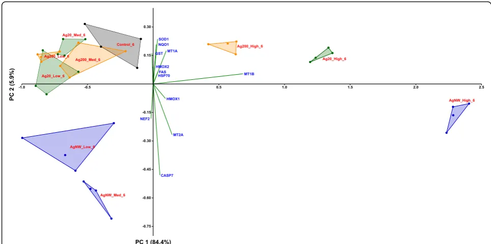
According to the PCA distribution, the effects related to the stress response were mainly dependent on the dose of AgNMs, with the highest dose treatment for each AgNMs inducing a distinctive pattern from the negative control group. Moreover, differences between the three AgNMs were observed at the highest tested dose, with AgNWs presenting the most distinctive pat-tern (Fig.7; Additional file4: Figure S3). From the differ-ential gene expression patterns, the genes coding for MTs (MT-1A, MT-1B and MT-2A) and HMOX-1 were the most important contributors to the existing variance in the data.
Expression of genes encoding proteins involved in the stress response in the basolateral compartment
Exposure to AgNMs induced small changes in the mRNA level of genes encoding stress response ele-ments in the basolateral compartment. A slight statis-tical increase in the mRNA of MT-2A was observed only for AgNWs, while in agreement with the effects observed on the apical side, AgNWs increased signifi-cantly the mRNA level of HMOX-1 at 6 h and 24 h post-exposure (Fig. 8). At 6 h post-exposure, Ag20 transiently up-regulated the mRNA level of Heat Shock Protein 70 (HSP70), a general marker for cellu-lar stress. In addition, exposure to the highest tested dose of Ag20, and AgNWs strongly down-regulated the transcription of Glutathione S-Transferase (GST) at 24 h post-exposure (Fig. 8). Regarding the mRNA level of genes encoding pro-apoptotic proteins,
a
b
Fig. 3Effects of Ag20, Ag200, AgNWs and Ag+(16μg/cm2) on LDH release at 6 h (a) respectively at 24 h post-exposure (b). Cells exposed to
vehicle served as negative control (Ctrl). Data are presented as the mean ± SD of at least three biological replicates. Data were expressed as the percentage of the LDH release from total LDH release (100%)
exposure to Ag20 up-regulated the level of FAS at 6 h post-exposure at all tested doses. An up-regulation of FAS and CASP7 was also observed after exposure to AgNWs at the dose of 5μg/cm2 at 6 h post-exposure (Fig. 8). PCA analysis did not reveal major differences in the data subsets at both time points evaluated (Additional file 5: Figure S4 and Additional file 6: Figure S5).
Expression of genes encoding proteins involved in the pro-inflammatory response in the apical compartment Similarly to what was observed for genes encoding pro-teins involved in the stress response, AgNWs displayed the highest ability to increase the mRNA level of pro-inflammatory molecules in the apical compartment. At 6 h post-exposure, AgNWs increased in a dose-dependent manner the mRNA level of the pro-inflam-matory interleukins 6 and 8(IL-6andIL-8), and of the
Intercellular Adhesion Molecule-1 (ICAM-1) and Vas-cular Cell Adhesion Molecule-1(VCAM-1)(Fig. 9). Ex-posure to Ag20 had also an impact on the mRNA level of VCAM-1 at 6 h post-exposure, increasing it at the two highest concentrations tested. Opposite to this, ICAM-1was down-regulated by Ag20 and Ag200 at 6 h post-exposure. At 24 h post-exposure, a stable down-regulation in the mRNA level ofVCAM-1was observed for Ag20 and Ag200 (Fig.9).
Considering that most of the cytokines, chemo-kines and adhesion molecules evaluated in this study are under the regulation of the transcription factor NF-kB, the mRNA level of NF-kB was inves-tigated. No significant effects were observed for any of the AgNMs tested at 6 h post-exposure, while at 24 h post-exposure, independently of the dose and type of AgNMs tested, a down-regulation was observed (Fig. 9).
c
d
b
a
Fig. 4Effects of Ag20, Ag200, AgNWs and Ag+(16μg/cm2) on cellular viability using Alamar Blue Assay at 6 h post-exposure on apical (a) and basolateral side (b), respectively at 24 h post exposure on apical (c) and basolateral side (d). Cells exposed to vehicle served as negative control (Ctrl). The data are presented as the mean ± SD of at least three biological replicates. Data were expressed relative to the negative control (100%)
The distinctive pro-inflammatory nature of AgNWs was further confirmed by PCA (Fig.10; Additional file7: Figure S6), with the most prominent differences being ob-served at 6 h post-exposure (Fig. 10). The most im-portant contributing factors in the spatial distribution were the genes encoding IL-6, IL-8,
VCAM-1 and ICAM-1. At 24 h post-exposure, the
differences between the AgNWs and the other two types of AgNMs were still observable to a lower ex-tent, with the same genes as for 6 h post-exposure behaving as the main contributors.
Expression of genes encoding proteins involved in the pro-inflammatory response in the basolateral compartment In parallel with the responses observed in the apical compartment, exposure to AgNWs up-regulated the mRNA level ofIL-6andIL-8in the basolateral compart-ment at both time points (Fig.11). Conversely, a repres-sion of IL-8 was observed after exposure to 0.05 and 0.5μg/cm2 Ag200 and 0.5μg/cm2 Ag20 (Fig. 11). Re-garding the mRNA level of the evaluated adhesion mole-cules, only AgNWs exposure induced significant alterations. Exposure to AgNWs strongly induced at 6 h
a
b
Fig. 5Induction of reactive oxygen species (ROS) at 6 h post-exposure. Effects of Ag20, Ag200 and AgNWs on apical (a) and basolateral side (b) after exposure to 0.05, 0.5 and 5μg/cm2. The data are presented as the mean ± SD of three biological replicates. Data were expressed relative to
the negative control (100%)
Fig. 6mRNA level of relevant stress markers in the apical compartment of the alveolar model at 6 h and 24 h post-exposure to Ag20, Ag200 and AgNWs
Fig. 7PCA analysis on the dataset of genes encoding stress response mediators at 6 h post-exposure in the apical compartment. The relative gene level (fold increase/decrease compared to negative control (gray)) of samples exposed to Ag20 (green), Ag200 (orange) and AgNWs (blue) at the three different doses (low = 0.05μg/cm2, medium = 0.5μg/cm2and high = 5μg/cm2) are represented on the scatter plot corresponding to PC1 and PC2
Fig. 8mRNA level of relevant stress markers in the basolateral compartment of the alveolar model at 6 h and 24 h post-exposure to Ag20, Ag200 and AgNWs
Fig. 9mRNA level of relevant inflammatory markers in the apical compartment of the alveolar model at 6 h and 24 h post-exposure to Ag20, Ag200 and AgNWs
Fig. 10PCA analysis on the dataset of genes encoding pro-inflammatory mediators at 6 h post-exposure in the apical compartment. The relative gene level (fold increase/decrease compared to negative control (gray)) of samples exposed to Ag20 (green), Ag200 (orange) and AgNWs (blue) at the three different doses (low = 0.05μg/cm2, medium = 0.5μg/cm2and high = 5μg/cm2) are represented on the scatter plot corresponding to
PC1 and PC2
post-exposure the mRNA level of all three adhesion molecules, while at 24 h post-exposure only ICAM-1 remained significantly up-regulated (Fig.11). The mRNA level of NF-kB was not modified after exposure to AgNMs (Fig.11).
The PCA analysis confirmed the distinctive pro-inflam-matory potential of AgNWs, with the genes encoding for the adhesion molecules E-SELECTIN, VCAM-1 and
ICAM-1 being the most important contributors
(Additional file 8: Figure S7 and Additional file 9: Figure S8).
Cytokine and chemokine secretion
The ability of the model to respond to LPS exposure by secretion of pro-inflammatory cytokines was first evalu-ated. The ALI exposure of the alveolar model to LPS in-creased in a dose-dependent manner the secretion of the pro-inflammatory cytokine IL-8 (Fig.12), without affect-ing the cellular viability (data not shown).
The quantification of the secreted interleukins in the cellular media from the basolateral compartment of the 3D tetra-culture alveolar model was performed after ex-posure to AgNWs, since only these AgNMs had an impact on the gene expression of pro-inflammatory cytokines. Ex-posure to AgNWs induced a dose-dependent increase in the secretion of pro−/anti-inflammatory messengers at both time points evaluated (Table 2). The lowest dose
tested (0.05μg/cm2) had a major impact at 6 h post-exposure, significantly increasing the expression of 6 out of 10 cytokines evaluated. Overall, after exposure to AgNWs, a more prominent biological response was ob-served at 6 h post-exposure, with an attenuation of the Fig. 11mRNA level of relevant inflammatory markers in the basolateral compartment of the alveolar model at 6 h and 24 h post-exposure to Ag20, Ag200 and AgNWs
Fig. 12Level of IL-8 (pg/mL) in cell culture undernatants of the tetra-culture after exposure to 0.625 and 6.25 µg/cm2LPS for 24
h. Data represent the mean of three biological replicates ± SD
Table 2 Level of secondary messengers in cell culture undernatants of the tetra-culture after exposure to 0.05, 0.5 and 5 μ g/cm 2 AgNWs for 6 and 24 h Cytokine Fun ction 6 h post-expos ure 24 h post -exposu re Neg_ control pg/mL 0.05 μ g/cm 2 pg/mL 0.5 μ g/cm 2 pg/mL 5 μ g/cm 2 pg/mL Neg_ control pg/mL 0.05 μ g/cm 2 pg/mL 0.5 μ g/cm 2 pg/mL 5 μ g/cm 2 pg/mL IL-6 Chem otactic and pro-i nflammat ory cyt okine; support s the growt h of B cell s 1.18 ± 0.10 6.22 ± 3.23 23.5 8 ± 1.71 * 27.98 ± 5.1 2 * 1.69 ± 0.26 1.76 ± 0.83 13.05 ± 2.4 6 * 34. 03 ± 0.80 * IL-8 C he m o ta ct ic an d p ro-in fla m m at o ry cy to ki n e 738 ± 33 2216 ± 233 * 2599 ± 33 * 2621 ± 60 * 1423 ± 74 1482 ± 447 2590 ± 22 * 2675 ± 57 * TNF -α Prom otes the inflammation proc ess by upr egulation of othe r pro -inflammatory cyt okines 0.83 ± 0.17 3.59 ± 1.34 8.51 ± 1.56 * 11.57 ± 3.2 5 * 1.47 ± 0.12 1.74 ± 0.42 5.44 ± 1.22 * 10. 70 ± 0.45 * IL-1 β Medi ator of the inflammatory respon se 1.69 ± 0.07 3.03 ± 0.53 * 5.13 ± 0.27 * 5.43 ± 0.59 * 3.56 ± 0.22 1.95 ± 0.5 9 * 4.78 ± 0.71 * 7.0 7 ± 0.41 * IL-2 Key function s in the immun e system , toleranc e and immu nity 0.17 ± 0.10 0.62 ± 0.40 * 1.65 ± 0.55 * 1.53 ± 0.52 * 0.34 ± 0.17 0.21 ± 0.21 1.17 ± 0.65 2.5 2 ± 0.46 * IL-4 Key regul ator in hum oral and adap tive im munity 0.006 ± 0.011 0.055 ± 0.0 12 * 0.15 3 ± 0.010 * 0.169 ± 0.0 42 * 0.006 ± 0.012 0.034 ± 0.021 0.135 ± 0.0 22 * 0.2 60 ± 0.024 * IL-12p 70 Con trols an d stim ulates the activit ies of natu ral killer cells 0.06 ± 0.02 0.35 ± 0.16 * 0.66 ± 0.11 * 0.92 ± 0.05 * 0.14 ± 0.04 0.14 ± 0.11 0.75 ± 0.15 * 1.4 0 ± 0.50 * IL-13 Key regul ator in hum oral and adap tive im munity proliferation 5.24 ± 0.22 11.94 ± 1.0 2 * 17.1 6 ± 2.05 * 17.65 ± 0.7 6 * 8.94 ± 0.67 6.52 ± 2.08 13.55 ± 1.3 1 * 18. 36 ± 2.01 * IL-10 Anti -inflammatory cyto kine; downre gulates the exp ression of Th1 cytoki nes 0.11 ± 0.03 1.44 ± 1.17 2.41 ± 0.79 * 2.56 ± 0.47 * 0.19 ± 0.04 0.45 ± 0.12 1.63 ± 0.59 * 2.5 7 ± 0.22 * IFN-γ Im m u n o st im u la to ry an d im m u n o m o d u la to ry ef fe ct s 0.13 ± 0.23 0.56 ± 0.59 3.12 ± 0.83 * 2.53 ± 1.52 * 0.28 ± 0.25 0.54 ± 0.59 2.24 ± 1.59 4.5 0 ± 0.90 * Data represent the mean of three biological replicates ± SD. Values statistically different from their control are in bold and marked by asterisk
pro-inflammatory effects being observed at 24 h post-ex-posure for the smallest and the intermediate dose. At the highest dose tested, an increased secretion was observed for all cytokines at both time points evaluated. The abso-lute values expressed in pg/mL are presented in Table2.
Nuclear translocation of the transcription factor NF-kB
The potential of AgNMs to induce the nuclear transloca-tion of NF-kB, one of the major molecular pathways with pro-inflammatory function was assessed as an early event at 4 h post-exposure. All three types of AgNMs clearly induced the nuclear translocation of NF-kB at the highest tested dose of 5μg/cm2 in the basolateral compartment (Fig. 13; Additional file 10: Figure S9 and Additional file11: Figure S10). In the case of AgNWs the nuclear translocation was ob-served also at the intermediate dose of 0.5μg/cm2 (Fig. 13).
Discussion
The high interest of industry to incorporate AgNMs in consumer products, including electronics and medical
products, and the imperative need for more realistic in vitro toxicity screening of nanomaterials has prompted the current study. We aimed to investigate the distinct-ive biological effects elicited by two different sizes of spherical silver particles (Ag20 and Ag200) and of PVP-coated silver nanowires (AgNWs) in a 3D tetra-culture in vitro model, representative for the alveo-lar barrier using the Vitrocell™Cloud System. The second objective was to investigate the influence of the AgNMs shape on biological responses. Only a few scientific arti-cles studied the biological effects of AgNWs, and to the best of our knowledge, none investigated the biological effects using an ALI system.
Stable suspensions were obtained following a pub-lished dispersion protocol [20]. The dissolution of Ag+ from the AgNMs due to the sonication process and the further incubation for 24 h was evaluated in order to ex-clude Ag+ as a confounding variable. The sonication procedure with or without subsequent 24 h incubation, did not impact the dissolution rate for any material, with Ag+representing less than < 0.2% of total Ag. Incubation with Poractant alfa, a surrogate of human lung
Fig. 13Potential of AgNWs to induce the nuclear translocation of NF-kB in endothelial cells at 4 h post-exposure. The alveolar model was exposed to AgNWs at the three concentrations studied. Cells exposed to vehicle served as Negative Control. Positive Control was exposed to Lipopolysaccharides (LPS) at a concentration of 10μg/mL for the same period of time. Cells were fixed and stained for NF-kB (green) and nuclei (blue)
surfactant in premature babies, had no effect on the dissolution kinetics of AgNMs. These results are in ac-cordance with recent studies that investigated the influ-ence of pulmonary surfactant and of other lung lining fluid components on the dissolution of AgNWs [38] and AgNPs [39]. Even though the concentration of free Ag+ was low in all conditions, the contribution of free Ag+to the biological effects observed cannot be fully excluded.
The in situ characterization of the nebulized AgNMs revealed changes in the agglomeration state of all ma-terials tested. The increase in size was also reported in previous studies and was attributed to the nebuli-zation process or to drying effects [30, 31]. The sprayed AgNWs maintained their needle-like shape after aerosolization.
The calculated deposition efficiencies after nebulisa-tion were dependent on the concentranebulisa-tion and the type of AgNMs sprayed. AgNWs displayed the lowest depos-ition efficiency, most probably due to their anisotropic shape, which may have interfered with the passage through the pores present in the membrane of the Aero-neb Pro® Aero-nebulizer. Deposition efficiencies for the AgNMs were lower than what was reported for solutions of substances [40,41], but in agreement with deposition efficiencies reported for other nanomaterials [31] using the Vitrocell™Cloud System or a similar exposure device (ALICE) [42,43]. The fact that the deposition of sprayed AgNMs was dependent on spraying conditions, as mem-brane pore size or ionic strength of the buffer used, is an important additional information.
All AgNMs decreased the cell viability of the 3D tetra-culture at 6 h post-exposure, with a recovery of the system being observed at 24 h. The results obtained from the viability assays indicated the hazard of AgNMs, and are in agreement with the current literature [44,45]. In the majority of studies using co-cultures, a higher re-silience after exposure to airborne chemicals and parti-cles when compared to mono-cultures was observed [46–48], reiterating the need for more relevant in vitro models to study the biological effects of chemicals.
Exposure to all three types of AgNMs induced the generation of ROS, as measured by the conversion of the ROS-sensitive dye DCFH-DA. The assay was per-formed on both the apical and endothelial side and more pronounced effects were found on the apical side. The generation of ROS, with the subsequent initiation of a pro-inflammatory response, is currently believed to be the mechanism of toxicity for many ENMs, including AgNMs [49–51].
A total number of 20 genes encoding proteins with anti-oxidant, pro-inflammatory and pro-apoptotic func-tion were selected in order to have a broad overview of the elicited biological responses, and to further discrim-inate between the AgNM specific responses. All three
AgNMs increased the mRNA level of the anti-oxidant HMOX-1 in the apical compartment at 6 h post-expos-ure, with the highest fold increase being observed for AgNWs. This up-regulation returned to basal levels for Ag20 and Ag200, while for AgNWs it remained at simi-lar levels at 24 h post-exposure. Moreover, exposure to AgNWs increased the mRNA level of HMOX-1 in the endothelial cells located on the basolateral side. HMOX-1 is an enzyme with anti-inflammatory and anti-oxidant function which catalyses the degradation of the pro-inflammatory free haeme, as well as the gener-ation of anti-inflammatory compounds, such as bilirubin and carbon monoxide [52]. Moreover, the Nrf2/ HMOX-1 axis has been shown to inhibit the NF-kB pathway and to reduce endothelial activation and dysfunc-tion by decreasing the expression of pro-inflammatory adhesion molecules on the surface of endothelial cells [53, 54]. The induction of anti-oxidant defence mech-anisms after exposure to AgNWs was supported by the increased mRNA levels of the three metallothio-neins (MTs) studied. MTs are small, cysteine-rich metal-binding proteins, which act as protective stress response elements by preserving homeostasis during exposure to heavy metals or oxidative stress insults [55]. Similar results were reported using various cell lines, including the respiratory cell line BEAS-2B, suggesting the universality of this defence mechanism after exposure to AgNMs [56–58]. Regarding the level of CASP7, a gene encoding an effector in the caspase cascade, only exposure to AgNWs had a statistically significant effect.
All AgNMs induced the nuclear translocation of the transcription factor NF-kB in endothelial cells. For AgNWs the nuclear translocation was visible starting from the intermediate dose while for the Ag20 and Ag200 it was visible only at the highest tested dose of 5μg/cm2. The nuclear translocation of NF-kB is an early event in the pro-inflammatory cascade by initiating the gene expression of multiple pro-inflammatory mediators [59]. Sustained nuclear translocation of NF-kB is an important hallmark in pathophysiological respiratory conditions such as asthma and chronic obstructive pul-monary disease [60, 61]. Even though all AgNMs in-duced the nuclear translocation of NF-kB at the highest tested dose, major differences in the mRNA level of genes encoding pro-inflammatory messengers were ob-served between AgNMs. Exposure to AgNWs induced an early increase in the mRNA level of adhesion mole-cules in both compartments, which returned to normal levels at 24 h post-exposure. Over-expression of adhesion molecules plays an important role in the perpetuation of chronic inflammatory processes in diseases such as ath-erosclerosis and asthma [62, 63]. The pro-inflammatory nature of AgNWs was further confirmed by the increased
levels of the genes encoding the pro-inflammatory IL-6 and IL-8. A dose- and time-dependent increase in the mRNA level of IL-6and IL-8was observed for AgNWs, while for Ag20 only the highest dose induced a signifi-cantly increased level of IL-6in the basolateral compart-ment at 6 h post-exposure. As the increased transcription or decreased degradation of pro-inflammatory genes does not necessarily translate into an actual pro-inflammatory response, the quantification of a battery of ten interleukins in cellular media from the basolateral compartment was performed (Table 2). Exposure to AgNWs induced a dose- and time-dependent increase in the secretion of all interleukins evaluated. A strong response was ob-served at 6 h post-exposure increasing the secretion of 6 out of 10 interleukins (IL-12p70, IL-13, IL-1β, IL-2, IL-4 and IL-8), starting from the lowest dose applied. The secretion of the other interleukins at 6 h post-exposure was statistically significant increased starting from the intermediate dose of 0.5μg/cm2. At 24 h post-exposure, the biological influence of 0.05μg/cm2 AgNWs was not different from the nega-tive control, except for IL-1β, which was significantly decreased. Moreover, for the majority of interleukins evaluated, at the lowest tested dose of AgNWs, higher absolute concentration values were observed at 6 h post-exposure than at 24 h post-exposure, suggesting the existence of a transient acute response after AgNWs exposure. Conversely, at 5μg/cm2 higher concentrations for each interleukin were observed at 24 h post-exposure. The ability of AgNWs to induce the secretion of pro-inflammatory mediators such as IL-8 and IL-1β was previously reported, albeit under submerged exposure and at least one order of magni-tude higher doses [64, 65].
Co-cultures of epithelial cells with immune cells have been shown to have an increased relevance in the evalu-ation of inhalable chemicals as they better preserve the inflammatory potential observed in vivo [22, 66, 67]. The bidirectional communication between epithelial and immune cells established by the direct cell contact or by soluble mediators is considered to be the main reason behind this increased relevance in the evaluation of the pro-inflammatory potential [22, 66, 67]. In the current study, the THP-1 monocytes were differentiated into macrophage-like cells by PMA treatment for 2 days with a subsequent 4 days of rest in medium without PMA. Based on the current literature and our experience, rested macrophages can respond better to stimuli then un-rested macrophages which are over-stimulated after the treatment with the differentiation factors and can no longer respond to other stimuli [68, 69]. Moreover, the basal inflammatory state of the tetra-culture by using rested macrophages was lower than in the previously de-scribed model [22]. The secretion of IL-4, IL-10 and
IL-13, three interleukins which are mainly representative for an anti-inflammatory or regulatory response, in-creased in the same manner as the other interleukins with a pro-inflammatory function [70]. This auto-regula-tory nature of the inflammaauto-regula-tory process is further supported by the detailed gene expression of pro-inflam-matory mediators observed. The early increase in the mRNA level of the pro-inflammatory cytokines and ad-hesion molecules at 6 h post-exposure was followed at 24 h by a return to basal level. In normal circum-stances, different subpopulations of macrophages with a pro-inflammatory (M1 subtype) or an anti-inflammatory phenotype (M2 subtype) are present at the alveolar level, and neither the M1 or M2 subtype is fully representative for the entire population of healthy macrophages in native tissue [71]. Moreover, these different macrophage pheno-types are present in the lung tissue during the initiation and resolution of the inflammatory processes [72]. The in-creased secretion of both anti-inflammatory and pro-in-flammatory cytokines observed in the current study highlights the capacity of the described in vitro model to reflect the intricate biological responses orchestrated by native respiratory epithelia.
Taking into account the overall results obtained, shape specific effects were observed, with AgNWs dis-playing the highest pro-inflammatory potential, while inducing the biological anti-oxidant defence mecha-nisms. In vivo and in vitro studies found that long fi-bres are more pathogenic than short ones, with a threshold for the induction of the pro-inflammatory re-sponses of up to 15μm in in vivo studies, and 5μm in in vitro studies [73–75]. High aspect ratio nanoparti-cles, including AgNWs have been shown to induce frustrated phagocytosis, due to the inability of macro-phages to engulf and further digest long fibres, this process being associated with a release of pro-inflammatory molecules which can cause extensive damage in the surrounding tissues and cells [73–76]. Even though we cannot attribute the higher pro-inflam-matory potential of AgNWs to frustrated phagocytosis as the AgNWs sprayed were less than 5μm in length, based on the results obtained in the dissolution experi-ments, the pro-inflammatory nature of AgNWs is due to their specific shape and not to the extra-cellular dis-solution of Ag+
. The increased intracellular dissolution of AgNWs due to their higher internalization or phys-ical penetration is a possible explanation for the more pronounced effects of AgNWs. The above-mentioned hypothesis is supported by a recent study describing the ability of AgNWs to enter the cells through a“ nee-dle-like”insertion mechanism [77]. Moreover, the
inter-nalized AgNWs were shown to be degraded
intracellularly and to release Ag+over several days after the internalization [77].
Conclusions
The proper characterization of the physicochemical proper-ties at the point of exposure is a critical issue when evaluat-ing the biocompatibility of ENMs as changes durevaluat-ing dispersion and exposure can occur. By directly exposing a complex 3D alveolar model to AgNMs on a large dynamic range (0.05–5μg/cm2) significant changes related to cyto-toxicity, anti-oxidant defence and pro-inflammatory re-sponses were observed. Starting from 0.05μg/cm2, a dose representative for an acute exposure in a high exposure oc-cupational scenario, AgNWs induced significant biological changes indicative for a pro-inflammatory response. Even though we tested the acute responses of doses where the highest one is in fact corresponding to lifetime exposure, long-term exposures using achievable acute doses are still needed in order to reveal the impact of AgNMs during chronic exposure.
Methods
Cell culture
The 3D-tetraculture alveolar model was assembled as published [26], with minor modifications. Millicell® cell culture inserts (surface area 0.3cm2; 1μm pore size; PET) were used to obtain the tetra-culture system as follows:
On day 1, human endothelial Ea.hy 926 cells were seeded on inverted inserts (3.6 × 10⁴ cells/insert). After 4 h, the inserts were returned to their normal orientation and human alveolar epithelial A549 cells were seeded (2.5 × 10⁴ cells/insert) in the apical compartment. Medium in the basolateral and apical compartment was adjusted to 900μL and 200μL, respectively. The cells were left for an additional 3 days to reach confluency on the insert membrane. On day 5, rested differentiated macrophages (RDM) derived from human THP-1 cell line (3.6 × 10⁴ cells/insert) and human mast cells HMC-1 (2.5 × 10⁴cells/insert) were added in the apical compartment. After a 4 h period in which the attach-ment of HMC-1 and RDM cells was achieved, the medium from the apical compartment was discarded and the complete alveolar model was left at air-liquid interface (ALI) for 24 h before the exposure to silver nanomaterials (AgNMs). The RDM were previously obtained by treating THP-1 cells for 2 days with phorbol-12-myristat-13-acetate (PMA) and another 4 days of “resting” by culturing the cells in medium without PMA. The final medium used for the tetra-culture contained only 1% FBS in order to limit the proliferation of HMC-1 cells. The complete media for mono-cultures and tetra-cultures are presented in Additional file 2: Table S2.
Particle characterization and preparation of dispersions
Two spherical silver particles with a diameter of 20 nm (Ag20) and 200 nm (Ag200), and PVP-coated
AgNWs (PL-AgW50-10 mg) with an average diameter of 40–50 nm and a length of up to 50μm were pur-chased as powders from PlasmaChem GmbH (Berlin, Germany). Brunauer–Emmett–Teller (BET) isotherm was used to obtain the specific surface area of the Ag20 and Ag200 in a dry state, while in the case of AgNWs, the analysis was not possible due to surface modifications as communicated by the provider. A standardized protocol was used to obtain stable sus-pensions for Ag20, Ag200 and AgNWs [20]. The size and zeta potential of the AgNMs suspensions (sus-pended in a mixture 1:1000 of PBS and double dis-tilled water (ddH2O)) were measured by dynamic light scattering (Zetasizer Nano Series, Malvern In-struments Ltd., Worcestershire, UK) during three independent measurements. For the preparation of the particle dispersions, the delivered acoustic power of the probe sonicator (UP200S) in a continuous mode and an amplitude of 50% was 3.75 J/s (Add-itional file 12: Figure S11), while the Critical Deliv-ered Sonication Energy (DSECR) for Ag20, Ag200 and AgNWs was 185 J/mL, 375 J/mL and 375 J/mL, re-spectively (Additional file 13: Figure S12). The stabil-ity of the particle suspensions was not assessed over longer periods of time, as fresh suspensions were prepared before each experiment.
Release of silver ions after sonication, and after the incubation with alveolar surfactant (Curosurf®)
Prior to exposure of the in vitro model and the evalu-ation of the biological responses elicited by AgNMs, the release of soluble silver due to the sonication process alone and after incubation with alveolar sur-factant was evaluated. In brief, suspensions of AgNMs at 5000μg/mL prepared in a mixture of PBS and ddH2O (1 to 1000) were sonicated to achieve the AgNM specific DSECR. Aliquots from these suspen-sions were taken immediately after the sonication and at 24 h post-sonication. For total silver species, an ali-quot from the well-dispersed suspension was digested in 65% HNO3 to obtain a homogenous silver solution which was further diluted in 1% HNO3to achieve the concentrations needed for ICP-MS measurement. For soluble silver quantification, aliquots from the well-dispersed suspension were centrifuged for 40 mins at 4000 g using centrifugal filter devices with a 3 kDa cut-off (Amicon ultra-4, Millipore, Ireland). The ultra-filtrates were further diluted in 1% HNO3 and the soluble silver species were evaluated by ICP-MS (Elan DRC-e, Perkin Elmer, Waltham, MA, USA).
In addition to the sonication process, which was pro-posed to have a significant impact on the dissolution of
AgNMs, the influence of alveolar surfactant on the leaching of Ag+ from the AgNMs was evaluated. Porac-tant alfa, a natural pulmonary surfacPorac-tant isolated from porcine lungs, with properties similar to human pul-monary surfactant was kindly donated by Chiesi Farma-ceutici (Parma, Italy). AgNMs were incubated for 24 h at 37 °C with Poractant alfa diluted 1:100 in ddH2O in order to simulate the interaction between the surfactant produced by the alveolar model and the sprayed AgNMs. Soluble silver species were further quantified as previously described.
Exposures at air-liquid interface to aerosols of AgNMs
The commercial available Vitrocell™Cloud System (Vitrocell™, Waldkirch, Germany) was used to mimic the inhalation exposure to AgNMs. The system is equipped with an Aeroneb Pro® vibrating membrane nebulizer, which generates a dense cloud of droplets with a median aerodynamic diameter of 4.0–6.0μm that are deposited at the bottom of the exposure chamber. Cells exposed to the vehicle (PBS and ddH2O 1:1000) served as negative controls.
Characterization of the deposited AgNMs
A total volume of 200μL of Ag20, Ag200 and AgNWs suspensions at concentrations of 50, 500 and 5000μg/ mL were sprayed to measure the deposition in terms of efficiency and uniformity. The above-mentioned con-centrations were selected based on a 50% deposition yield reported for a similar exposure device (ALICE system), in order to obtain a theoretical deposited dose of 0.05, 0.5 and 5μg/cm2. The deposition efficiencies and the uniformity of depositions were assessed during three independent exposures for each AgNM tested. In each exposure run, 5 surrogate inserts filled with 1 mL of ddH2O were exposed to the AgNM aerosols, and the total content of the inserts was retrieved in metal-free tubes for further analysis. The quantification of the de-posited mass per cm2 was further done by measuring total silver species with ICP-MS.
The uniformity of the deposition was further evalu-ated by Helium Ion Microscopy (HIM), Secondary Ion Mass spectrometry (SIMS) and Enhanced Darkfield Mi-croscopy (CytoViva®). HIM consists in scanning the sample with a finely focussed He+ion beam of sub-nm resolution and detecting secondary electrons to gener-ate images. In contrast to conventional Scanning Elec-tron Microscopy (SEM) using an elecElec-tron beam to scan the sample, HIM provides better contrast details due to smaller beam-sample interaction volumes, better depth of field and the possibility to image insulating samples without prior deposition of a conductive film. In addition, the HIM used for this study was equipped with a SIMS system developed by LIST. The integrated
HIM-SIMS instrument hence allows acquiring both high-resolution structural information and elemental/ chemical information from the same field of view [33, 34]. The secondary electron images were obtained with a helium beam at a beam energy of 20 keV and a beam intensity of 0.5 pA. The SIMS images were acquired with a neon beam at beam energy of 20 keV and beam intensity of 2 pA.
Suspensions of AgNMs were sprayed using the Vitrocell™ Cloud System on glass coverslips for the high-resolution dark field microscopy, while for HIM-SIMS analysis silicon wafers were used.
Cytotoxicity and metabolic impairment
Alamar Blue (AB) and Lactate Dehydrogenase Assays (LDH) were used to assess a possible cytotoxic effect at 6 h and 24 h post-exposure. For AB assay, AgNMs exposed inserts were washed with PBS and further in-cubated with 250μL of medium containing 400μM resazurin for 1 h at 37 °C and 5% CO2. The assay was performed on both apical and basolateral sides. Ali-quots of 100μL were taken from both compartments and transferred into a 96 well plate to measure the fluorescence intensity. The values were expressed as relative values compared to negative control.
For the LDH assays, a commercially available kit from Promega® was used according to the supplier’s manual. Briefly, after AgNMs exposure, 100μL of medium from the basolateral compartment was incu-bated with 100μL LDH-substrate mix for 10 mins with the subsequent addition of 50μL of stop solu-tion. For the positive control, cells were previously lysed using Triton X-100 and maximum LDH release was quantified in a similar manner. Values were expressed as a percentage of the maximum LDH re-lease. Fluorescence readings for both assays were done using a multi-mode microplate reader in emis-sion mode (ex: 560 nm, em: 590 nm; Tecan, Austria).
Generation of reactive oxygen species
The oxidative stress induced by the Ag20, Ag200 and AgNWs was determined at 6 h post-exposure using the ROS sensitive dye 2′,7′-Dichlorofluorescin diace-tate (DCFH-DA). Briefly, at 6 h post-exposure, cells were loaded for 30 min with 300μL of 40μM DCFH-DA diluted in PBS in both compartments. The DCFH-DA excess was washed with PBS and cells were lysed in both compartments with 1% sodium do-decyl sulfate (Biorad). 100μL from each lysate were pipetted in a 96 black well plate and the fluorescent signal was measured with a multiplate reader (ex: 485 nm, em: 535 nm; Tecan, Austria).
The biological impact of ROS on reduced and oxidized glutathione ratio
The ratio between reduced glutathione (GSH) and oxi-dized glutathione (GSSG) was measured using the com-mercially available kit GSH/GSSG-Glo® Assay from Promega, according to the manufacturer’s instruction. Briefly, at 6 h post-exposure, cell inserts were washed gently with PBS and 50μL of Total Glutathione Lysis Reagent or Oxidized Glutathione Lysis Reagent were added on both sides of the inserts, followed by a 5 min centrifugation at 600 g. 20μL of supernatant was trans-ferred to a white 96 well microplate and incubated for 30 mins with 50μL of Luciferin Generation Reagent at room temperature (RT) in the dark. After the incubation time, 100μL of Luciferin Detection reagent was added and the luminescence was read after 15 min using a multi-mode microplate reader.
Real-time reverse transcriptase polymerase chain reaction (real-time RT-PCR) of pro-inflammatory and stress response markers
At 6 h and 24 h post-exposures, cells were washed twice with PBS and total RNA was isolated from the apical and basolateral compartments with RNeasy Mini Kit (Qiagen, Leusden, Netherlands) following manufacturer’s guidelines. Nanodrop ND1000 spectrophotometer (Thermo Scientific, Villebon-sur-Yvette, France) was used to assess the concentration and purity of the extracted RNA. Integ-rity of the extracted RNA was controlled using the Agilent 2100 Bioanalyzer (Agilent Technologies, Diegem, Belgium), with all samples displaying RIN (RNA integrity number) values above 8. cDNA were prepared from 1μg of RNA using the following reagents: Protoscript II reverse tran-scriptase, murine RNase inhibitor (New England Biolabs, Ipswich, MA, US), dNTPs (Promega) and random primers (Invitrogen, Carlsbad, NM, USA) according to manufac-turer’s protocol. qRT-PCR was performed on a ViiA 7 Real-Time PCR System (Thermo-Fisher, Waltham, MA, USA) using the Takyon low ROX SYBR MasterMix dTTP Blue Kit (Eurogentec, Liege, Belgium). No-Template and genomic DNA controls were added in each plate to exclude possible contamination from the used reagents, and with genomic DNA. Three technical replicates were performed for each sample. Four reference genes B2M, HPRT1, YWHAZandSDHAwere validated as stable candidates be-tween experimental samples using GeNorm in the Bioga-zelle qBase PLUS software. The relative gene expression was calculated based on theΔΔCT method using the Bioga-zelle qbase PLUS software 2.5. The list of primers (Eurogentec Liege, Belgium) for the reference and interest genes is summarized in Additional file 2: Table S3. A biological significant impact on gene expression was considered when values were statistically different (P< 0.05, Mann-Whitney) and when the fold change relative
to the negative control was below or above the arbitrary threshold of 1.5.
Cytokine and chemokine secretion
The ability of the model to increase the secretion of pro-inflammatory cytokines after exposure to lipopoly-saccharide (LPS) was evaluated. The alveolar model was exposed directly at the ALI to 0.625 and 6.25μg/cm2 LPS and the level of IL-8, a known pro-inflammatory cytokine, was measured from the cellular undernatants at 24 h post-exposure. The measurement was done using a Quantikine ELISA assay kit (R&D systems, Minneap-olis MN, USA), according to the manufacturer’s instruc-tions. The absorbance was measured using a Synergy 2 Multi-Mode Microplate Reader (BioTek).
At 6 h and 24 h post-exposure to AgNWs, aliquots of media were taken from the basolateral compartment and stored at −80 °C until analysis. The concentrations of IL-1β, IL-2, IL-4, IL-6, IL-8, IL-10, IL-12p70, IL-13, IFN-γ and TNF-α were measured using a V-PLEX pro-inflammatory Panel 1 Human kit according to the manufacturer’s instructions (Meso Scale Diagnostics, Rockville, US). Quantification of the inflammatory cyto-kines was performed on a MSD QuickPlex SQ 120 (Meso Scale Diagnostics) following the manufacturer’s protocols.
Confocal microscopy
At 4 h post-exposure, the inserts were washed twice with PBS, and fixed for 10 min using 4% buffered paraformaldehyde at RT. After fixation, cells were washed and incubated with 10% bovine serum albu-min (BSA) in PBS (w/v).
For the nuclear translocation of NF-kB, the endothelial cells from the basolateral compartment were incubated for 60 min, in the dark, at RT with the primary antibody for NF-kB (ab16502; Abcam, UK) diluted 1:1000 in 1% BSA in PBS (w/v). A secondary antibody conjugated with the fluorophore DyLight633 (AS09 633; Agrisera, Vännas, Sweden) was used. Tetra-cultures exposed for the same period of time to 10μg/mL Lipopolysaccha-rides (LPS) were used as positive control.
For a comparison before and after exposure to AgNWs, a staining similar to the one described pre-viously for NF-kB translocation was used on the ap-ical part of the alveolar model. The macrophage population was stained using the primary antibody for CD11b (AM32402PU-N - ACRIS) diluted 1:10 and a secondary antibody conjugated with the fluor-ophore AlexaFluor488. The cellular membranes were counterstained with cell mask deep red (C10046; Invitrogen, Leusden, the Netherlands) and nuclei with Hoechst 33342.
A Zeiss LSM 880 with airyscan with an inverted Zeiss microscope (Axiovert 200 M, Lasers: HeNe 633 nm, HeNe 543 nm, Ar 488 nm and Diode 405 nm; Zeiss, Jena, Germany) was used for image acquisition. Image processing and visualization was performed using the Zeiss Software ZEN 2011.
Statistics
All data are presented as the mean ± standard deviation (SD) of at least three biological replicates unless other-wise specified. Statistical analysis was done using Sigma-Plot v13.0 software. A one-way analysis of variance (ANOVA) with a subsequent Dunnett’s post-hoc test was performed. Differences between values were consid-ered statistically significant when P< 0.05(*). For princi-pal component analysis (PCA) the free software Past3 (https://folk.uio.no/ohammer/past/) was used.
Additional files
Additional file 1:Figure S1.Secondary electron image of pristine AgNWs (bar scale 500 nm) obtained on the Helium Ion Microscopy– Secondary Ion Mass Spectrometry (HIM-SIMS) instrument. Samples were deposited on a Silicon wafer. (PDF 135 kb)
Additional file 2:Table S1.Intra- and inter-exposure variability calculated based on the deposited quantities of Ag20, Ag200 and AgNWs during three independent exposures (Exp_1, Exp_2 and Exp_3), using the Vitrocell™Cloud System.Table S2.Complete media for mono-cultures and tetra-culture.
Table S3.List of primer sequences used for qRT-PCR experiments on the apical and basolateral side of the alveolar model. (DOCX 23 kb)
Additional file 3:Figure S2.The alveolar model was exposed to AgNWs at the highest tested concentration (5μg/cm2). Cells exposed to vehicle served as Negative Control. Cells were fixed and stained for cellular membranes (red) and nuclei (blue). Macrophage population was stained with CD11b (green). (PDF 3939 kb)
Additional file 4:Figure S3.PCA analysis on the dataset of genes encoding stress response mediators at 24 h post-exposure in the apical compartment. The relative gene level (fold increase/decrease compared to negative control (gray)) of samples exposed to Ag20 (green), Ag200 (orange) and AgNWs (blue) at the three different doses (low = 0.05μg/ cm2, medium = 0.5μg/cm2and high = 5μg/cm2) are represented on the scatter plot corresponding to PC1 and PC2. (PDF 30 kb)
Additional file 5:Figure S4.PCA analysis on the dataset of genes encoding stress response mediators at 6 h post-exposure in the basal compartment. The relative gene level (fold increase/decrease compared to negative control (gray)) of samples exposed to Ag20 (green), Ag200 (orange) and AgNWs (blue) at the three different doses (low = 0.05μg/ cm2, medium = 0.5μg/cm2and high = 5μg/cm2) are represented on the scatter plot corresponding to PC1 and PC2. (PDF 29 kb)
Additional file 6:Figure S5.PCA analysis on the dataset of genes encoding stress response mediators at 24 h post-exposure in the basal compartment. The relative gene level (fold increase/decrease compared to negative control (gray)) of samples exposed to Ag20 (green), Ag200 (orange) and AgNWs (blue) at the three different doses (low = 0.05μg/ cm2, medium = 0.5μg/cm2and high = 5μg/cm2) are represented on the scatter plot corresponding to PC1 and PC2. (PDF 21 kb)
Additional file 7:Figure S6.PCA analysis on the dataset of genes encoding pro-inflammatory mediators at 24 h post-exposure in the apical compartment. The relative gene level (fold increase/decrease compared to negative control (gray)) of samples exposed to Ag20 (green), Ag200 (orange) and AgNWs (blue) at the three different doses (low = 0.05μg/
cm2, medium = 0.5μg/cm2and high = 5μg/cm2) are represented on the
scatter plot corresponding to PC1 and PC2. (PDF 22 kb)
Additional file 8:Figure S7.PCA analysis on the dataset of genes encoding pro-inflammatory mediators at 6 h post-exposure in the basal compartment. The relative gene level (fold increase/decrease compared to negative control (gray)) of samples exposed to Ag20 (green), Ag200 (orange) and AgNWs (blue) at the three different doses (low = 0.05μg/ cm2, medium = 0.5μg/cm2and high = 5μg/cm2) are represented on the
scatter plot corresponding to PC1 and PC2. (PDF 21 kb)
Additional file 9:Figure S8.PCA analysis on the dataset of genes encoding pro-inflammatory mediators at 24 h post-exposure in the basal compartment. The relative gene level (fold increase/decrease compared to negative control (gray)) of samples exposed to Ag20 (green), Ag200 (orange) and AgNWs (blue) at the three different doses (low = 0.05μg/cm2, medium = 0.5μg/cm2and high = 5μg/cm2)
are represented on the scatter plot corresponding to PC1 and PC2. (PDF 21 kb)
Additional file 10:Figure S9.Potential of Ag20 to induce the nuclear translocation of NF-kB in endothelial cells at 4 h post-exposure. The alveolar model was exposed to Ag20 at the three concentrations studied. Cells exposed to vehicle served as Negative Control. Positive Control was exposed to Lipopolysaccharides (LPS) at a concentration of 10μg/mL for the same period of time. Cells were fixed and stained for NF-kB (green) and nuclei (blue). (PDF 1722 kb)
Additional file 11:Figure S10.Potential of Ag200 to induce the nuclear translocation of NF-kB in endothelial cells at 4 h post-exposure. The alveolar model was exposed to Ag200 at the three concentrations studied. Cells exposed to vehicle served as Negative Control. Positive Control was exposed to Lipopolysaccharides (LPS) at a concentration of 10μg/mL for the same period of time. Cells were fixed and stained for NF-kB (green) and nuclei (blue). (PDF 1773 kb)
Additional file 12:Figure S11.Calorimetric calibration of the UP200S sonicator. 50 mL of ddH2O were sonicated at amplitude of 50% in a
continuous mode up to 4800 s and the difference in temperature was measured. The plot representing the difference in temperature (ΔT) as a function of the sonication period (Δt) was constructed, and from the first linear portion, the delivered acoustic power (W = Joule/Second) was calculated. (PDF 26 kb)
Additional file 13:Figure S12.Determination of Critical Delivered Sonication Energy (DSECR) for Ag20, Ag200 and AgNWs. The plot is
presenting the mean hydrodynamic diameter as a function of the total delivered sonication energy (DSE). Hydrodynamic diameter was measured by DLS. (PDF 19 kb)
Acknowledgements
The authors thank An Jacobs (VITO) for the excellent assistance during the practical work of the study and to Urszula Szalaj from the Institute of High Pressure Physics of the Polish Academy of Science for help in BET analysis. Curosurf® was kindly provided by Chiesi Farmaceutici (Parma, Italy). IF was supported by the ERASMUS Programme and LIST. The contribution of EM, TS and SC was made possible by the LIST financed internal project IMPERIS (2017-2018). ACG, TS, and IN were able to contribute thanks to financial support obtained from the European Commission under Grant Agreement N° 604602 of the FP7 project FutureNanoNeeds and from VITO. Parts of the molecular biology methods were developed within the Fonds National de la Recherche (FNR) project NANION (FNR/12/SR/4009651). JNA and TW have received funding from the European Union’s Horizon 2020 research and innovation programme under grant agreement No 720964 (project npSCOPE,www.npscope.eu).
Funding
IF was supported by the ERASMUS Programme and LIST. The contribution of EM, TS and SC was made possible by the LIST financed internal project IMPERIS (2017–2018). ACG, TS, and IN were able to contribute thanks to financial support obtained from the European Commission under Grant Agreement N° 604602 of the FP7 project FutureNanoNeeds and from VITO. Parts of the molecular biology methods were developed within the Fonds National de la Recherche (FNR) project NANION (FNR/12/SR/
4009651). JNA and TW have received funding from the European Union’s Horizon 2020 research and innovation programme under grant agreement No 720964 (project npSCOPE,www.npscope.eu).
Availability of data and materials
The datasets used and/or analysed during the current study are available from the corresponding author on reasonable request.
Authors’contributions
IF planned and performed the experiments at the ALI, performed the data evaluation and produced all figures and Tables. AC trained IF in the use of the 3D model and contributed later to the experiments. SC planned the molecular biological part of the work and supervised the related bioinformatics approaches. EM supervised the exposure studies and provided guidance for the staining for the LSCM. TS supervised the planning of experiments and work at the LCSM. IN contributed to the planning of endpoints and their measurement. JZ performed the ICP-MS analysis. JNA and TW performed the HIM-SIMS ana-lyses. BK and AP were involved in the graphical representation and the statistical evaluation of the data. FL supervised the general planning and the reporting of results. ACG supervised the daily work and general experimen-tal planning. All authors commented critically on earlier drafts of the manuscript. All authors read and approved the final manuscript.
Ethics approval and consent to participate
The need for ethics approval or consent to participate is not applicable.
Consent for publication
The need for consent to publish is not applicable.
Competing interests
The authors declare that they have no competing interests.
Publisher’s Note
Springer Nature remains neutral with regard to jurisdictional claims in published maps and institutional affiliations.
Author details
1Toxicology Department, Faculty of Pharmacy, Iuliu Hațieganu University of Medicine and Pharmacy, Cluj-Napoca, Romania.2Environmental Research and Innovation (ERIN) Department, Luxembourg Institute of Science and Technology, Belvaux, Luxembourg.3Health Unit, Flemish Institute for Technological Research (VITO NV), Mol, Belgium.4Material Research and Technology (MRT) Department, Luxembourg Institute of Science and Technology, Belvaux, Luxembourg.5Faculty of Medicine, University of Information Technology and Management in Rzeszow, Sucharskiego 2, Rzeszow, Poland.6Centre for Radiobiology and Biological Dosimetry, Institute of Nuclear Chemistry and Technology, Dorodna 16, Warszawa, Poland.
Received: 23 July 2018 Accepted: 6 March 2019
References
1. Marchesan S, Prato M. Nanomaterials for (Nano) medicine. ACS Publications; 2012. 2. The Nanodatabase [Internet]. DTU Environment, the Danish Ecological
Council and Danish Consumer Council. 2007 [cited 2018 May 4]. Available from:http://nanodb.dk/
3. Prabhu S, Poulose EK. Silver nanoparticles: mechanism of antimicrobial action, synthesis, medical applications, and toxicity effects. Int nano Lett. 2012;2(1):32.
4. Tran QH, Le A-T. Silver nanoparticles: synthesis, properties, toxicology, applications and perspectives. Adv Nat Sci Nanosci Nanotechnol. 2013; 4(3):33001.
5. Van De Groep J, Spinelli P, Polman A. Transparent conducting silver nanowire networks. Nano Lett. 2012;12(6):3138–44.
6. Liu C-H, Yu X. Silver nanowire-based transparent, flexible, and conductive thin film. Nanoscale Res Lett. 2011;6(1):75.
7. Kuhlbusch TAJ, Asbach C, Fissan H, Göhler D, Stintz M. Nanoparticle exposure at nanotechnology workplaces: a review. Part Fibre Toxicol. 2011;8(1):22.
8. Yang W, Peters JI, Williams RO III. Inhaled nanoparticles—a current review. Int J Pharm. 2008;356(1–2):239–47.
9. Sung JH, Ji JH, Park JD, Yoon JU, Kim DS, Jeon KS, et al. Subchronic inhalation toxicity of silver nanoparticles. Toxicol Sci. 2008;108(2):452–61.
10. Kim JS, Sung JH, Ji JH, Song KS, Lee JH, Kang CS, et al. In vivo genotoxicity of silver nanoparticles after 90-day silver nanoparticle inhalation exposure. Saf Health Work. 2011;2(1):34–8.
11. Ji JH, Jung JH, Kim SS, Yoon J-U, Park JD, Choi BS, et al. Twenty-eight-day inhalation toxicity study of silver nanoparticles in Sprague-Dawley rats. Inhal Toxicol. 2007;19(10):857–71.
12. Hyun J-S, Lee BS, Ryu HY, Sung JH, Chung KH, Yu IJ. Effects of repeated silver nanoparticles exposure on the histological structure and mucins of nasal respiratory mucosa in rats. Toxicol Lett. 2008;182(1–3):24–8. 13. Johnston HJ, Hutchison G, Christensen FM, Peters S, Hankin S, Stone V. A
review of the in vivo and in vitro toxicity of silver and gold particulates: particle attributes and biological mechanisms responsible for the observed toxicity. Crit Rev Toxicol. 2010;40(4):328–46.
14. Suliman Y, Omar A, Ali D, Alarifi S, Harrath AH, Mansour L, et al. Evaluation of cytotoxic, oxidative stress, proinflammatory and genotoxic effect of silver nanoparticles in human lung epithelial cells. Environ Toxicol. 2015;30(2):149–60. 15. Park E-J, Yi J, Kim Y, Choi K, Park K. Silver nanoparticles induce cytotoxicity
by a Trojan-horse type mechanism. Toxicol Vitr. 2010;24(3):872–8. 16. Lee YS, Kim DW, Lee YH, Oh JH, Yoon S, Choi MS, et al. Silver nanoparticles
induce apoptosis and G2/M arrest via PKCζ-dependent signaling in A549 lung cells. Arch Toxicol. 2011;85(12):1529–40.
17. Gliga AR, Skoglund S, Wallinder IO, Fadeel B, Karlsson HL. Size-dependent cytotoxicity of silver nanoparticles in human lung cells: the role of cellular uptake, agglomeration and ag release. Part Fibre Toxicol. 2014;11(1):11. 18. Foldbjerg R, Dang DA, Autrup H. Cytotoxicity and genotoxicity of silver
nanoparticles in the human lung cancer cell line, A549. Arch Toxicol. 2011; 85(7):743–50.
19. Lacroix G, Koch W, Ritter D, Gutleb AC, Larsen ST, Loret T, et al. Air–liquid Interface in vitro models for respiratory toxicology research: consensus workshop and recommendations. Appl Vitr Toxicol. 2018;4(2):91–106. 20. DeLoid GM, Cohen JM, Pyrgiotakis G, Demokritou P. Preparation,
characterization, and in vitro dosimetry of dispersed, engineered nanomaterials. Nat Protoc. 2017;12(2):355.
21. Müller L, Gasser M, Raemy DO, Herzog F, Brandenberger C, Schmid O, et al. Realistic exposure methods for investigating the interaction of nanoparticles with the lung at the air-liquid interface in vitro. Insciences J. 2011;1(1):30–64. 22. Klein SG, Cambier S, Hennen J, Legay S, Serchi T, Nelissen I, et al. Endothelial
responses of the alveolar barrier in vitro in a dose-controlled exposure to diesel exhaust particulate matter. Part Fibre Toxicol. 2017;14(1):7. 23. Brandenberger C, Rothen-Rutishauser B, Mühlfeld C, Schmid O, Ferron GA,
Maier KL, et al. Effects and uptake of gold nanoparticles deposited at the air–liquid interface of a human epithelial airway model. Toxicol Appl Pharmacol. 2010;242(1):56–65.
24. Kim JS, Peters TM, O’Shaughnessy PT, Adamcakova-Dodd A, Thorne PS. Validation of an in vitro exposure system for toxicity assessment of air-delivered nanomaterials. Toxicol Vitr. 2013;27(1):164–73. 25. Fizeșan I, Cambier S, Moschini E, Chary A, Pop A, Kiss B, et al. In vitro cellular
models, a resourceful tool in respiratory toxicology. Farmacia. 2018;66(4):573–80. 26. Klein S, Serchi T, Hoffmann L, Blomeke B, Gutleb A. An improved 3D
tetraculture system mimicking the cellular organisation at the alveolar barrier to study the potential toxic effects of particles on the lung. Part Fibre Toxicol. 2013;10(1):31.
27. Fizeșan I, Chary A, Cambier S, Moschini E, Serchi T, Nelissen I, et al. Responsiveness assessment of a 3D tetra-culture alveolar model exposed to diesel exhaust particulate matter. Toxicol Vitr. 2018;53:67–79.
28. Ji Z, Wang X, Zhang H, Lin S, Meng H, Sun B, et al. Designed synthesis of CeO2 nanorods and nanowires for studying toxicological effects of high aspect ratio nanomaterials. ACS Nano. 2012;6(6):5366–80.
29. Wang X, Xia T, Ntim SA, Ji Z, George S, Meng H, et al. Quantitative techniques for assessing and controlling the dispersion and biological effects of multiwalled carbon nanotubes in mammalian tissue culture cells. ACS Nano. 2010;4(12):7241–52.
30. Graczyk H, Bryan LC, Lewinski N, Suarez G, Coullerez G, Bowen P, et al. Physicochemical characterization of nebulized superparamagnetic iron oxide nanoparticles (SPIONs). J Aerosol Med Pulm Drug Deliv. 2015;28(1):43–51. 31. Durantie E, Vanhecke D, Rodriguez-Lorenzo L, Delhaes F, Balog S, Septiadi D, et
al. Biodistribution of single and aggregated gold nanoparticles exposed to the human lung epithelial tissue barrier at the air-liquid interface. Part Fibre Toxicol. 2017;14(1):49.