Effect of hypoxia-inducible factor-1α induction by CoCl
2on
breast cancer cells survival: influence of cytochrome-c and
survivin
Abstrak
Latar belakang: Jaringan tumor biasanya mengalami hipoksia karena terganggunya suplai oksigen ke daerah tersebut. Respons adaptasi terhadap keadaan hipoksia jaringan kanker diperantarai oleh (HIF-1α). HIF-1α meningkatkan ekspresi protein pro-apoptosis, sitokrom-c dan meningkatkan ekspresi anti-apoptosis survivin. Dalam penelitian ini akan dipelajari peran HIF-1α terhadap ketahanan hidup sel kanker payudara melalui pengaturan pro-apoptosis sitokrom-c dan anti-apoptosis survivin.
Metode: Cell line kanker payudara T47D diinduksi dengan CoCl2, kemudian dipanen dan dilakukan analisis terhadap ekspresi HIF-1α, sitokrom-c, survivin dan viabilitas sel.
Hasil: Induksi HIF-1α dengan CoCl2 menyebabkan peningkatan protein dan mRNA HIF-1α, protein sitokrom-c dan mRNA survivin, namun tidak menyebabkan perubahan viabilitas sel.
Kesimpulan: Induksi HIF-1α tidak menyebabkan perubahan pada viabilitas sel kanker payudara T47D akibat pengaturan keseimbangan antara ekspresi pro-apoptosis sitokrom-c dan anti-pro-apoptosis survivin.
Abstract
Background: Tumor tissue usually became hypoxic due to disruption of oxygen supply. Adaptation response to hypoxia is mediated by transcription factor, hypoxia-
inducible factor-1α (HIF-1α). HIF-1α signaling is known
to increase the expression of pro-apoptotic protein cytochrome-c, and anti- apoptotic survivin. In this study
we wanted to analyze the role of HIF-1α on breast cancer
cells survival through pro-apoptosis cytohrome-c and anti-apoptosis survivin regulation.
Methods: Breast cancer cell lines T47D were induced
by CoCl2 then harvested to analyze the expression of HIF-1α, protein cytochrome-c, mRNA survivin and cell
viabilities.
Results: HIF-1α induction by CoCl2 causes the increase of protein and mRNA of HIF-1α, cytochrome-c protein, and survivin mRNA, but does not cause the changes in
cell viability.
Conclusion: HIF-1α induction have no effects on
breast cancer cell line T47D viabilities due to the
balance regulation between pro-apoptosis expression
cytochrome-c and anti-apoptosis survivin.
Keywords: apoptosis, cell viability, cytochrome-c, hypoxia-inducible factor-1α, survivin pISSN: 0853-1773 • eISSN: 2252-8083 • http://dx.doi.org/10.13181/mji.v23i3.933 • Med J Indones. 2014;23:139-46
Correspondence author: Septelia I. Wanandi, septelia.inawati@ui.ac.id
Basic Medical Research
Copyright @ 2014 Authors. This is an open access article distributed under the terms of the Creative Commons Attribution-NonCommercial-ShareAlike 4.0 International License (http://creativecommons.org/licenses/by-nc-sa/4.0/), which permits unrestricted non-commercial use, distribution, and reproduction in any medium, provided the original author and source are properly cited.
Reni Paramita,1 Mohamad Sadikin,2 Noorwati Sutandyo,3 Septelia I. Wanandi4 1 Doctoral student in Biomedical Study, Faculty of Medicine, Universitas Indonesia, Jakarta, Indonesia
2 Department of Biochemistry and Molecular Biology, Faculty of Medicine, Universitas Indonesia, Jakarta, Indonesia 3 Department of Internal Medicine, Faculty of Medicine, Universitas Indonesia, Jakarta, Indonesia
The incidence of breast cancer was ranked the
highest of cancer in Indonesia in 2012.1 Cancer develops if apoptosis is disturbed and cell growth is faster and live longer than normal. In otherwords, tumor growth is supported by the ability to avoid
apoptosis.2 The tumor blood vessels are structurally and functionally abnormal causing temporarily
irregular of blood flow which leads to hypoxic environment in the tumor tissue, and finally induce the increase of hypoxia-inducible factor-1α (HIF-1α)
protein stability in various tumor tissues.3
HIF-1α is a transcription factor of many target
genes that functions in hypoxia adaptation response.
Protein HIF-1 has two sub-units, HIF-α and β. HIF-1α domain contains basic-Helix-Loop-Helix (bHLH) at the N-terminal, two domains Per-ARNT-Sim (PAS), an oxygen-dependent degradation domain (ODD) which mediates the stability of HIF-1 dependent oxygen, and a transactivation domain (TAD) at the C-terminal (C-TAD).4
The presence of oxygen causes prolyl hydroxylase
(PHD) enzyme to catalyze the hydroxylation of proline residues 402 and 564 in the ODD domain of HIF-1α protein in cytoplasm.5 The hydroxylated HIF-α is recognized by von Hippel Lindau (VHL) protein thus bind to it. The HIF-α protein that binds to vHL can be degraded through ubiquitination.4 During hypoxic condition, the hydroxylation of HIF-1α protein does not occur due to the lack of oxygen as a substrate for PHD thus increasing the stability of HIF-1α in the cytoplasm.6 The stabilized form of HIF-1α protein will translocate from the cytoplasm to the nucleus, where it heterodimerizes with HIF-1β.The HIF-α/HIF-β complex binds to hypoxic responsive elements (HREs) sequence located in the upstream of the target genes of HIF-α protein
leading to induction of the expression of target genes
which play some roles in tumor growth, such as erythropoietin (EPO), vascular endothelial growth factor (VEGF), glucose transporter-1, 3 ( GLU1, 3) and glucose transporter 1 (GLUT-1), carbonic anhydrase-9 (CA IX) and lactate dehydrogenase-A
(LDHA).7
Besides increasing the survival of tumor cells in
hypoxic conditions, HIF-1α is also involved in
apoptosis of tumor cells. Ardyanto found that the
induction of HIF-1α by CoCl2 for 6 hours in gastric carcinoma cell line increased the HIF-1α protein
expression leading to decreased and increased in apoptosis and proliferation, respectively.8 HIF-1α
protein can affect the release of cytochrome-c through
the p53 pathway and BNIP3.9 Various signals that induce apoptosis will converge in mitochondrial as a releaser of cytochrome-c and then will activate the
caspase cascade that leads to apoptosis.
Several studies have shown that HIF-1α can inhibit apoptosis pathway by increasing the expression of
inhibitors of apoptosis proteins such as survivin.10 Peng, et al11 found that HIF-1α protein can activate the transcription of survivin gene in breast cancer cells by binding directly to the promoters of genes survivin.Survivin inhibits the conversion of pro-caspase 9 to the active form. It has been demonstrated that survivin has become a target for cancer
treatment, in which, inhibition of its expression was shown to increase the effectiveness of chemotherapy
and chemoradiation.12
Induction of HIF-1α by CoCl2 with various
concentrations has been performed on variety of cells, including tumor cells.Cobalt can inactivate
PHD by binding to the iron binding site of PHD because of its structure similar to the iron (Fe2+). The result is that HIF-1α does not undergo hydroxylation by PHD and is not degraded by ubiquitin, which resulted in increased stability of HIF-1α.13 The aim of this study is to analyze the role of HIF-1α on
breast cancer cells survival through pro-apoptosis cytochrome-c and anti-apoptosis regulation.
METHODS
Cell culture and treatments
Breast cancer cell line T47D was used as a sample
because it represents most of the breast cancer characteristics, i.e. derived from ductal carcinoma breast cancer cells, estrogen and progesteron receptor positive, and p53 mutan. T47D cells
were induced by CoCl2 then we examined the expression of HIF-1α, cell viability, cytochrome-c
and survivin.
HIF induction
T47D cells were grown in 24-well plate with a number of 2.5 X 10⁴ cells in each well and maintained in 1 mL of medium Dulbecco’s modified Eagle’s medium (DMEM) containing 10% fetal bovine serum (FBS), 100 U/mL penicillin, 100
ug/mL streptomycin, and 292 ug/mL L-glutamine
80% confluent, the medium was discarded and then the cells were induced with CoCl2 250 µM or
500 µM dissolved in DMEM without FBS for 2, 6, 24 or 48 hours. T47D cells with the treatment but without CoCl2 induction was used as a control.
After induction by CoCl2, cells were harvested and were analyzed for HIF expression, cell viability and
cytochrome-c.
ELISA HIF-1α
Cells that have been induced by CoCl2 were examined for HIF-1α protein by ELISA using Human/Mouse HIF-1α Total Immunoassay (R&D Systems) reagen kit.
HIF-α mRNA and survivin mRNA RT-PCR
RNA samples were isolated and amplified using SYBR-Green kit (BioRad). The HIF-1α primer sequences used were 5’-CCA GCA ACT TGA TGA TGA GG-3 ‘(forward) and 5’-TTG ATT GAG TGC AGG GTC AG-3’ (reverse) (designed by Dr.dr. Novi Silvia Hardiany, MBiomed) whereas Survivin primer sequences used were 5’-GCC AGA CAT TGA AGA CGA CCC GGA-3 ‘(forward) and 5’-CGC CGG TCG ATG GCA ACT TT-3’ (reverse).14
Cytochrome-c analysis
Cytochrome-c concentration was analyzed using sandwich ELISA technique by Human Cytochrome-c Immunoassay kit (Quantikine ®).
Viability assay by MTS method
The number of living cells after induction with CoCl2 was measured by MTS method using CellTiter 96R Aqueous Non-Radioactive Cell Proliferation Assay (a) (Promega). Reagent used is a solution of a substance containing tetrazolium [3-(4,5-dimethylthiazol-2-yl)-5-(3carboxymethoxyphenyl)-2 - (4-sulfophenyl)-2H-tetrazolium, inner salt; MTS(a)] and a metabolic binding reagent (phenazines methosulfate; PMS).
Statistical analysis
The results of each measurement were analyzed using ANOVA method for significance and with Pearson
correlation test for correlation analysis. Both of them
were analyzed using statistical program SPSS 11.5. Statistical significance was set at p < 0.05.
RESULTS
T47D cultures
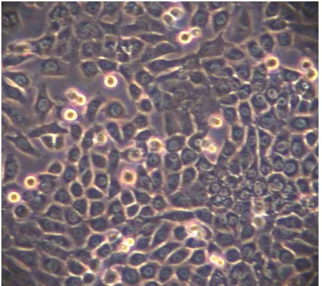
T47D cells obtained from Dr.rer.nat. Marselina
Irasonia Tan, MS (School of Life Sciences, ITB) was grown and propagated in DMEM medium with 10% FBS (Figure 1).
Figure 1. T47D cells were cultured in DMEM medium
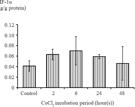
Optimization of CoCl2 incubation
Optimization of CoCl2 induction was performed on T47D cells. T47D cells were induced by 500 µM
CoCl2 for 2, 6, 24, and 48 hours to determine the
most optimal time for incubation. After induction
with various period of time, it was concluded that the concentration of HIF-1α protein were highest in
six hours after incubation. CoCl2 incubation more
than six hours showed a decreased in HIF-1α protein concentrations (Figure 2). Based on this result, the induction of HIF-1α were conducted by six hour
incubation of 500 µM CoCl2.
Protein HIF-1α analysis
HIF-1α protein concentration increased in line with
increased concentrations of CoCl2 given. However there is no significant difference between the ratio of the concentration of HIF-1α induced by 250 µM and 500
µM CoCl2 with the control sample (Figure 3). Strong positive correlation was found between the increase
in the concentration of CoCl2 and the concentration
0 0.02 0.04 0.06 0.08 0.1 0.12
Control 2 6 24 48
HIF-1α concentration (ng) per mg protein
CoCl2 incubation period (hour(s))
Figure 2. Optimation of CoCl2 incubation time. T47D cells
were induced by CoCl2 500 µM for 2, 6, 24, and 48 hours to
determine the most optimal incubation time. It was concluded that the increase in HIF-1α protein concentrations were highest
after induction for six hours
0 0.5 1 1.5 2 2.5 3
Control 250 µM 500 µM
Ratio of HIF-1α
concentration of against control
CoCl2 concentration
Analysis of HIF-1α mRNA
mRNA expression of HIF-1α of T47D cells were increased in line with the increased concentrations
of CoCl2 (Figure 4). However, the mRNA expression of HIF-1α was shown no significant difference between control and CoCl2-induced cells. Strong
positive significant correlations (Pearson, r = 0.998) was found between the increase of HIF-1α protein concentrations and mRNA expression of HIF-1α. Strong positive correlation was also found between HIF-1α protein concentrations expression although it was not significant (Pearson, r = 0,976).
Cytochrome-c analysis
Cytochrome-c concentration increased in line with Figure 3. Effect of CoCl2 induction on HIF-1α protein
concentration. CoCl2 induction led to an increase in HIF-1α protein concentration in T47D cells though it was not significant (p > 0.05)
0 1 2 3 4 5 6 7
Control 250 µM 500 µM
The ratio of normalized HIF-1αmRNA expression
CoCl2 concentration
*
*
Figure 4. Effect of CoCl2 induction on HIF-1α mRNA and
protein expression. CoCl2 induction led to an increase in HIF-1α mRNA expression of T47D cells. There is significant difference of HIF-1α mRNA expression between control and
cells induced by 500 µM CoCl2 (t-test, p = 0.004). Symbol ‘*’ shows statistically significant vs control
0 0.5 1 1.5 2 2.5 3 3.5
Control 250 µM 500 µM
The ratio of cytochrome-c concentration against control
CoCl2 concentration
increased concentrations of CoCl2. However, the concentration of cytochrome-c T47D were not reach statistically difference between control cells
and T47D cells-induced by 250 and 500 µM CoCl2
(t-test, p > 0.1) (Figure 5). There was strong positive correlation between the concentration of CoCl2 and
cytochrome-c (Pearson, r = 0.998).
Survivin expression analysis
Survivin mRNA expression was increased with
increased concentrations of CoCl2. Significant difference was found between the expression of survivin mRNA of control cells and cells induced by
250 µM CoCl2 (t-test, p = 0.005) and 500 µM CoCl2,
respectively (t-test, p = 0.024) (Figure 6).
Figure 5. The influence of the CoCl2 induction on cytochrome-c.
CoCl2 induction led to an increase in cytochrome-c concentration of T47D cells. No significant difference of cytochrome-c concentration between control (without induction) and T47D
cells-induced by CoCl2 (t-test, p > 0.1) HIF-1α (ng/g protein) 0.12 0.10 0.08 0.06 0.04 0.02 0
Control 2 6 24 48
CoCl2 incubation period (hour(s))
Ratio of HIF-1α
concentration against control 3 2.5 2 1.5 1 0.5 0
Control 250 µM 500 µM
CoCl2 concentration
Ratio of normalized HIF-1α mRNA expression 7 6 5 4 3 2 1 0
Control 250 µM 500 µM
CoCl2 concentration
Ratio of cytochrome-c con
-centration against control 3.5 3 2.5 2 1.5 1 0.5 0
Control 250 µM 500 µM
CoCl2 concentration
0 0.51 1.52 2.53 3.5 4 4.5 5
Control 250 µM 500 µM
Ratio of normalized survivin mRNA expression
CoCl2 concentration
*
*
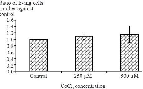
Analysis of cell viability
T47D cells’ viability after induction with CoCl2 did
not show any differences. There is no significant difference of viability ratios between control cells and
cells induced by CoCl2 (t-test, p > 0.05) (Figure 7). Figure 6. Effect of CoCl2 induction on survivin mRNA
expression. CoCl2 induction leads to increased expression of survivin mRNA in T47D cells. Significant difference was found between the expression of survivin mRNA of T47D control cells (without induction) and T47D cells induced by
CoCl2 250 µM. Symbol ‘*’ shows statistically significant vs
control
0.000 0.200 0.400 0.600 0.800 1.000 1.200 1.400 1.600
Control 250 µM 500 µM
Ratio of living cells number against control
CoCl2 concentration
Figure 7. CoCl2 induction effect on cell viability. There was no significant difference between viability ratios of control cells
(without induction) and T47D cells induced by CoCl2 250 and
500 µM (t-test, p > 0.05)
Correlation between HIF and cytochrome-c, survivin and cell viability
There was strong positive correlation between HIF-1α protein and the cytochrome-c (Pearson, r = 0.985)
in T47D cells induced by CoCl2. Strong positive
correlation was obtained between survivin mRNA expression and HIF-1α protein (Pearson, r = 0.999).
In addition, there was a strong positive correlation between survivin mRNA expression and HIF-1α mRNA expression (Pearson, r = 0.994). There was no significant correlation between HIF-1α protein
concentration and cell viability.
DISCUSSION
CoCl2 induction, HIF-1α, and cytochrome-c
From the present study, we found a strong positive correlations between the increase of
CoCl2 concentration and HIF-1α protein as well as mRNA expression in T47D cells. These results were in consistent with the study conducted by Hendrawan, et al15 which found that the gene expression of HIF-1α myocardial cells increased gradually in line with the duration of hypoxia and reached a peak at day 21. We also found a strong positive relationship between the concentration of HIF-1α protein and of cytochrome-c in the
cytoplasm of T47D cells induced by CoCl2, and
between the expression of HIF-1α mRNA and
the concentration of cytochrome-c. These results
are consistent with the study of Ardyanto which show that the apoptotic index of myocardial cells increases with duration of hypoxia, although there is no significant association between increased expression of HIF-1α with the increase in the
apoptotic index.16
HIF-1α protein can induce cell apoptotic through two pathways, the p53 pathway and Bcl-2/ adenovirus E1B 19 kDa-interacting protein 3 (BNIP3) pathway. P53 protein is a tumor
suppressor protein and a transcription factor of
various pro-apoptotic genes. P53 protein can be
induced by many toxic substances, including gamma radiation, ultraviolet, genotoxic drugs
and oxidative stress. Meanwhile, BNIP3 is a pro-apoptotic protein of Bcl-2 proteins family which
have only BH3 domain.9
HIF-1α protein can increase the stability of p53
protein. The increase of p53 protein stability thus inhibit the anti-apoptotic proteins Bcl-2 and
Bcl-XL so that the pro-apoptotic proteins Bax and Bak can be freely increase the permeability
of mitochondrial cell membrane. The increased of mitochondrial cell membrane permeability
will increase the cytochrome-c removal
from mitochondrial intermembrane space to cytoplasm.9
Ratio of normalized survivin mRNA
expression 5 4.5 4 3.5 3 2.5 2 1.5 1 0.5 0
Control 250 µM 500 µM
CoCl2 concentration
Ratio of living cells
number against control
1.6
1.4 1.2 1.0 0.8
0.6
0.4 0.2 0.0
Control 250 µM 500 µM
CoCl2 concentration *
Although p53 was mutated in most cancers, HIF-1α protein can induce apoptosis through BNIP3 pathway. HIF-1α protein can bind to hypoxia response element (HRE) sequence in the promoter region of BNIP3 gene to increase BNIP3 gene
expression.17 The increase of BNIP3 expression would inhibit anti-apoptosis Bcl-2 and Bcl-XL so that the pro-apoptotic protein Bax and Bak
can induce the removal of cytochrome-c from mitochondrial intermembrane space to cytoplasm.
The cytochrome-c in the cytoplasm will bind
to Apaf-1 protein to form apoptosome body.
Apoptosome body will then activate caspase-9, and the active caspase-9 will activate the apoptotic executor caspase-3. Once activated, the apoptotic executor caspase-3 will induce apoptosis.9
Despite causing an increase in pro-apoptotic protein cytochrome-c in cytoplasm, the excessive
expression of HIF-1α in breast carcinoma also strongly associated with poor clinical prognosis. The increased expression of HIF-1α in breast cancer also associated with increased in tumor progression which characterized by excessive vascularization, tumor invasion, tumor size,
tumor metastasis, tumor stage and degree of histopatologic.18
HIF-1α and survivin
There is strong positive correlation between HIF-1α protein concentration and survivin mRNA
expression in this study. We also found strong
positive correlation between HIF-1α mRNA expression and survivin mRNA expression.
Strong positive correlation between the expression of HIF-1α and mRNA survivin expression showed that HIF-1α play a role in increasing the expression of survivin which can
cause the inhibition of apoptosis leading to the
increase of tumor growth. Peng, et al11 found that HIF-1α protein can activate the transcription of survivin gene in breast cancer cell line MCF-7
by binding directly to the promoters of survivin genes.The increase of HIF-1α gene expression
can increase the promoter activity of survivin in
gene in breast cancer cell line MCF-7, but not in normal breast cell line MCF-10A. The results of
this study indicate that the increased expression
of HIF-1α gene is associated with increased
expression of survivin in breast cancer cell line T47D.
Expression of survivin appears to have a very large correlation with the expression of Bcl-2 (anti-apoptotic protein) and the decreased (anti-apoptotic
index in breast cancer.19 Increased survivin expression is also associated with an increased risk
of recurrences, locoregional lymph node invasion and metastasis.10 Survivin can inhibit caspase-9 activation by cytochrome-c. The consequence of
this inhibition on caspase-9 activation is that the
apoptotic executor caspase-3 will be stopped so that the apoptosis will be inhibited.10
The inhibition of HIF-1α mRNA in lung
adenocarcinoma cell line and colorectal carcinoma
cell line downregulates the expression of survivin mRNA.20 It is also found that inhibition of survivin in vitro and in vivo suppressed the potential growth
tumor and increased the sensitivity of tumor cells to chemotherapy agents and also inhibited the occurrence of angiogenesis. Inhibition of survivin
mRNA expression also halted the growth of mice
lymphoma tumor cells.21
It has been known that the increased stability of HIF affects mainly the increase in survivin gene
expression compared to the release of cytochrome-c
into the cytoplasm. Inhibition of HIF is expected
to suppress anti-apoptotic protein survivin so that
apoptotic will continue. Inhibition of HIF-1α would
also suppress the release of cytochrome-c into the
cytoplasm which causes apoptosis does not occur so that cancer cells will continue to proliferate.
This cancer cells proliferation can be inhibited by administration of cytotoxic chemotherapy drugs.22 Sekarutami23 have shown that patients with cervical cancer who received combination of chemoradiotherapy plus hypoxia response modifier therapy (KON) showed the highest response (90.9%) compared to patients who only received chemoradiotherapy (69.2%) or radiotherapy plus KON (63.6%).
HIF and cell viability
Induction of HIF-1α in this study did not affect
the viability of T47D cells. Analysis about the
role of HIF-1α towards the pro-apoptotic protein
cytochrome-c and anti-apoptotic survivin in
previous studies were conducted in separate studies. In addition, the role of HIF-1α toward
pro and anti-apoptotic proteins has never been studied on T47D breast cancer cells line, but
study integrates the two factors: the role of HIF-1α towards pro-apoptotic cytochrome-c and
anti-apoptosis in the same cells, ie breast cancer cells
line T47D. Thus, the role of HIF-1α protein with respect to the balance regulation between pro-apoptotic and anti-pro-apoptotic was directly visible
from no differences in cells viabilities before and
after HIF-1α induction.
The increasing of pro-apoptotic protein
cytochrome-c after induction of HIF-1α by
CoCl2 administration in this study succeeded in inhibiting the increase of T47D breast cancer cells
viabilities. Meanwhile, the increasing of survivin after induction of HIF-1α by CoCl2 was suceeded
to inhibit T47D cells apoptosis. The end result of pro-apoptotic protein cytochrome-c and
anti-apoptotic survivin expression regulation by HIF-1α is the balance of apoptotic pathway, as shown
by the absence of differences in the T47D cells
viabilities before and after HIF-1α induction by
CoCl2.
Based on the above results, it can be concluded that CoCl2 induction caused an increase of HIF-1α that were correlated with the increased of pro-apoptotic cytochrome-c protein as well as anti-apoptotic survivin mRNA level. Therefore, the induction of HIF-1α by CoCl2 did not cause changes in breast cancer cell viability.
In conclusion, this study shown that HIF-1α
induction have no effects on breast cancer cell line T47D viabilities because of the balance regulation
between pro-apoptosis expression cytochrome-c
and anti-apoptosis survivin. Therefore, our results motivate careful assessment of the possibility of
HIF-1α as a target therapy in cancer.
Conflicts of interest
All authors have nothing to disclose.
REFERENCES
1. International Agency for Research on Cancer (IARC) [Internet]. Breast Cancer: Estimated Incidence, Mortality and Prevalence Worldwide in 2012. World Health Organization 2012. Available from: http://globocan.iarc.fr/
Pages/fact_sheets_cancer.aspx
2. Okazaki T, Ebihara S, Asada M, Yamanda S, Niu K, Arai H. Erythropoietin promotes the growth of tumors lacking
its receptor and decreases survival tumor-bearing mice by
enhancing angiogenesis. Neoplasia. 2008;10(9):932-9.
3. Hill R, Bristow RG. The scientific basis of radiotherapy. In: Tannock IF, Hill RP, Bristow RG, Harrington L,
editors. The basic science of oncology. 4th ed. Boston: Mc Graw Hill; 2005. p. 298.
4. Patel SA, Simon MC. Biology of hypoxia-inducible
factor-2α in development and disease. Cell Death Differ. 2008;15(4):628-34.
5. Fandrey J, Gorr TA, Gassmann M. Regulating cellular oxygen sensing by hydroxylation. Cardiovasc Res. 2006;71(4):642-51.
6. Mole DR. Iron homesotasis and its interaction with prolyl hydroxylases. Antioxid Redox Signal. 2010;12(4):445-58. 7. Ke Q, Costa M. Hypoxia-inducible factor-1 (HIF-1). Mol
Pharmacol. 2006;70(5):1469-80.
8. Ardyanto TD, Osaki M, Nagahama Y, Yamaga K, Maeta N, Tamura T, et al. Down-regulation of cobalt-induced HIF-1α expression correlates with cell proliferation and apoptosis in human gastric carcinoma cells. Oncol Report. 2008;19(2):339-43.
9. Greijer AE, van der Wal E. The role of hypoxia inducible factor 1 (HIF-1) in hypoxia induced apoptosis. J Clin Pathol. 2004;57(10):1009-14.
10. Mita AC, Mita MM, Nawrocki ST, Giles FJ. Survivin: key regulator of mitosis and apoptosis and novel target for cancer therapeutics. Clin Cancer Res. 2008;14(16):5000-5.
11. Peng X, Karna P, Cao Z, Jiang B, Zhou M, Yang L. Cross-talk between epidermal growth factor receptor and HIF-1 signal pathways increases resistance to apoptosis
by upregulating survivin gene expression. J Biol Chem.
2006;281(36):25903-14.
12. Li F and Ling X. Survivin study: an update of ‘what is the next wave’? J Cell Physiol. 2006;208(3):476-86.
13. Maxwell P, Salnikow K. HIF-1: An oxygen and metal
responsive transcription factor. Cancer Biol Ther.
2004;3(1):29-35.
14. Aulia G. Modulasi stress oksidatif pada ketahanan hidup sel punca kanker payudara (CD24-/CD44+) melalui induksi rotenone: tinjauan pada ekspresi manganese superoxide dismutase [tesis]. Jakarta: Universitas Indonesia; 2012.
Indonesian.
15. Hendrawan S, Jusman SWA, Ferdinal F, Prijanti AR, Wanandi SI, Sadikin M. Expression of hypoxia inducible factor-1α (HIF-1α) gene and apoptosis in the
heart induced by systemic hypoxia. Med J Indones.
2009;18(2):97-101.
16. Ardyanto TD, Osaki M, Tokuyasu N, Nagahama Y,
Ito H. CoCl2-induced HIF-1α expression correlates
with proliferation and apoptosis in MKN-1 cells: A possible role for the PI3K/Akt pathway. Int J Oncol. 2006;29(3):549-55.
17. Kothari S, Cizeau J, McMillan-Ward E, Israels SJ, Bailes M, Ens K, et al. BNIP3 plays a role in
hypoxic cell death in human epithelial cells that is
inhibited by growth factors EGF and IGF. Oncogene 2003;22(30):4734-44.
18. Galanis A, Pappa A, Giannakakis A, Lanitis E, Dangaj D, Sandaltzopoulos R. Reactive oxygen species and HIF-1 signalling in cancer. Cancer Lett. 2008;266(1):12-20. 19. N, Triaspolitica. "Kanker Payudara: Informasi, Penyebab,
<https://nanyadongdok.blogspot.com/2017/06/kanker-20. Wu XY, Fu ZX, Wang XH. Effect of hypoxia-inducible factor 1-α on Survivin in colorectal cancer. Mol Med Rep. 2010;3(3):409-15.
21. Kanwar JR, Shen WP, Kanwar RK, Berg RW, Krissansen GW. Effects of survivin antagonists on growth of
established tumors and B7-1 immunogene therapy. J Natl
Cancer Inst. 2001;93(20):1541-52.
22. Brizel DM. Chemical modifiers of radiation response. In:
Perez CA, Brady LW, editors. Principles and practice of
radiation oncology. 5th ed. Philadelphia: Lippincott-Raven Publisher; 2008. p. 611-19.
![Hydrogen bonding and π–π stacking interactions in tris(1,10 phenanthroline κ2N,N′)nickel(II) bis{[1 tert butylimidazole 2(3H) thione κS]trichloridonickelate(II)} acetonitrile disolvate](data:image/gif;base64,R0lGODlhAQABAIAAAP///wAAACH5BAEAAAAALAAAAAABAAEAAAICRAEAOw==)